Revision 9
Cell Signaling Technology

Orders: 877-616-CELL (2355) [email protected]

Support: 877-678-TECH (8324)

Web: [email protected] cellsignal.com

3 Trask LaneDanversMassachusetts01923USA
For Research Use Only. Not for Use in Diagnostic Procedures.
Applications:

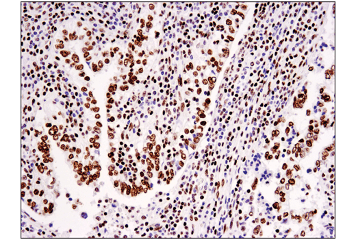
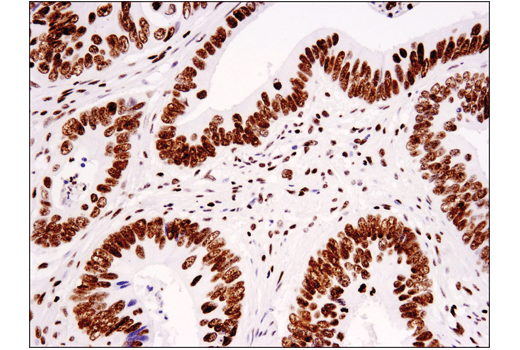
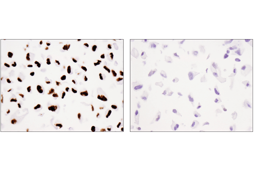
WB, IHC-P, IF-IC, FC-FP, ChIP, ChIP-seq, C&R

REACTIVITY:

H M R Mk

SENSITIVITY:

Endogenous

MW (kDa):

17

Source/Isotype:

Rabbit IgG

UniProt ID:

#P68431

Entrez-Gene Id:

8350

Product Information

Product Usage Information

For optimal ChIP and ChIP-seq results, use 10 μl of antibody and 10 μg of chromatin (approximately 4 x 10⁶ cells) per IP. This antibody has been validated using SimpleChIP® Enzymatic Chromatin IP Kits.

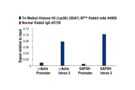
The CUT&RUN dilution was determined using CUT&RUN Assay Kit #86652.
Application Dilution
Western Blotting 1:1000
Immunohistochemistry (Paraffin) 1:50 - 1:200
Immunofluorescence (Immunocytochemistry) 1:25600 - 1:102400
Flow Cytometry (Fixed/Permeabilized) 1:50 - 1:200
Chromatin IP 1:50
Chromatin IP-seq 1:50
CUT&RUN 1:50

Storage

Supplied in 10 mM sodium HEPES (pH 7.5), 150 mM NaCl, 100 µg/ml BSA, 50% glycerol and less than 0.02% sodium azide. Store at –20°C. Do not aliquot the antibody.

For a carrier free (BSA and azide free) version of this product see product #53775.

Specificity / Sensitivity

Tri-Methyl-Histone H3 (Lys36) (D5A7) XP® Rabbit mAb detects endogenous levels of histone H3 only when tri-methylated on Lys36. The antibody does not cross-react with non-methylated, mono-methylated, or di-methylated Lys36. In addition, the antibody does not cross-react with histone H3 methylated at Lys4, Lys9, Lys27 or histone H4 methylated at Lys20.

Species Reactivity:

Human, Mouse, Rat, Monkey

Species predicted to react based on 100% sequence homology

Hamster, Chicken, D. melanogaster, Xenopus, Zebrafish, Bovine

Source / Purification

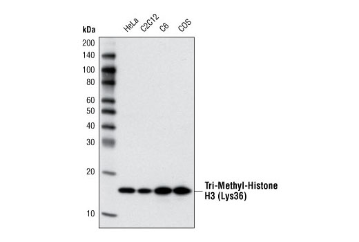
Monoclonal antibody is produced by immunizing animals with a synthetic peptide corresponding to the amino terminus of histone H3 in which Lys36 is tri-methylated.

Background

The nucleosome, made up of four core histone proteins (H2A, H2B, H3, and H4), is the primary building block of chromatin. Originally thought to function as a static scaffold for DNA packaging, histones have now been shown to be dynamic proteins, undergoing multiple types of post-translational modifications, including acetylation, phosphorylation, methylation, and ubiquitination (1). Histone methylation is a major determinant for the formation of active and inactive regions of the genome and is crucial for the proper programming of the genome during development (2,3). Arginine methylation of histones H3 (Arg2, 17, 26) and H4 (Arg3) promotes transcriptional activation and is mediated by a family of protein arginine methyltransferases (PRMTs), including the co-activators PRMT1 and CARM1 (PRMT4) (4). In contrast, a more diverse set of histone lysine methyltransferases has been identified, all but one of which contain a conserved catalytic SET domain originally identified in the Drosophila Su(var)3-9, Enhancer of zeste, and Trithorax proteins. Lysine methylation occurs primarily on histones H3 (Lys4, 9, 27, 36, 79) and H4 (Lys20) and has been implicated in both transcriptional activation and silencing (4). Methylation of these lysine residues coordinates the recruitment of chromatin modifying enzymes containing methyl-lysine binding modules such as chromodomains (HP1, PRC1), PHD fingers (BPTF, ING2), tudor domains (53BP1), and WD-40 domains (WDR5) (5-8). The discovery of histone demethylases, such as PADI4, LSD1, JMJD1, JMJD2, and JHDM1, has shown that methylation is a reversible epigenetic marker (9).

  1. Peterson, C.L. and Laniel, M.A. (2004) Curr Biol 14, R546-51.
  2. Kubicek, S. et al. (2006) Ernst Schering Res Found Workshop, 1-27.
  3. Lin, W. and Dent, S.Y. (2006) Curr Opin Genet Dev 16, 137-42.
  4. Lee, D.Y. et al. (2005) Endocr Rev 26, 147-70.
  5. Daniel, J.A. et al. (2005) Cell Cycle 4, 919-26.
  6. Shi, X. et al. (2006) Nature 442, 96-9.
  7. Wysocka, J. et al. (2006) Nature 442, 86-90.
  8. Wysocka, J. et al. (2005) Cell 121, 859-72.
  9. Trojer, P. and Reinberg, D. (2006) Cell 125, 213-7.

Species Reactivity

Species reactivity is determined by testing in at least one approved application (e.g., western blot).

Western Blot Buffer

IMPORTANT: For western blots, incubate membrane with diluted primary antibody in 5% w/v BSA, 1X TBS, 0.1% Tween® 20 at 4°C with gentle shaking, overnight.

Applications Key

WB: Western Blotting IHC-P: Immunohistochemistry (Paraffin) IF-IC: Immunofluorescence (Immunocytochemistry) FC-FP: Flow Cytometry (Fixed/Permeabilized) ChIP: Chromatin IP ChIP-seq: Chromatin IP-seq C&R: CUT&RUN

Cross-Reactivity Key

H: human M: mouse R: rat Hm: hamster Mk: monkey Vir: virus Mi: mink C: chicken Dm: D. melanogaster X: Xenopus Z: zebrafish B: bovine Dg: dog Pg: pig Sc: S. cerevisiae Ce: C. elegans Hr: horse GP: Guinea Pig Rab: rabbit All: all species expected

Trademarks and Patents

Cell Signaling Technology is a trademark of Cell Signaling Technology, Inc.
SignalStain is a registered trademark of Cell Signaling Technology, Inc.
XP is a registered trademark of Cell Signaling Technology, Inc.
All other trademarks are the property of their respective owners. Visit cellsignal.com/trademarks for more information.

限制使用

除非 CST 的合法授书代表以书面形式书行明确同意,否书以下条款适用于 CST、其关书方或分书商提供的书品。 任何书充本条款或与本条款不同的客书条款和条件,除非书 CST 的合法授书代表以书面形式书独接受, 否书均被拒书,并且无效。

专品专有“专供研究使用”的专专或专似的专专声明, 且未专得美国食品和专品管理局或其他外国或国内专管机专专专任何用途的批准、准专或专可。客专不得将任何专品用于任何专断或治专目的, 或以任何不符合专专声明的方式使用专品。CST 专售或专可的专品提供专作专最专用专的客专,且专用于研专用途。将专品用于专断、专防或治专目的, 或专专售(专独或作专专成)或其他商专目的而专专专品,均需要 CST 的专独专可。客专:(a) 不得专独或与其他材料专合向任何第三方出售、专可、 出借、捐专或以其他方式专专或提供任何专品,或使用专品制造任何商专专品,(b) 不得复制、修改、逆向工程、反专专、 反专专专品或以其他方式专专专专专品的基专专专或技专,或使用专品开专任何与 CST 的专品或服专专争的专品或服专, (c) 不得更改或专除专品上的任何商专、商品名称、徽专、专利或版专声明或专专,(d) 只能根据 CST 的专品专售条款和任何适用文档使用专品, (e) 专遵守客专与专品一起使用的任何第三方专品或服专的任何专可、服专条款或专似专专

Revision 9
#4909

Tri-Methyl-Histone H3 (Lys36) (D5A7) XP® Rabbit mAb

Western Blotting Image 1: Tri-Methyl-Histone H3 (Lys36) (D5A7) XP® Rabbit mAb Expand Image
使用 Tri-Methyl-Histone H3 (Lys36) (D5A7) XP® Rabbit mAb 对不同细胞系的提取物进行蛋白质印迹分析。
Immunohistochemistry Image 1: Tri-Methyl-Histone H3 (Lys36) (D5A7) XP® Rabbit mAb Expand Image
使用 Tri-Methyl-Histone H3(K36) (D5A7) XP(R) Rabbit mAb,对石蜡包埋的人乳腺浸润性乳头状癌组织进行免疫组织化学分析。
Immunohistochemistry Image 2: Tri-Methyl-Histone H3 (Lys36) (D5A7) XP® Rabbit mAb Expand Image
使用 Tri-Methyl-Histone H3(K36) (D5A7) XP(R) Rabbit mAb 对石蜡包埋的人结肠癌细胞进行免疫组织化学分析。
Immunohistochemistry Image 3: Tri-Methyl-Histone H3 (Lys36) (D5A7) XP® Rabbit mAb Expand Image
使用 Tri-Methyl-Histone H3(K36) (D5A7) XP(R) Rabbit mAb 对石蜡包埋的 786-O 细胞沉淀物(左图,阳性)或 A498 细胞沉淀物(右图,阴性)进行免疫组织化学分析。注意,A498 细胞系出现 SETD2 突变。
Immunohistochemistry Image 4: Tri-Methyl-Histone H3 (Lys36) (D5A7) XP® Rabbit mAb Expand Image
在存在非甲基肽(左图)或 K36 三甲基化肽(右图)的情况下,使用 Tri-Methyl-Histone H3 (K36) (D5A7) XP(R) Rabbit Antibody,对石蜡包埋的人卵巢浆液性乳头状癌细胞进行免疫组织化学分析。
Immunohistochemistry Image 5: Tri-Methyl-Histone H3 (Lys36) (D5A7) XP® Rabbit mAb Expand Image
使用 Tri-Methyl-Histone H3(K36) (D5A7) XP(R) Rabbit mAb,对石蜡包埋的人类前列腺癌进行免疫组织化学分析。
Immunofluorescence Image 1: Tri-Methyl-Histone H3 (Lys36) (D5A7) XP® Rabbit mAb Expand Image
使用 Tri-Methyl-Histone H3 (Lys36) (D5A7) XP® Rabbit mAb(绿色)和 COX IV (4D11-B3-E8) Mouse mAb #11967(红色)对 Hela 细胞进行共聚焦免疫荧光分析。
Flow Cytometry Image 1: Tri-Methyl-Histone H3 (Lys36) (D5A7) XP® Rabbit mAb Expand Image
使用 Tri-Methyl-Histone H3 (Lys36) (D5A7) XP® Rabbit mAb(实线)或浓度匹配的 Rabbit (DA1E) mAb IgG XP® Isotype Control #3900(虚线)对 A498 细胞(蓝色)和 786-O 细胞(绿色)进行流式细胞分析。Anti-rabbit IgG (H+L), F(ab')2 Fragment (Alexa Fluor® 488 Conjugate) #4412 用作二抗。
Chromatin Immunoprecipitation Image 1: Tri-Methyl-Histone H3 (Lys36) (D5A7) XP® Rabbit mAb Expand Image
使用 SimpleChIP® Plus Enzymatic Chromatin IP Kit (Magnetic Beads) #9005 对 HeLa 细胞中提取的交联染色质,在加入 Tri-Methyl-Histone H3 (Lys36) (D5A7) XP® Rabbit mAb 后进行染色质免疫沉淀分析。使用 SimpleChIP® ChIP-seq DNA Library Prep Kit for Illumina® #56795 制成 DNA 库。图中显示与 ACTG1/γ-Actin 之间的结合,ACTG1/γ-Actin 是一种已知的 H3K36me3 靶标基因(参见包含 ChIP-qPCR 数据的其他图)。
Chromatin Immunoprecipitation Image 2: Tri-Methyl-Histone H3 (Lys36) (D5A7) XP® Rabbit mAb Expand Image
使用 SimpleChIP® Plus Enzymatic Chromatin IP Kit (Magnetic Beads) #9005 对 HeLa 细胞中提取的交联染色质,在加入 Tri-Methyl-Histone H3 (Lys36) (D5A7) XP® Rabbit mAb 后进行染色质免疫沉淀分析。使用 SimpleChIP® ChIP-seq DNA Library Prep Kit for Illumina® #56795 制成 DNA 库。该图显示在染色体 17(上图)和 H3K36me3 的已知靶基因 ACTG1/γ-Actin(下图)内结合(参见包含 CUT&RUN-qPCR 数据的其他结果图)。
Chromatin Immunoprecipitation Image 3: Tri-Methyl-Histone H3 (Lys36) (D5A7) XP® Rabbit mAb Expand Image
使用 SimpleChIP® Plus Enzymatic Chromatin IP Kit (Magnetic Beads) #9005 对 HeLa 细胞的交联染色质与  Tri-Methyl-Histone H3 (Lys36) (D5A7) XP® Rabbit mAb 或 Normal Rabbit IgG #2729 进行染色质免疫沉淀分析。使用 SimpleChIP® Human γ-Actin Promoter Primers #5037、SimpleChIP® Human γ-Actin Intron 3 Primers #5047、SimpleChIP® Human GAPDH Promoter Primers #4471 和 SimpleChIP® Human GAPDH Intron 2 Primers #4478,通过实时 PCR 对富集的 DNA 进行定量。将每份样品中免疫沉淀的 DNA 的量表现为相对于所输入染色质总量(等于 1)的信号。
CUT and RUN Image 1: Tri-Methyl-Histone H3 (Lys36) (D5A7) XP® Rabbit mAb Expand Image
使用 CUT&RUN Assay Kit #86652 对 HeLa 细胞和 Tri-Methyl-Histone H3 (Lys36) (D5A7) XP® Rabbit mAb 进行 CUT&RUN 检测。使用 DNA Library Prep Kit for Illumina (ChIP-seq, CUT&RUN) #56795 制备 DNA 文库。该图显示在 H3K36me3 的已知靶基因 JTB 内的结合(参见包含 CUT&RUN-qPCR 数据的其他结果图)。
CUT and RUN Image 2: Tri-Methyl-Histone H3 (Lys36) (D5A7) XP® Rabbit mAb Expand Image
使用 CUT&RUN Assay Kit #86652 对 HeLa 细胞和 Tri-Methyl-Histone H3 (Lys36) (D5A7) XP® Rabbit mAb 进行 CUT&RUN 检测。使用 DNA Library Prep Kit for Illumina (ChIP-seq, CUT&RUN) #56795 制备 DNA 文库。该图显示在 ATCG1 基因(上图)和 H3K36me3 的已知靶基因 JTB(下图)内结合(参见包含 CUT&RUN-qPCR 数据的其他结果图)。
CUT and RUN Image 3: Tri-Methyl-Histone H3 (Lys36) (D5A7) XP® Rabbit mAb Expand Image
使用 CUT&RUN Assay Kit #86652 对 Hela 细胞和 Tri-Methyl-Histone H3 (Lys36) (D5A7) XP® Rabbit mAb 或 Rabbit (DA1E) mAb IgG XP® Isotype Control (CUT&RUN) #66362 进行 CUT&RUN 检测。使用 human JTB exon 1 和 SimpleChIP® Human α Satellite Repeat Primers #4486,通过实时 PCR 对富集的 DNA 进行定量分析。将每份样品中免疫沉淀的 DNA 的量表现为相对于所输入染色质总量(等于 1)的信号。
Product Image 1: Tri-Methyl-Histone H3 (Lys36) (D5A7) XP® Rabbit mAb Expand Image
使用肽 ELISA 检测 Tri-Methyl-Histone H3 (Lys36) (D5A7) XP® Rabbit mAb 特异性。图表显示在不同竞争肽的浓度不断升高的情况下,抗体与预包被三甲基化组蛋白 H3 (Lys36) 肽的结合情况。如图所示,只有三甲基化组蛋白 H3 (Lys36) 肽在与抗体的结合占优势。