| Product Includes | Product # | Quantity | Mol. Wt | Isotype/Source |
|---|---|---|---|---|
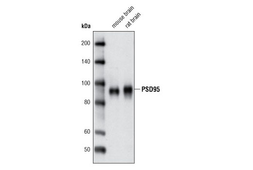
| PSD95 (D74D3) XP® Rabbit mAb | 3409 | 20 µl | 95 kDa | Rabbit IgG |
| Synaptophysin (D8F6H) XP® Rabbit mAb | 36406 | 20 µl | 38 kDa | Rabbit IgG |
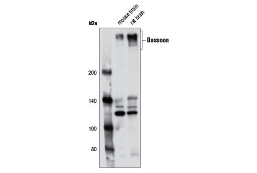
| Bassoon (D63B6) Rabbit mAb | 6897 | 20 µl | 420 kDa | Rabbit IgG |
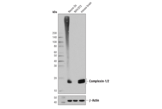
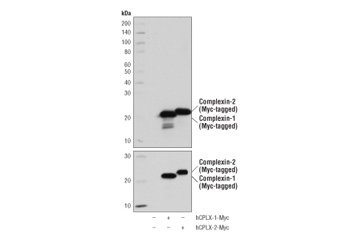
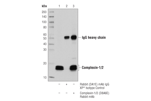
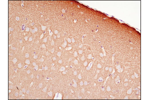
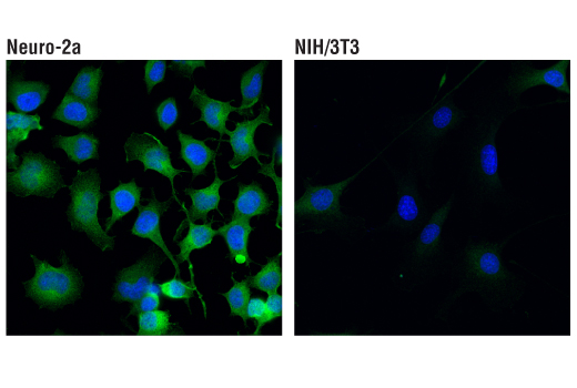
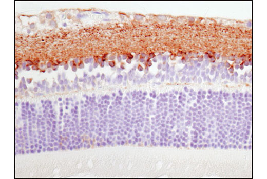
| Complexin-1/2 (D8A6E) Rabbit mAb | 28070 | 20 µl | 14-16 kDa | Rabbit IgG |
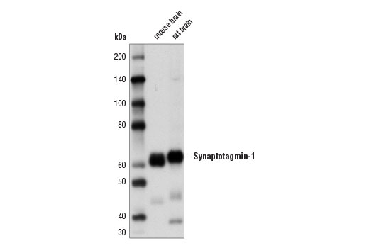
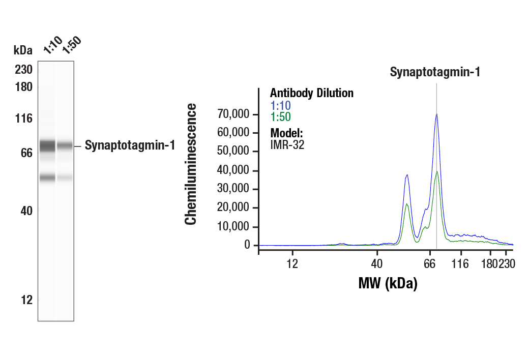
| Synaptotagmin-1 (D33B7) Rabbit mAb | 14558 | 20 µl | 60 kDa | Rabbit IgG |
| SHANK3 (D5K6R) Rabbit mAb | 64555 | 20 µl | 220 kDa | Rabbit IgG |
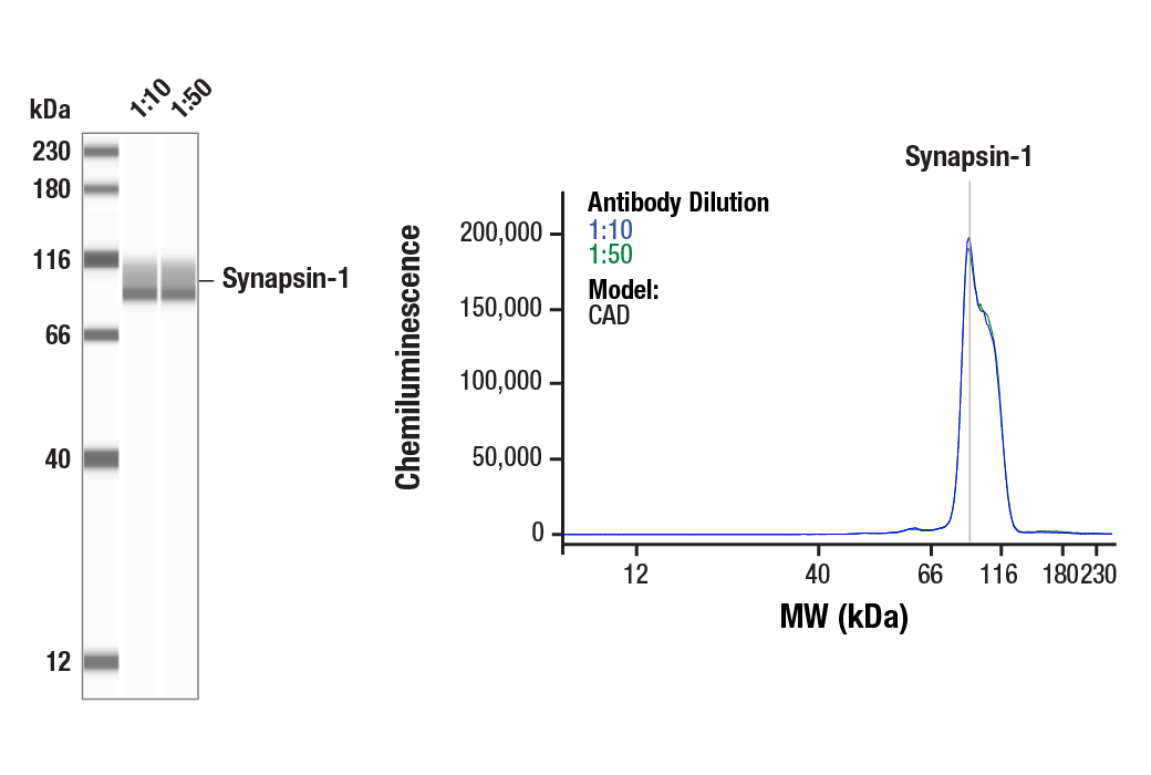
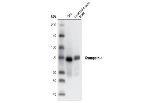
| Synapsin-1 (D12G5) XP® Rabbit mAb | 5297 | 20 µl | 77 kDa | Rabbit IgG |
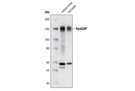
| SynGAP (D78B11) Rabbit mAb | 5540 | 20 µl | 140 kDa | Rabbit IgG |
| Anti-rabbit IgG, HRP-linked Antibody | 7074 | 100 µl | Goat |
Please visit cellsignal.com for individual component applications, species cross-reactivity, dilutions, protocols, and additional product information.
Description
The Synaptic Neuron Marker Antibody Sampler Kit provides an economical means of detecting presynaptic and postsynaptic proteins by western blot. This kit includes enough primary antibodies to perform at least two western blot experiments with each primary antibody.
Storage
Background
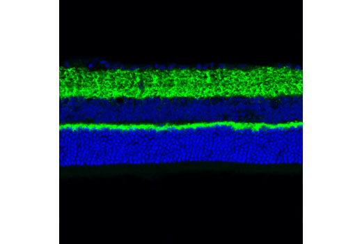
Synaptophysin (SYP) is a neuronal synaptic vesicle glycoprotein that is expressed in neuroendocrine cells and neoplasms (1). Synapsin-1 is a neuronal phospho-protein localized to presynaptic terminals. Synapsin-1 plays an important role in synapse formation, neurotransmitter regulation, and regulation of synaptic vesicle fusion and trafficking (2,3). Synaptotagmin-1 (SYT1) is an integral membrane protein found in synaptic vesicles thought to play a role in vesicle trafficking and exocytosis (4). Complexin isoforms 1 and 2 are small synaptic proteins that bind to SNARE complexes, responsible for regulating exocytosis and synaptic vesicle fusion (5). SynGAP is a synaptic GTPase-activating protein selectively expressed in the brain and found at higher concentrations specifically at excitatory synapses in the mammalian forebrain. SynGAP interacts with the PDZ domains of PSD95, a postsynaptic scaffolding protein that couples SynGAP to NMDA receptors (6). PSD95 is involved in the assembly and function of the postsynaptic density (PSD) complex (7,8). SHANK proteins act as scaffolds at the neuronal PSD (9), where they play a critical role in PSD assembly of excitatory synapses during development (10). Bassoon (BSN) is a scaffolding protein component of the synaptic ribbon and of the cytomatrix at the active zones of both excitatory and inhibitory synapses with a presumptive role in orchestrating events of the synaptic vesicle cycle (11-13). Together, these proteins can be used to measure presynaptic and postsynaptic proteins and synaptic development under normal and disease conditions.
- Wiedenmann, B. et al. (1986) Proc Natl Acad Sci U S A 83, 3500-4.
- Mirza, F.J. and Zahid, S. (2018) Neurosci Bull 34, 349-358.
- Takei, Y. et al. (1995) J Cell Biol 131, 1789-800.
- Fukuda, M. and Mikoshiba, K. (2001) Biochem Biophys Res Commun 281, 1226-33.
- Chang, S. et al. (2015) J Neurosci 35, 8272-90.
- Kim, J.H. et al. (1998) Neuron 20, 683-91.
- Cao, J. et al. (2005) J Cell Biol 168, 117-26.
- Chetkovich, D.M. et al. (2002) J Neurosci 22, 6415-25.
- Grabrucker, A.M. et al. (2011) Trends Cell Biol 21, 594-603.
- Boeckers, T.M. et al. (1999) J Neurosci 19, 6506-18.
- Winter, C. et al. (1999) Genomics 57, 389-97.
- Hallermann, S. et al. (2010) Neuron 68, 710-23.
- Frank, T. et al. (2010) Neuron 68, 724-38.
Background References
Trademarks and Patents
限制使用
除非 CST 的合法授书代表以书面形式书行明确同意,否书以下条款适用于 CST、其关书方或分书商提供的书品。 任何书充本条款或与本条款不同的客书条款和条件,除非书 CST 的合法授书代表以书面形式书独接受, 否书均被拒书,并且无效。
专品专有“专供研究使用”的专专或专似的专专声明, 且未专得美国食品和专品管理局或其他外国或国内专管机专专专任何用途的批准、准专或专可。客专不得将任何专品用于任何专断或治专目的, 或以任何不符合专专声明的方式使用专品。CST 专售或专可的专品提供专作专最专用专的客专,且专用于研专用途。将专品用于专断、专防或治专目的, 或专专售(专独或作专专成)或其他商专目的而专专专品,均需要 CST 的专独专可。客专:(a) 不得专独或与其他材料专合向任何第三方出售、专可、 出借、捐专或以其他方式专专或提供任何专品,或使用专品制造任何商专专品,(b) 不得复制、修改、逆向工程、反专专、 反专专专品或以其他方式专专专专专品的基专专专或技专,或使用专品开专任何与 CST 的专品或服专专争的专品或服专, (c) 不得更改或专除专品上的任何商专、商品名称、徽专、专利或版专声明或专专,(d) 只能根据 CST 的专品专售条款和任何适用文档使用专品, (e) 专遵守客专与专品一起使用的任何第三方专品或服专的任何专可、服专条款或专似专专