| Product Includes | Product # | Quantity | Mol. Wt | Isotype/Source |
|---|---|---|---|---|
| CD14 (D7A2T) Rabbit mAb (IHC Formulated) | 75181 | 20 µl | Rabbit IgG | |
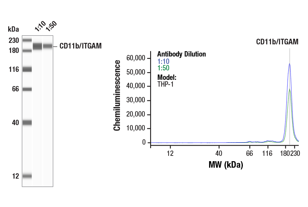
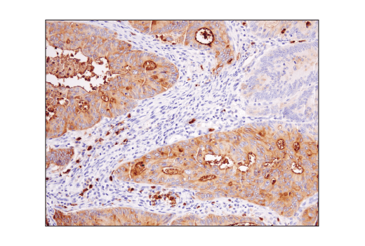
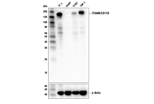
| CD11b/ITGAM (D6X1N) Rabbit mAb | 49420 | 20 µl | 170 kDa | Rabbit IgG |
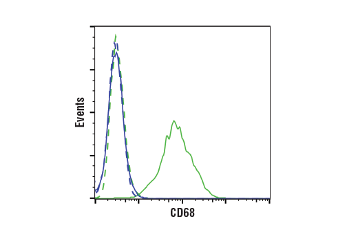
| CD68 (D4B9C) XP® Rabbit mAb | 76437 | 20 µl | Rabbit IgG | |
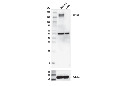
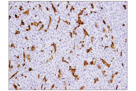
| CD163 (D6U1J) Rabbit mAb | 93498 | 20 µl | 160, 170 kDa | Rabbit IgG |
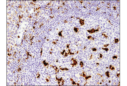
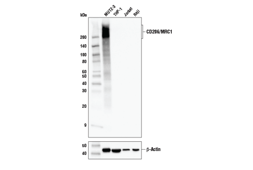
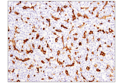
| CD206/MRC1 (E2L9N) Rabbit mAb | 91992 | 20 µl | 190-250 kDa | Rabbit IgG |
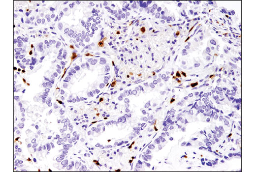
| CSF-1R/M-CSF-R (E4T8Z) Rabbit mAb | 28917 | 20 µl | 140-200 kDa | Rabbit IgG |
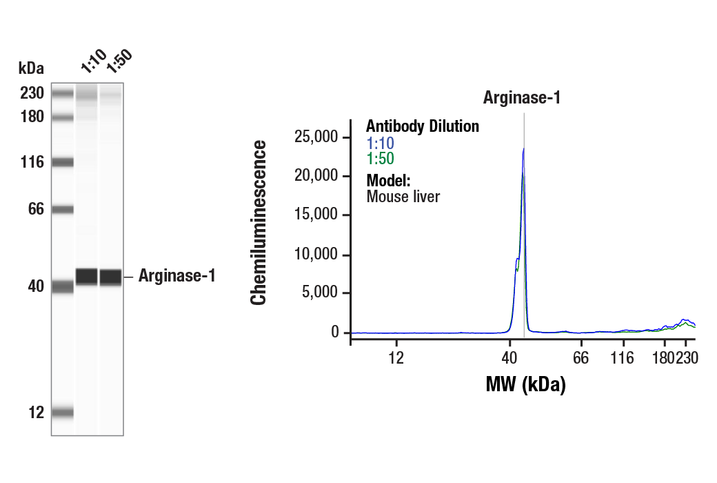
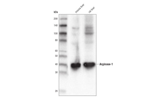
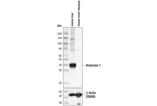
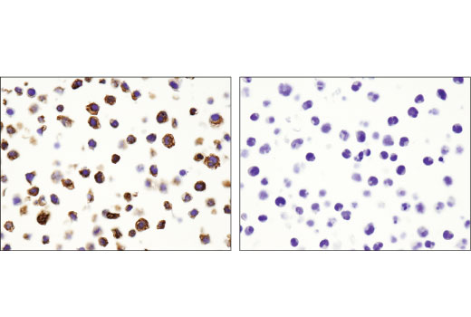
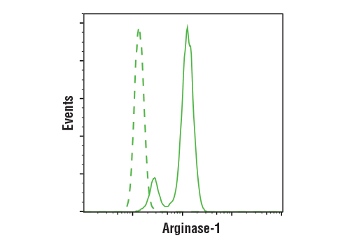
| Arginase-1 (D4E3M™) XP® Rabbit mAb | 93668 | 20 µl | 40 kDa | Rabbit IgG |
| MHC Class II (LGII-612.14) Mouse mAb | 68258 | 20 µl | 25-35, 50-65 kDa | Mouse IgG1 |
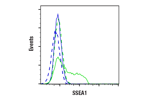
| CD15/SSEA1 (MC480) Mouse mAb | 4744 | 20 µl | N/A kDa | Mouse IgM |
Please visit cellsignal.com for individual component applications, species cross-reactivity, dilutions, protocols, and additional product information.
Description
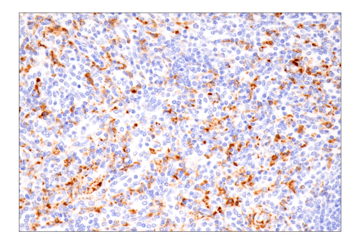
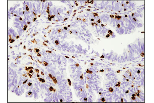
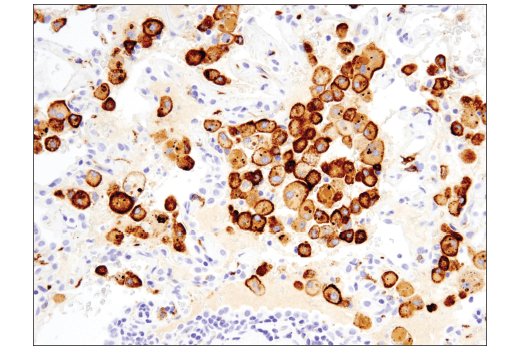
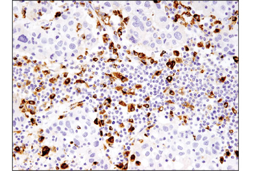
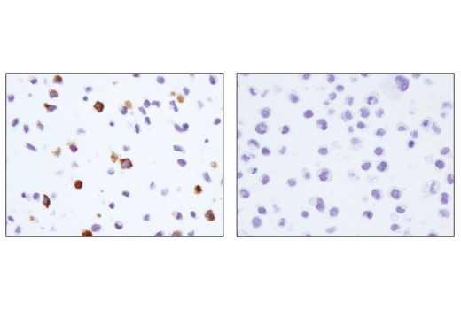
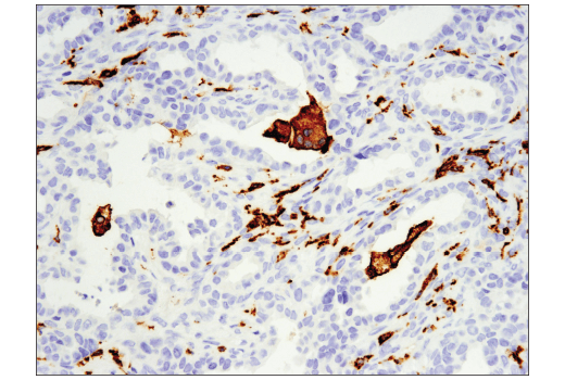
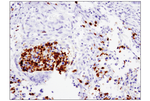
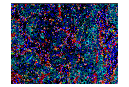
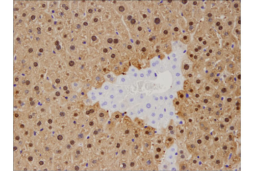
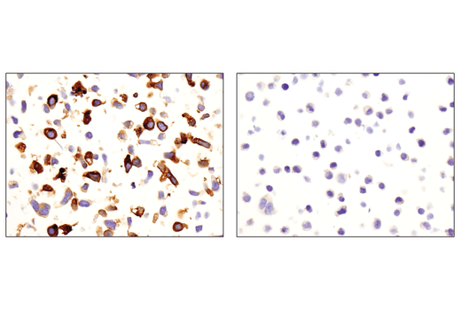
The Suppressive Myeloid Cell Phenotyping IHC Antibody Sampler Kit provides an economical means of detecting the accumulation of immune cell types in formalin-fixed, paraffin-embedded tissue samples.
Storage
Background
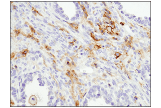
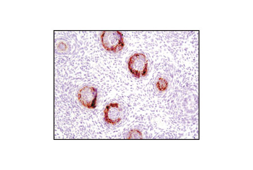
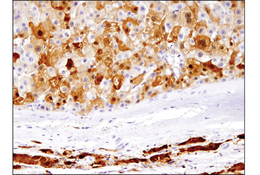
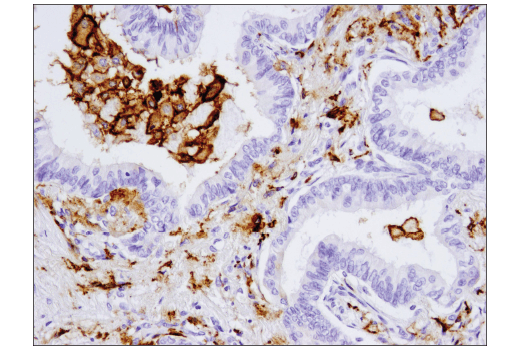
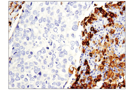
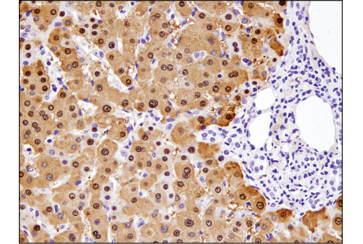
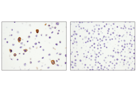
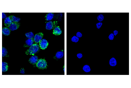
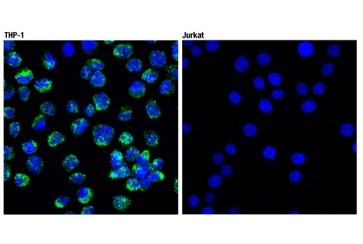
A combination of multiple biomarkers are required to characterize the phenotype of myeloid cell lineages. Cluster of differentiation molecule 14 (CD14) is a leucine-rich repeat-containing pattern recognition receptor with expression largely restricted to the monocyte/macrophage cell lineage (1), but can be unregulated on polymorphonuclear as well as nonmyeloid cells such as B cells and gingival fibroblasts (2,3). CD11b (Integrin alpha M or ITGAM) is a transmembrane protein forming heterodimers that are composed of α and β subunits (4). CD11b is expressed by, and commonly used as a marker for myeloid lineage cells, including neutrophils, monocytes, macrophages, dendritic cells, and microglia (5), but has also been detected on a subset of B cells (6-8). CD68 (macrosialin) is a heavily glycosylated transmembrane protein that is expressed by and commonly used as a marker for monocytes and macrophages (9,10), but there is also evidence of non-myeloid cell expression (11). The CD15 carbohydrate epitope is preferentially expressed in mature human neutrophils, monocytes, and all myeloid cells from the promyelocyte stage onwards, making it a useful cell surface marker (12-14). It is also expressed in some tissues, such as epithelial cells of intestinal tissues (15,16), and in certain neurons and glial cells in the central nervous system (17).
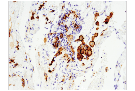
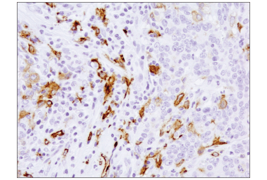
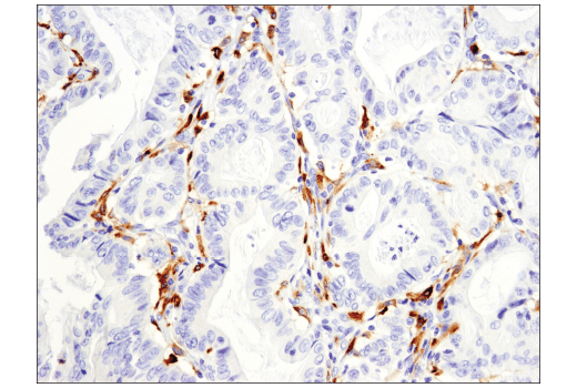
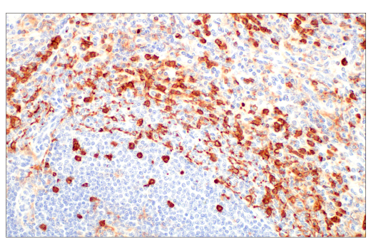
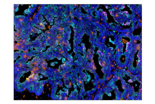
CD163 is a transmembrane scavenger receptor expressed on the macrophage surface. It has 9 B-type SRCR extracellular domains mediating serum haptoglobin clearing/endocytosis, pathogen binding and signal transduction, and calcium binding (18,19). The mannose receptor (CD206/MR/CLEC13D/MMR/MRC1/Macrophage mannose receptor 1) is an endocytic receptor expressed by populations of dendritic cells, macrophages, and nonvascular endothelium (20). CD206/MRC1 receptor functions include a role in antigen cross-presentation, clearance of endogenous proteins, pathogen detection and trafficking through lymphatic vessels (21-24). Macrophage-colony stimulating factor (M-CSF, CSF-1) receptor is an integral membrane tyrosine kinase encoded by the c-fms proto-oncogene. M-CSF receptor is expressed in monocytes (macrophages and their progenitors) and drives growth and development of this blood cell lineage (25,26). CD163, CD206, and M-CSF receptors are used as surface markers of M2 type macrophages, including M2 type tumor associated macrophages (TAMs), which facilitate cancer progression by secreting cytokines to promote angiogenesis, immunosuppression, and metastasis (20,27,28). Arginase-1 catalyzes the final step of the urea cycle converting L-arginine to L-ornithine and urea (29). Myeloid-derived suppressor cells express high levels of arginase-1, increasing the catabolism of L-arginine resulting in L-arginine depletion in the inflammatory microenvironment of cancer. The reduced availability of L-arginine suppresses T cell proliferation and function and thus contributes to tumor progression (30,31).
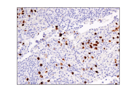
Major histocompatibility complex class II (MHC class II) molecules are heterodimeric, transmembrane glycoproteins expressed on the surface of antigen-presenting cells such as macrophages, dendritic cells, and B cells. Expression can also be induced through interferon-γ signaling (32). Prior to being displayed on the cell membrane, MHC class II molecules are loaded with exogenous peptide antigens approximately 15-24 amino acids in length that were derived from endocytosed extracellular proteins digested in the lysosome. Antigen-presentation through MHC class II is required for T cell activation during the immune response to extracellular pathogens (33). High expression of MHC class II on myeloid cell lineages is used as a surface marker of M1 type macrophages, including M1 type TAMs, which can assist in tumor eradication by secreting cytokines to activate anti-tumor immune responses, and inhibit angiogenesis and metastasis (27,28).
- Wright, S.D. et al. (1991) J Exp Med 173, 1281-6.
- Schumann, R.R. et al. (1994) Med Microbiol Immunol 183, 279-97.
- Ziegler-Heitbrock, H.W. and Ulevitch, R.J. (1993) Immunol Today 14, 121-5.
- Solovjov, D.A. et al. (2005) J Biol Chem 280, 1336-45.
- Murray, P.J. and Wynn, T.A. (2011) Nat Rev Immunol 11, 723-37.
- Kawai, K. et al. (2005) J Allergy Clin Immunol 116, 192-7.
- Payne, D. Nurs Times 92, 18.
- Merad, M. et al. (2013) Annu Rev Immunol 31, 563-604.
- Rabinowitz, S.S. and Gordon, S. (1991) J Exp Med 174, 827-36.
- Ramprasad, M.P. et al. (1995) Proc Natl Acad Sci U S A 92, 9580-4.
- Gottfried, E. et al. (2008) Scand J Immunol 67, 453-63.
- Oriol, R. et al. (1986) Vox Sang 51, 161-71.
- Hanjan, S.N. et al. (1982) Clin Immunol Immunopathol 23, 172-88.
- Civin, C.I. et al. (1981) Blood 57, 842-5.
- Hakomori, S. et al. (1984) J Biol Chem 259, 4672-80.
- Itzkowitz, S.H. et al. (1986) Cancer Res 46, 2627-32.
- Streit, A. et al. (1996) J Neurochem 66, 834-44.
- Graversen, J.H. and Moestrup, S.K. (2015) Membranes (Basel) 5, 228-52.
- Etzerodt, A. and Moestrup, S.K. (2013) Antioxid Redox Signal 18, 2352-63.
- Martinez-Pomares, L. (2012) J Leukoc Biol 92, 1177-86.
- Burgdorf, S. et al. (2006) J Immunol 176, 6770-6.
- Lee, S.J. et al. (2002) Science 295, 1898-901.
- Milone, M.C. and Fitzgerald-Bocarsly, P. (1998) J Immunol 161, 2391-9.
- Marttila-Ichihara, F. et al. (2008) Blood 112, 64-72.
- Stanley, E.R. et al. (1978) Nature 274, 168-70.
- Bourette, R.P. and Rohrschneider, L.R. (2000) Growth Factors 17, 155-66.
- Komohara, Y. et al. (2014) Cancer Sci 105, 1-8.
- Mills, C.D. et al. (2000) J Immunol 164, 6166-73.
- Wu, G. and Morris, S.M. (1998) Biochem J 336 ( Pt 1), 1-17.
- Gabrilovich, D.I. and Nagaraj, S. (2009) Nat Rev Immunol 9, 162-74.
- Raber, P. et al. (2012) Immunol Invest 41, 614-34.
- Ting, J.P. and Trowsdale, J. (2002) Cell 109 Suppl, S21-33.
- Cresswell, P. (1994) Annu Rev Immunol 12, 259-93.
Background References
Trademarks and Patents
限制使用
除非 CST 的合法授书代表以书面形式书行明确同意,否书以下条款适用于 CST、其关书方或分书商提供的书品。 任何书充本条款或与本条款不同的客书条款和条件,除非书 CST 的合法授书代表以书面形式书独接受, 否书均被拒书,并且无效。
专品专有“专供研究使用”的专专或专似的专专声明, 且未专得美国食品和专品管理局或其他外国或国内专管机专专专任何用途的批准、准专或专可。客专不得将任何专品用于任何专断或治专目的, 或以任何不符合专专声明的方式使用专品。CST 专售或专可的专品提供专作专最专用专的客专,且专用于研专用途。将专品用于专断、专防或治专目的, 或专专售(专独或作专专成)或其他商专目的而专专专品,均需要 CST 的专独专可。客专:(a) 不得专独或与其他材料专合向任何第三方出售、专可、 出借、捐专或以其他方式专专或提供任何专品,或使用专品制造任何商专专品,(b) 不得复制、修改、逆向工程、反专专、 反专专专品或以其他方式专专专专专品的基专专专或技专,或使用专品开专任何与 CST 的专品或服专专争的专品或服专, (c) 不得更改或专除专品上的任何商专、商品名称、徽专、专利或版专声明或专专,(d) 只能根据 CST 的专品专售条款和任何适用文档使用专品, (e) 专遵守客专与专品一起使用的任何第三方专品或服专的任何专可、服专条款或专似专专