WB, W-S, IHC-P, IF-IC, FC-FP
H M R Mk B Pg
Endogenous
90
Rabbit IgG
#P51812
6197
Product Information
Product Usage Information
| Application | Dilution |
|---|---|
| Western Blotting | 1:1000 |
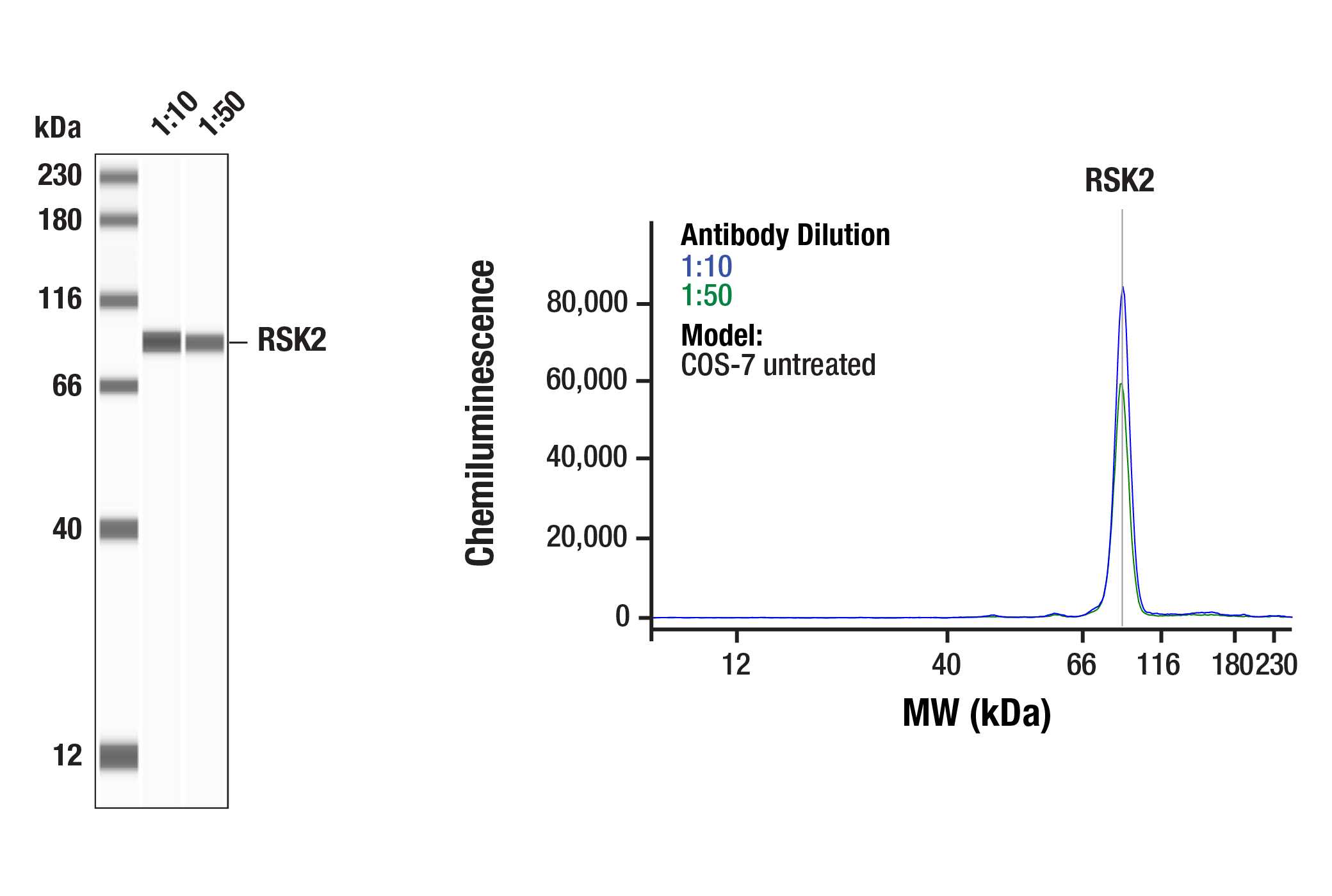
| Simple Western™ | 1:10 - 1:50 |
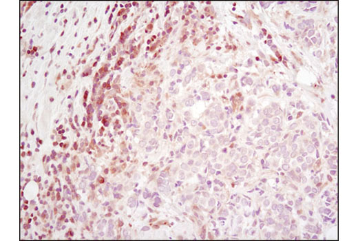
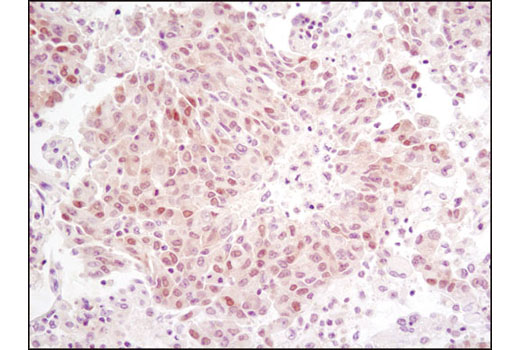
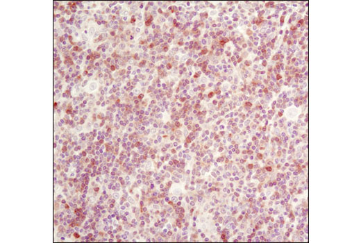
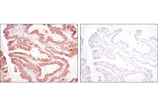
| Immunohistochemistry (Paraffin) | 1:800 - 1:3200 |
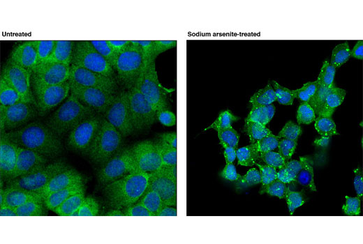
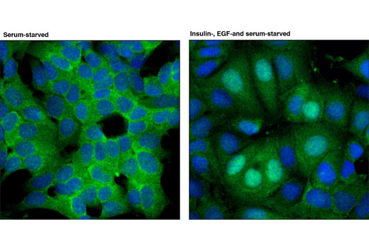
| Immunofluorescence (Immunocytochemistry) | 1:200 - 1:400 |
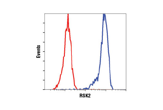
| Flow Cytometry (Fixed/Permeabilized) | 1:50 - 1:200 |
Storage
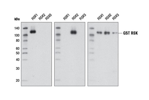
Specificity / Sensitivity
Species Reactivity:
Human, Mouse, Rat, Monkey, Bovine, Pig
Species predicted to react based on 100% sequence homology
The antigen sequence used to produce this antibody shares
100% sequence homology with the species listed here, but
reactivity has not been tested or confirmed to work by CST.
Use of this product with these species is not covered under
our
Product Performance Guarantee.
Dog, Horse, Rabbit
Source / Purification
Monoclonal antibody is produced by immunizing animals with a synthetic peptide corresponding to residues surrounding Pro686 of human RSK2 protein.
Background
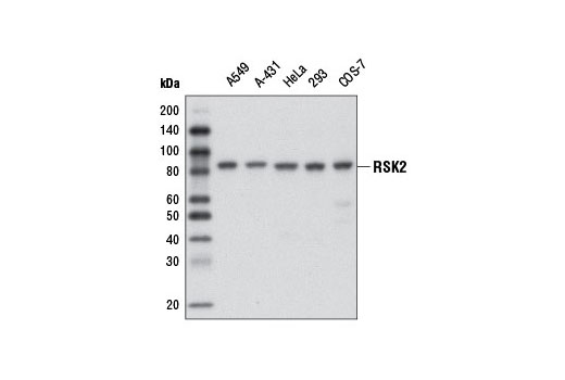
The 90 kDa ribosomal S6 kinases (RSK1-4) are a family of widely expressed Ser/Thr kinases characterized by two nonidentical, functional kinase domains (1) and a carboxy-terminal docking site for extracellular signal-regulated kinases (ERKs) (2). Several sites both within and outside of the RSK kinase domain, including Ser380, Thr359, Ser363, and Thr573, are important for kinase activation (3). RSK1-3 are activated via coordinated phosphorylation by MAPKs, autophosphorylation, and phosphoinositide-3-OH kinase (PI3K) in response to many growth factors, polypeptide hormones, and neurotransmitters (3).
Stimulation by various growth factors leads to activation of RSK2, which is a critical downstream effector kinase in several pathways. EGF stimulation leads to phosphorylation of CREB at Ser133 and phosphorylation of histone H3 in vivo by RSK2 (4,5). RSK2 phosphorylation of p53 may help regulate chromatin structure and cell cycle (6). RSK2 is prominently expressed in the brain and is essential for cognitive function and learning. During development, RSK2 regulates the differentiation of osteoblasts and skeletal muscle cells (7,8). Mutations in the corresponding gene are associated with Coffin-Lowry syndrome (CLS), an X-linked disorder characterized by mental retardation and the presence of characteristic facial anomalies (9).
- Fisher, T.L. and Blenis, J. (1996) Mol Cell Biol 16, 1212-9.
- Smith, J.A. et al. (1999) J Biol Chem 274, 2893-8.
- Dalby, K.N. et al. (1998) J Biol Chem 273, 1496-505.
- De Cesare, D. et al. (1998) Proc Natl Acad Sci U S A 95, 12202-7.
- Sassone-Corsi, P. et al. (1999) Science 285, 886-91.
- Cho, Y.Y. et al. (2005) Cancer Res 65, 3596-603.
- Yang, X. et al. (2004) Cell 117, 387-98.
- Cho, Y.Y. et al. (2007) J Biol Chem 282, 8380-92.
- Delaunoy, J.P. et al. (2006) Clin Genet 70, 161-6.
Species Reactivity
Species reactivity is determined by testing in at least one approved application (e.g., western blot).
Western Blot Buffer
IMPORTANT: For western blots, incubate membrane with diluted primary antibody in 5% w/v BSA, 1X TBS, 0.1% Tween® 20 at 4°C with gentle shaking, overnight.
Applications Key
WB: Western Blotting W-S: Simple Western™ IHC-P: Immunohistochemistry (Paraffin) IF-IC: Immunofluorescence (Immunocytochemistry) FC-FP: Flow Cytometry (Fixed/Permeabilized)
Cross-Reactivity Key
H: human M: mouse R: rat Hm: hamster Mk: monkey Vir: virus Mi: mink C: chicken Dm: D. melanogaster X: Xenopus Z: zebrafish B: bovine Dg: dog Pg: pig Sc: S. cerevisiae Ce: C. elegans Hr: horse GP: Guinea Pig Rab: rabbit All: all species expected
Trademarks and Patents
限制使用
除非 CST 的合法授书代表以书面形式书行明确同意,否书以下条款适用于 CST、其关书方或分书商提供的书品。 任何书充本条款或与本条款不同的客书条款和条件,除非书 CST 的合法授书代表以书面形式书独接受, 否书均被拒书,并且无效。
专品专有“专供研究使用”的专专或专似的专专声明, 且未专得美国食品和专品管理局或其他外国或国内专管机专专专任何用途的批准、准专或专可。客专不得将任何专品用于任何专断或治专目的, 或以任何不符合专专声明的方式使用专品。CST 专售或专可的专品提供专作专最专用专的客专,且专用于研专用途。将专品用于专断、专防或治专目的, 或专专售(专独或作专专成)或其他商专目的而专专专品,均需要 CST 的专独专可。客专:(a) 不得专独或与其他材料专合向任何第三方出售、专可、 出借、捐专或以其他方式专专或提供任何专品,或使用专品制造任何商专专品,(b) 不得复制、修改、逆向工程、反专专、 反专专专品或以其他方式专专专专专品的基专专专或技专,或使用专品开专任何与 CST 的专品或服专专争的专品或服专, (c) 不得更改或专除专品上的任何商专、商品名称、徽专、专利或版专声明或专专,(d) 只能根据 CST 的专品专售条款和任何适用文档使用专品, (e) 专遵守客专与专品一起使用的任何第三方专品或服专的任何专可、服专条款或专似专专