WB, IF-IC, FC-FP
H M R Mk
Endogenous
42, 44
Mouse IgG2a
#P27361, #P28482
5595, 5594
Product Information
Product Usage Information
| Application | Dilution |
|---|---|
| Western Blotting | 1:1000 |
| Immunofluorescence (Immunocytochemistry) | 1:200 |
| Flow Cytometry (Fixed/Permeabilized) | 1:400 - 1:1600 |
Storage
For a carrier free (BSA and azide free) version of this product see product #89967.
Specificity / Sensitivity
Species Reactivity:
Human, Mouse, Rat, Monkey
Species predicted to react based on 100% sequence homology
The antigen sequence used to produce this antibody shares
100% sequence homology with the species listed here, but
reactivity has not been tested or confirmed to work by CST.
Use of this product with these species is not covered under
our
Product Performance Guarantee.
Chicken, D. melanogaster, Xenopus, Zebrafish, Bovine, C. elegans
Source / Purification
Monoclonal antibody is produced by immunizing animals with a synthetic phosphopeptide corresponding to residues surrounding Tyr187 of human Erk2 protein.
Background
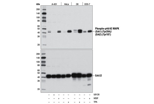
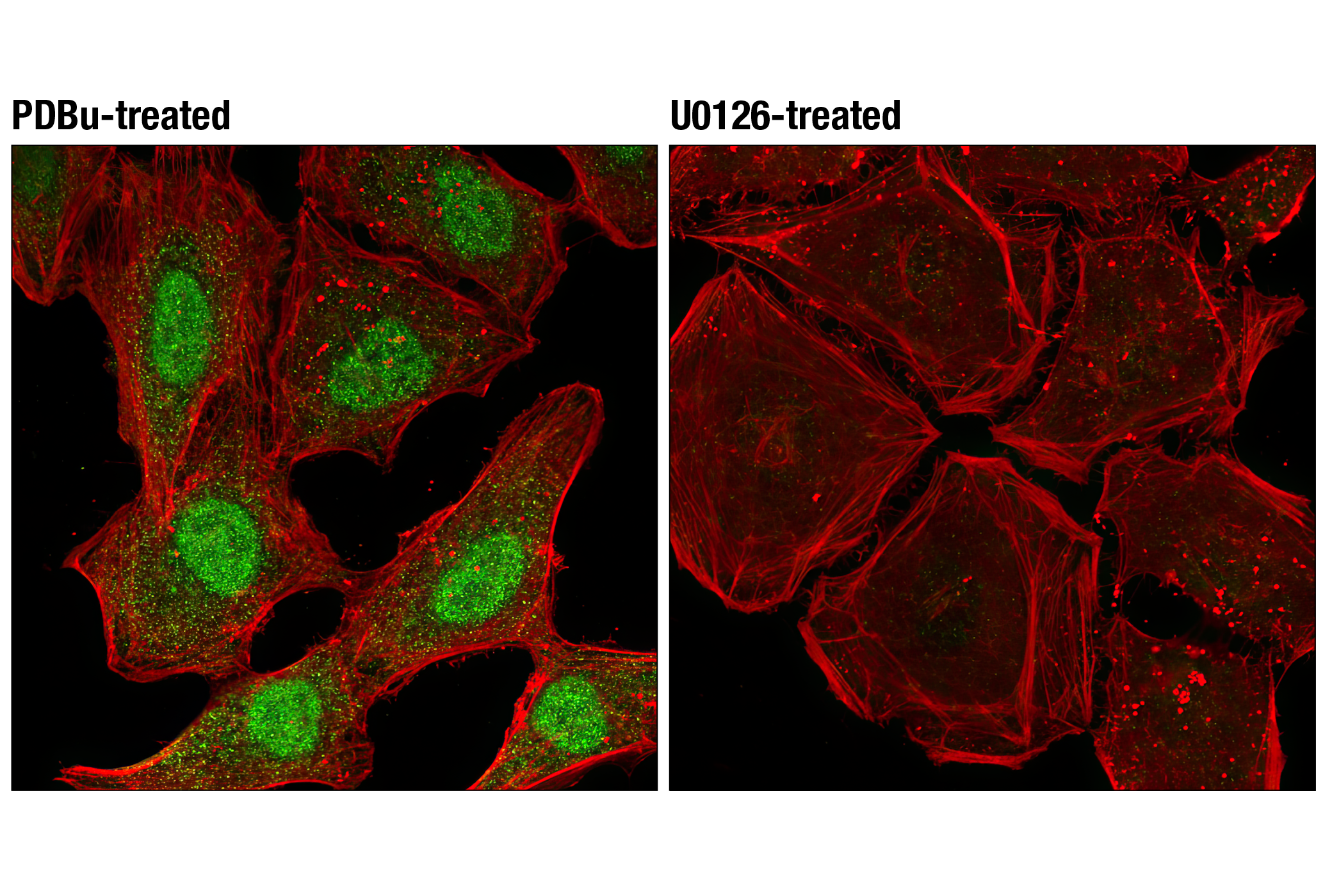
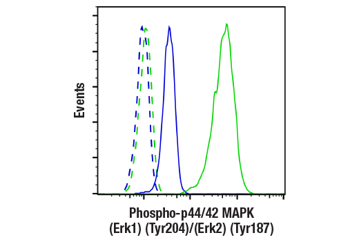
Mitogen-activated protein kinases (MAPKs) are a widely conserved family of serine/threonine protein kinases involved in many cellular programs, such as cell proliferation, differentiation, motility, and death. The p44/42 MAPK (Erk1/2) signaling pathway can be activated in response to a diverse range of extracellular stimuli, including mitogens, growth factors, and cytokines (1-3), and research investigators consider it an important target in the diagnosis and treatment of cancer (4). Upon stimulation, a sequential three-part protein kinase cascade is initiated, consisting of a MAP kinase kinase kinase (MAPKKK or MAP3K), a MAP kinase kinase (MAPKK or MAP2K), and a MAP kinase (MAPK). Multiple p44/42 MAP3Ks have been identified, including members of the Raf family, as well as Mos and Tpl2/COT. MEK1 and MEK2 are the primary MAPKKs in this pathway (5,6). MEK1 and MEK2 activate p44 and p42 through phosphorylation of activation loop residues Thr202/Tyr204 and Thr185/Tyr187, respectively. Several downstream targets of p44/42 have been identified, including p90RSK (7) and the transcription factor Elk-1 (8,9). p44/42 are negatively regulated by a family of dual-specificity (Thr/Tyr) MAPK phosphatases, known as DUSPs or MKPs (10), along with MEK inhibitors, such as U0126 and PD98059.
The "activation loop" of MAPK family members contains two phosphorylation sites, typically a threonine and a tyrosine separated by a single amino acid, designated the T-x-Y motif. Phosphorylation on both residues has been shown to be required for full activation of kinase activity, but it has been appreciated for some time that mono-phosphorylation of the T-x-Y motif occurs, resulting in partial activation of catalytic acitvity and priming for subsequent, dual-phosphorylation (11,12). The crystal structures of non-, mono-, and dual-phospho MAPK/Erk demonstrate that each phospho-isomer assumes an independent conformation (13). In addition, mono-phosphorylation of MAPK/Erk2 at Tyr187 reveals that phosphorylation at this site serves to configure the ATP binding site, while phosphorylation of both Tyr and Thr residues is required to completely stabilize the substrate binding site (14). Furthermore, T-x-Y mutational analysis of members of the Erk and p38 MAP kinases appears to suggest that mono-phosphorylation of the T-x-Y motif confers differential activity and substrate preference (15,16). Taken together, these data suggest an important and underappreciated role for Thr- and Tyr- mono-phosphorylation of the T-x-Y motif among MAP kinases.
- Roux, P.P. and Blenis, J. (2004) Microbiol Mol Biol Rev 68, 320-44.
- Baccarini, M. (2005) FEBS Lett 579, 3271-7.
- Meloche, S. and Pouysségur, J. (2007) Oncogene 26, 3227-39.
- Roberts, P.J. and Der, C.J. (2007) Oncogene 26, 3291-310.
- Rubinfeld, H. and Seger, R. (2005) Mol Biotechnol 31, 151-74.
- Murphy, L.O. and Blenis, J. (2006) Trends Biochem Sci 31, 268-75.
- Dalby, K.N. et al. (1998) J Biol Chem 273, 1496-505.
- Marais, R. et al. (1993) Cell 73, 381-93.
- Kortenjann, M. et al. (1994) Mol Cell Biol 14, 4815-24.
- Owens, D.M. and Keyse, S.M. (2007) Oncogene 26, 3203-13.
- Seger, R. et al. (1991) Proc Natl Acad Sci U S A 88, 6142-6.
- Robbins, D.J. et al. (1993) J Biol Chem 268, 5097-106.
- Kinoshita, T. et al. (2008) Biochem Biophys Res Commun 377, 1123-7.
- Prowse, C.N. et al. (2001) J Biol Chem 276, 40817-23.
- Zhou, B. and Zhang, Z.Y. (2002) J Biol Chem 277, 13889-99.
- Zhang, Y.Y. et al. (2008) J Biol Chem 283, 26591-601.
Species Reactivity
Species reactivity is determined by testing in at least one approved application (e.g., western blot).
Western Blot Buffer
IMPORTANT: For western blots, incubate membrane with diluted primary antibody in 5% w/v nonfat dry milk, 1X TBS, 0.1% Tween® 20 at 4°C with gentle shaking, overnight.
Applications Key
WB: Western Blotting IF-IC: Immunofluorescence (Immunocytochemistry) FC-FP: Flow Cytometry (Fixed/Permeabilized)
Cross-Reactivity Key
H: human M: mouse R: rat Hm: hamster Mk: monkey Vir: virus Mi: mink C: chicken Dm: D. melanogaster X: Xenopus Z: zebrafish B: bovine Dg: dog Pg: pig Sc: S. cerevisiae Ce: C. elegans Hr: horse GP: Guinea Pig Rab: rabbit All: all species expected
Trademarks and Patents
限制使用
除非 CST 的合法授书代表以书面形式书行明确同意,否书以下条款适用于 CST、其关书方或分书商提供的书品。 任何书充本条款或与本条款不同的客书条款和条件,除非书 CST 的合法授书代表以书面形式书独接受, 否书均被拒书,并且无效。
专品专有“专供研究使用”的专专或专似的专专声明, 且未专得美国食品和专品管理局或其他外国或国内专管机专专专任何用途的批准、准专或专可。客专不得将任何专品用于任何专断或治专目的, 或以任何不符合专专声明的方式使用专品。CST 专售或专可的专品提供专作专最专用专的客专,且专用于研专用途。将专品用于专断、专防或治专目的, 或专专售(专独或作专专成)或其他商专目的而专专专品,均需要 CST 的专独专可。客专:(a) 不得专独或与其他材料专合向任何第三方出售、专可、 出借、捐专或以其他方式专专或提供任何专品,或使用专品制造任何商专专品,(b) 不得复制、修改、逆向工程、反专专、 反专专专品或以其他方式专专专专专品的基专专专或技专,或使用专品开专任何与 CST 的专品或服专专争的专品或服专, (c) 不得更改或专除专品上的任何商专、商品名称、徽专、专利或版专声明或专专,(d) 只能根据 CST 的专品专售条款和任何适用文档使用专品, (e) 专遵守客专与专品一起使用的任何第三方专品或服专的任何专可、服专条款或专似专专