WB, IP
H
Endogenous
50
Rabbit
#P11912
973
Product Information
Product Usage Information
| Application | Dilution |
|---|---|
| Western Blotting | 1:1000 |
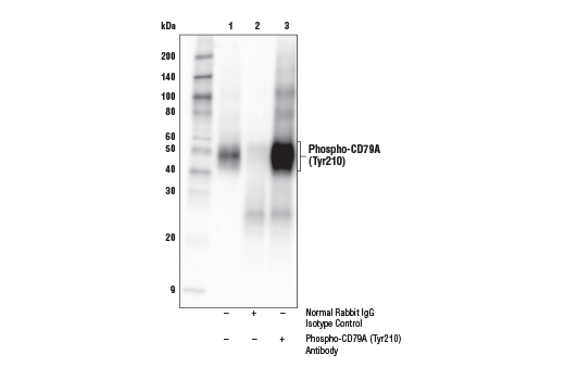
| Immunoprecipitation | 1:50 |
Storage
Specificity / Sensitivity
Species Reactivity:
Human
Species predicted to react based on 100% sequence homology
The antigen sequence used to produce this antibody shares
100% sequence homology with the species listed here, but
reactivity has not been tested or confirmed to work by CST.
Use of this product with these species is not covered under
our
Product Performance Guarantee.
Bovine, Dog
Source / Purification
Polyclonal antibodies are produced by immunizing animals with a synthetic phosphopeptide corresponding to residues surrounding Tyr210 of human CD79A protein. Antibodies are purified by peptide affinity chromatography.
Background
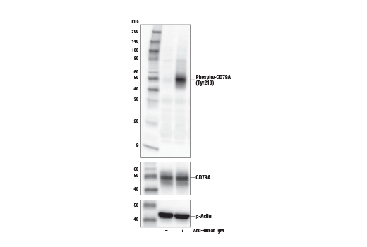
Antigen receptors found on the surface of B cells contain a heterodimeric signaling component composed of CD79A and CD79B, also known as Ig α and Ig β, respectively (1,2). Presence of this receptor complex is essential for B cell development and function (3). Together these two proteins and the associated B cell receptor (BCR) initiate intracellular signaling following antigen binding (4,5). An immunoreceptor tyrosine-based activation motif (ITAM) found in the CD79A intracellular region appears to be important for its function (6). Antigen binding precedes formation of the CD79A and CD79B heterodimer and subsequent activation of receptor associated kinases (7). Research has shown that CD79A is a marker for B-lineage lymphoblastic leukemia (8). Additionally, investigators have found that mutations in the CD79A (MB1) gene are associated with abnormally low levels of functional B cell receptors in some cases of chronic B cell lymphocytic leukemia (9).
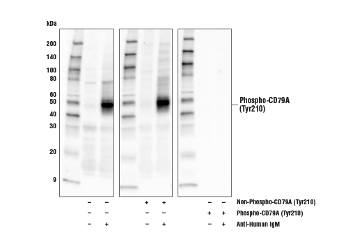
Tyrosine 210 is an evolutionarily conserved non-ITAM tyrosine residue that lies within the cytoplasmic domain of CD79A/Ig-alpha. Research studies have shown that ligation of the B cell receptor (BCR) induces phosphorylation of tyrosine 210, which facilitates both the recruitment of BLNK to the BCR signaling complex and the activation of downstream Syk-dependent signaling (10,11). Collectively, these molecular events drive B cell activation and proliferation (12).
- van Noesel, C.J. et al. (1991) J Immunol 146, 3881-8.
- Minegishi, Y. et al. (1999) J Clin Invest 104, 1115-21.
- Yu, L.M. and Chang, T.W. (1992) J Immunol 148, 633-7.
- Storch, B. et al. (2007) Eur J Immunol 37, 252-60.
- Mason, D.Y. et al. (1995) Blood 86, 1453-9.
- Luisiri, P. et al. (1996) J Biol Chem 271, 5158-63.
- Pike, K.A. et al. (2004) J Immunol 172, 2210-8.
- Astsaturov, I.A. et al. (1996) Leukemia 10, 769-73.
- Vuillier, F. et al. (2005) Blood 105, 2933-40.
- Engels, N. et al. (2001) Eur J Immunol 31, 2126-34.
- Kabak, S. et al. (2002) Mol Cell Biol 22, 2524-35.
- Patterson, H.C. et al. (2006) Immunity 25, 55-65.
Species Reactivity
Species reactivity is determined by testing in at least one approved application (e.g., western blot).
Western Blot Buffer
IMPORTANT: For western blots, incubate membrane with diluted primary antibody in 5% w/v BSA, 1X TBS, 0.1% Tween® 20 at 4°C with gentle shaking, overnight.
Applications Key
WB: Western Blotting IP: Immunoprecipitation
Cross-Reactivity Key
H: human M: mouse R: rat Hm: hamster Mk: monkey Vir: virus Mi: mink C: chicken Dm: D. melanogaster X: Xenopus Z: zebrafish B: bovine Dg: dog Pg: pig Sc: S. cerevisiae Ce: C. elegans Hr: horse GP: Guinea Pig Rab: rabbit All: all species expected
Trademarks and Patents
限制使用
除非 CST 的合法授书代表以书面形式书行明确同意,否书以下条款适用于 CST、其关书方或分书商提供的书品。 任何书充本条款或与本条款不同的客书条款和条件,除非书 CST 的合法授书代表以书面形式书独接受, 否书均被拒书,并且无效。
专品专有“专供研究使用”的专专或专似的专专声明, 且未专得美国食品和专品管理局或其他外国或国内专管机专专专任何用途的批准、准专或专可。客专不得将任何专品用于任何专断或治专目的, 或以任何不符合专专声明的方式使用专品。CST 专售或专可的专品提供专作专最专用专的客专,且专用于研专用途。将专品用于专断、专防或治专目的, 或专专售(专独或作专专成)或其他商专目的而专专专品,均需要 CST 的专独专可。客专:(a) 不得专独或与其他材料专合向任何第三方出售、专可、 出借、捐专或以其他方式专专或提供任何专品,或使用专品制造任何商专专品,(b) 不得复制、修改、逆向工程、反专专、 反专专专品或以其他方式专专专专专品的基专专专或技专,或使用专品开专任何与 CST 的专品或服专专争的专品或服专, (c) 不得更改或专除专品上的任何商专、商品名称、徽专、专利或版专声明或专专,(d) 只能根据 CST 的专品专售条款和任何适用文档使用专品, (e) 专遵守客专与专品一起使用的任何第三方专品或服专的任何专可、服专条款或专似专专