Revision 8
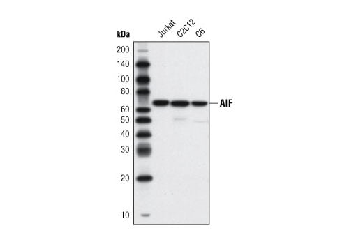
Western blot analysis of extracts from Jurkat, C2C12, and C6 cells using AIF (D39D2) XP® Rabbit mAb.
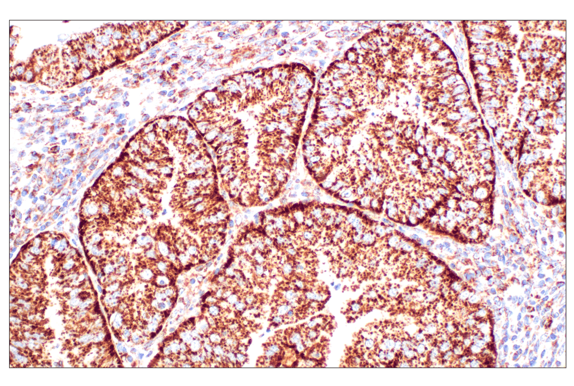
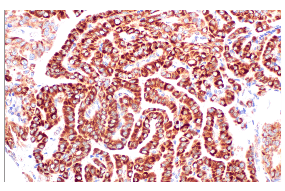
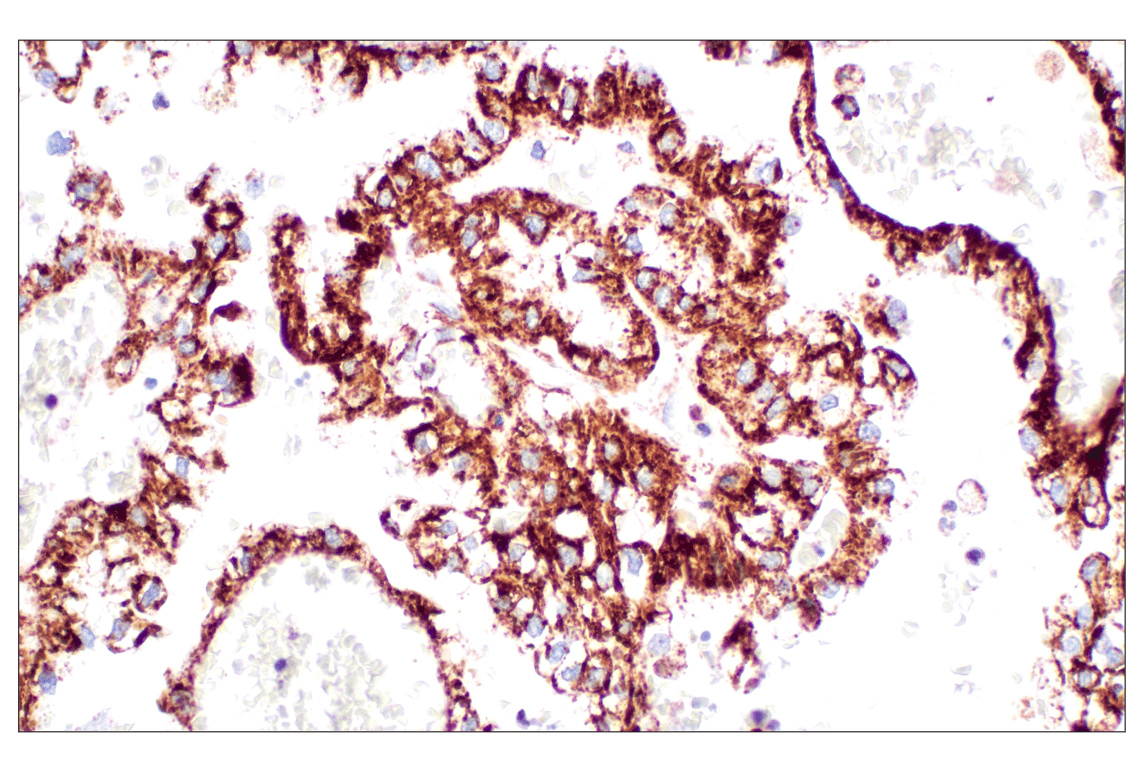
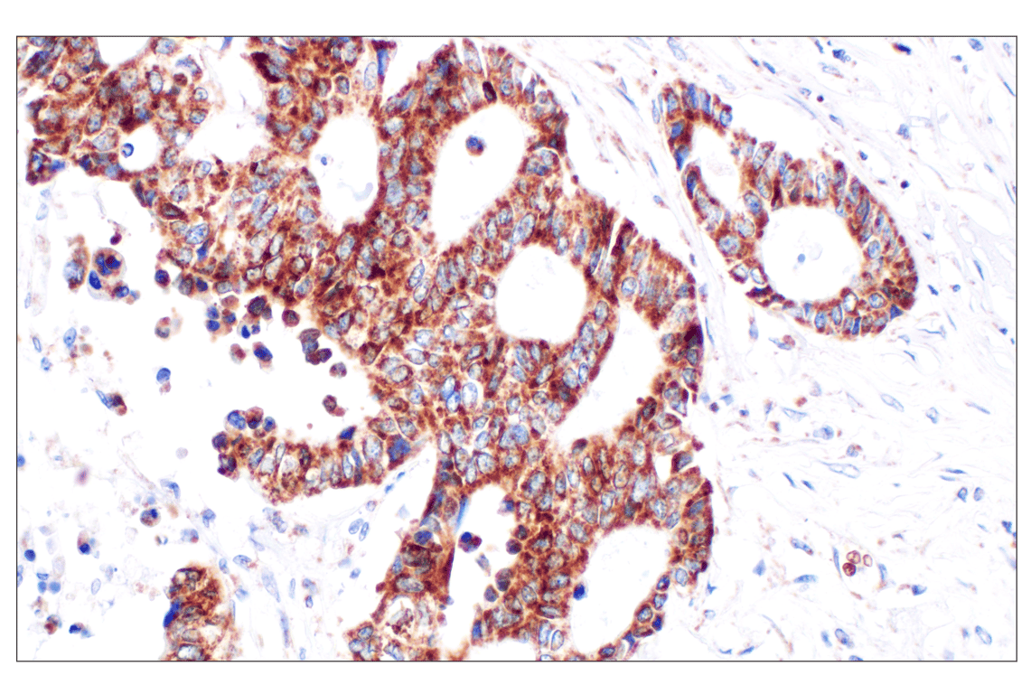
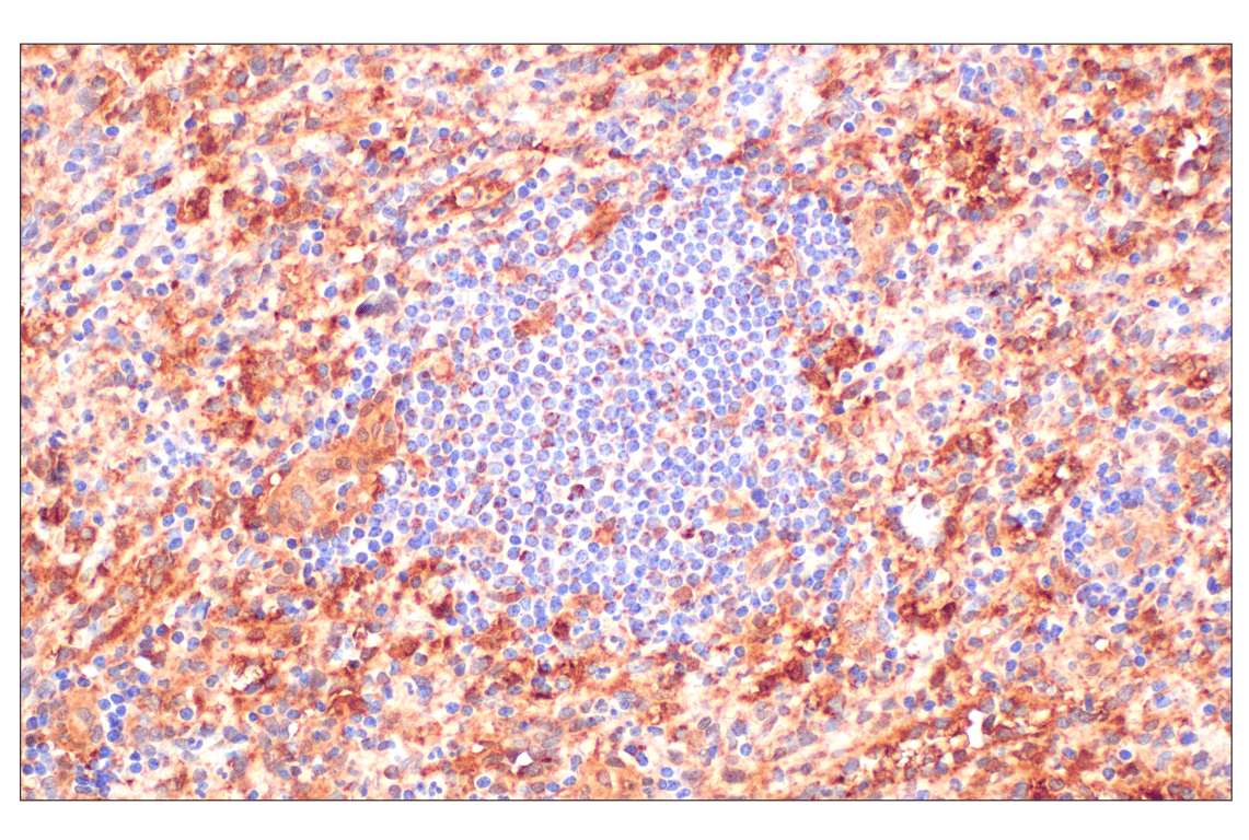
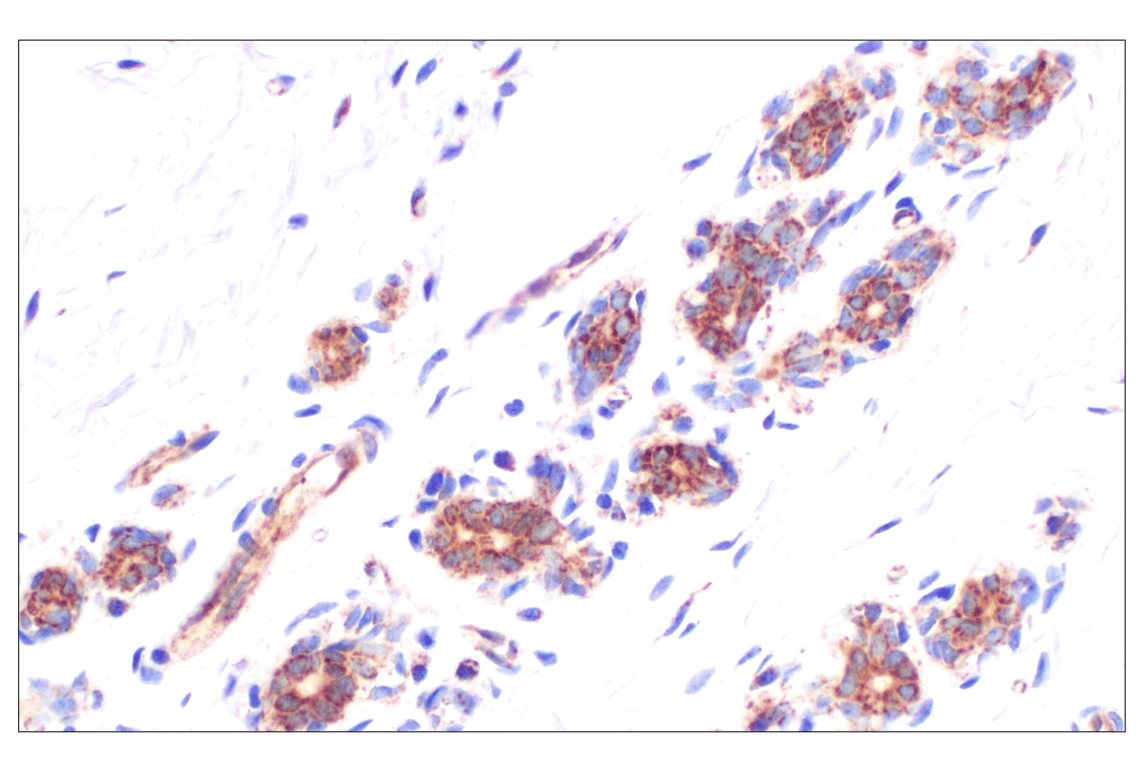
Immunohistochemical analysis of paraffin-embedded human endometrioid adenocarcinoma using AIF (D39D2) XP® Rabbit mAb.
After the primary antibody is bound to the target protein, a complex with HRP-linked secondary antibody is formed. The LumiGLO® is added and emits light during enzyme catalyzed decomposition.
Orders: 877-616-CELL (2355) • [email protected] • Support: 877-678-TECH (8324) • [email protected] •
Web:
cellsignal.com For Research Use Only. Not for Use in Diagnostic Procedures.
Revision 8
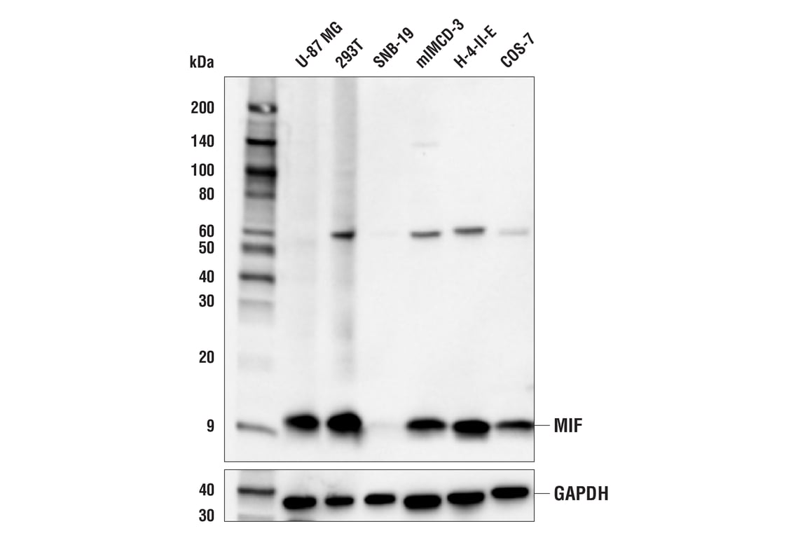
Western blot analysis of extracts from various cell lines using MIF (E8S8H) Rabbit mAb (upper) and GAPDH (D16H11) XP® Rabbit mAb #5174 (lower). The absence of MIF protein in SNB-19 cells is consistent with mRNA expression profiles reported in public bioinformatic databases, confirming specificity of the antibody for MIF.
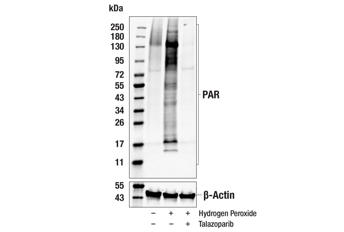
Western blot analysis of extracts from HeLa cells untreated (-), treated (+) with hydrogen peroxide (0.5 mM, 5 min), or treated with Parp inhibitor Talazoparib #80577 (100 nM, 3 hr) prior to hydrogen peroxide treatment (0.5 mM, 5 min), using Poly/Mono-ADP Ribose (D9P7Z) Rabbit mAb (upper) or B-Actin (13E5) Rabbit mAb #4970 (lower) and detected with Anti-rabbit IgG (H+L) (DyLight 680 Conjugate) #5366.
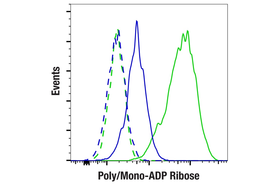
Flow cytometric analysis of HeLa cells treated with Talazoparib #80577 (100 nM, 3 hr) then hydrogen peroxide (500 mM, 5 min) (blue), or treated with hydrogen peroxide alone (500 mM, 5 min) (green), using Poly/Mono-ADP Ribose (D9P7Z) Rabbit mAb (solid lines) or concentration-matched Rabbit (DA1E) mAb IgG XP® Isotype Control #3900 (dashed lines). Anti-rabbit IgG (H+L), F(ab')2 Fragment (Alexa Fluor® 488 Conjugate) #4412 was used as a secondary antibody.
Orders: 877-616-CELL (2355) • [email protected] • Support: 877-678-TECH (8324) • [email protected] •
Web:
cellsignal.com For Research Use Only. Not for Use in Diagnostic Procedures.
Revision 8
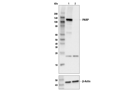
Western blot analysis of extracts from control HEK293 cells (lane 1) or PARP knockout HEK293 cells (lane 2) using PARP (46D11) Rabbit mAb #9532 (upper) or β-actin (13E5) Rabbit mAb #4970 (lower). The absence of signal in the PARP knockout HEK293 cells confirms the specificity of the antibody for PARP.
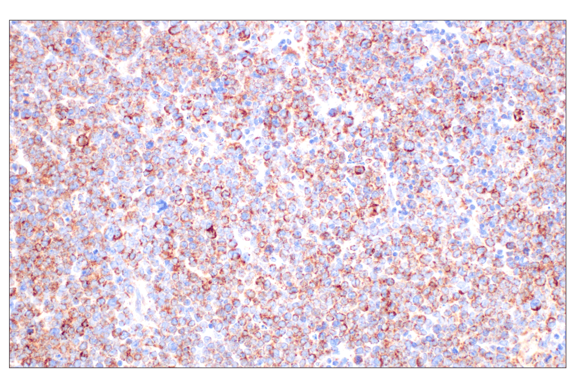
Immunohistochemical analysis of paraffin-embedded human papillary thyroid carcinoma using AIF (D39D2) XP® Rabbit mAb.
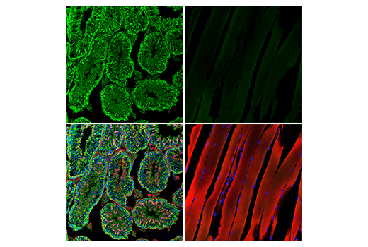
Confocal immunofluorescent analysis of mouse testis (left) and skeletal muscle (right) using MIF (E8S8H) Rabbit mAb (green). Actin filaments were labeled with DyLight™ 554 Phalloidin #13054 (red). Samples were mounted in ProLong® Gold Antifade Reagent with DAPI #8961 (blue).
Orders: 877-616-CELL (2355) • [email protected] • Support: 877-678-TECH (8324) • [email protected] •
Web:
cellsignal.com For Research Use Only. Not for Use in Diagnostic Procedures.
Revision 8
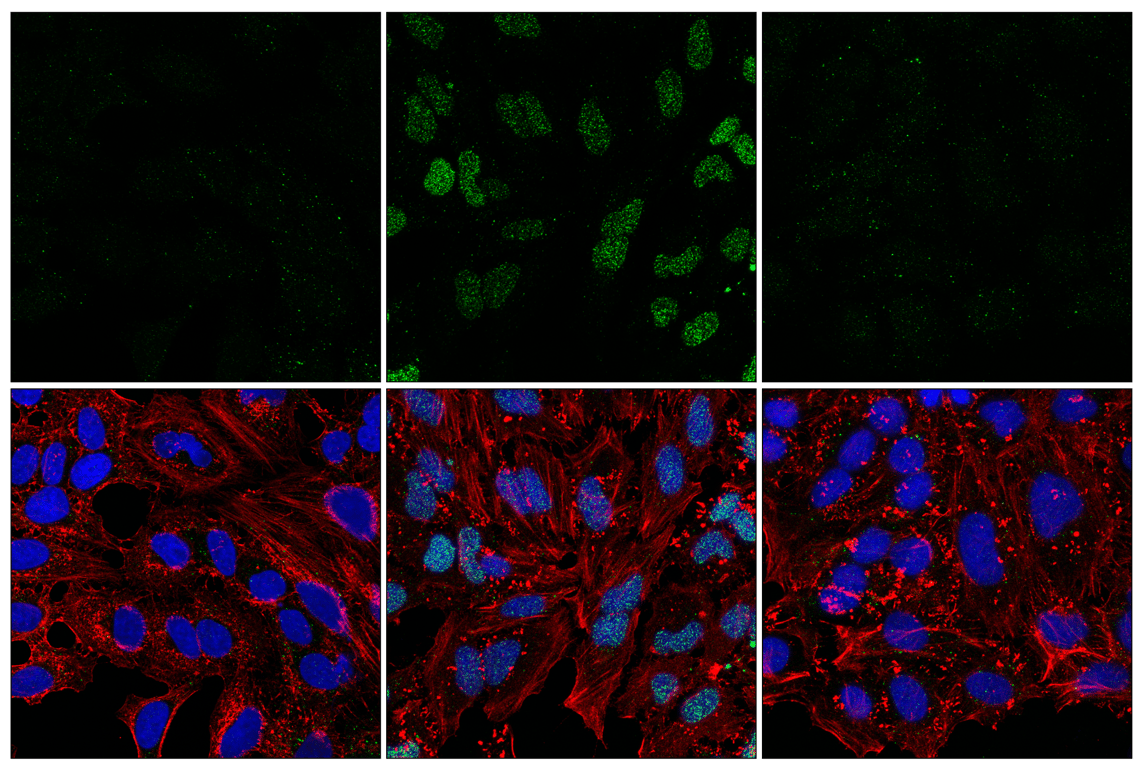
Confocal immunofluorescent analysis of HeLa cells, either untreated (left), treated with hydrogen peroxide (500 mM, 5 min; middle), or treated with PARP inhibitor Talazoparib #80577 (100 nmol/L, 3 hr) followed by hydrogen peroxide (500 mM, 5 min; right), using Poly/Mono-ADP Ribose (D9P7Z) Rabbit mAb (green), β-Actin (8H10D10) Mouse mAb #3700 (red), and DAPI #4083 (blue).
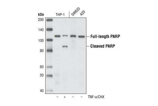
Western blot analysis of extracts from THP-1 cells, untreated or treated with TNF-α and cycloheximide as well as control extracts from SW620 and A20 cell lines, using PARP (46D11) Rabbit mAb.
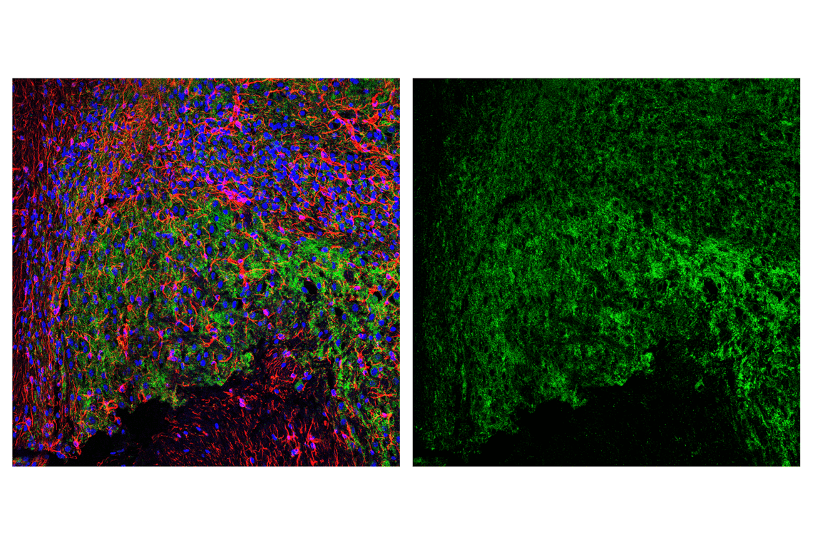
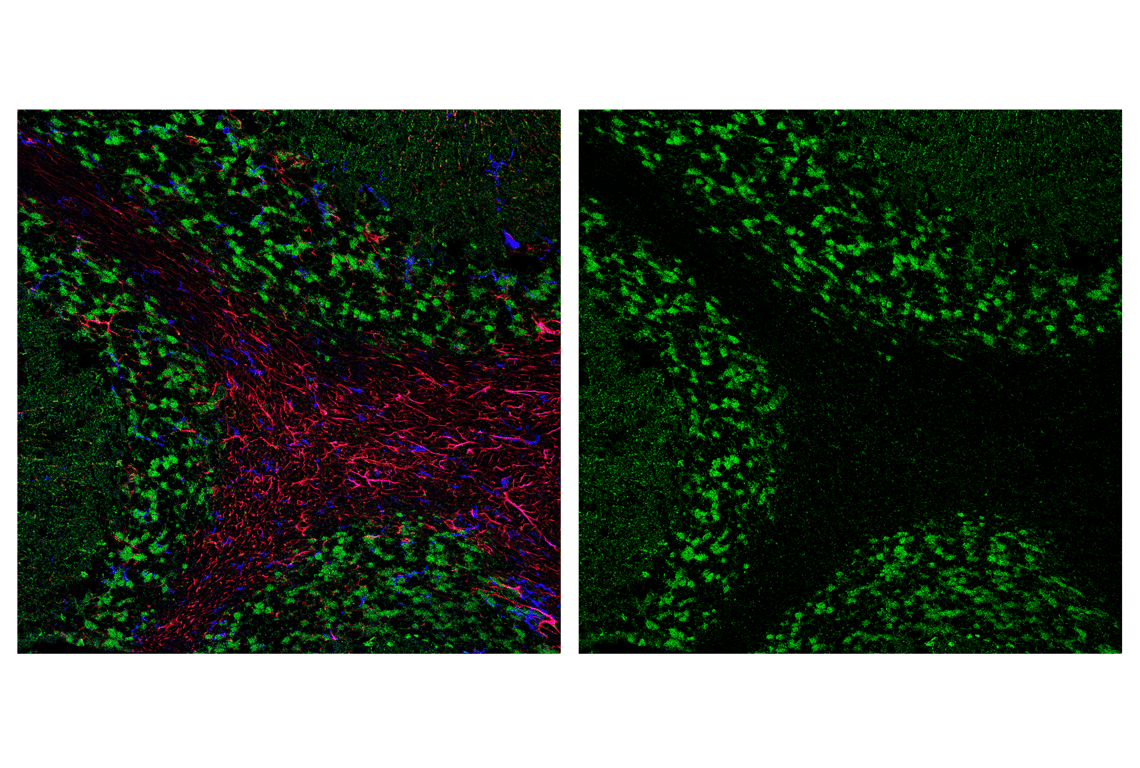
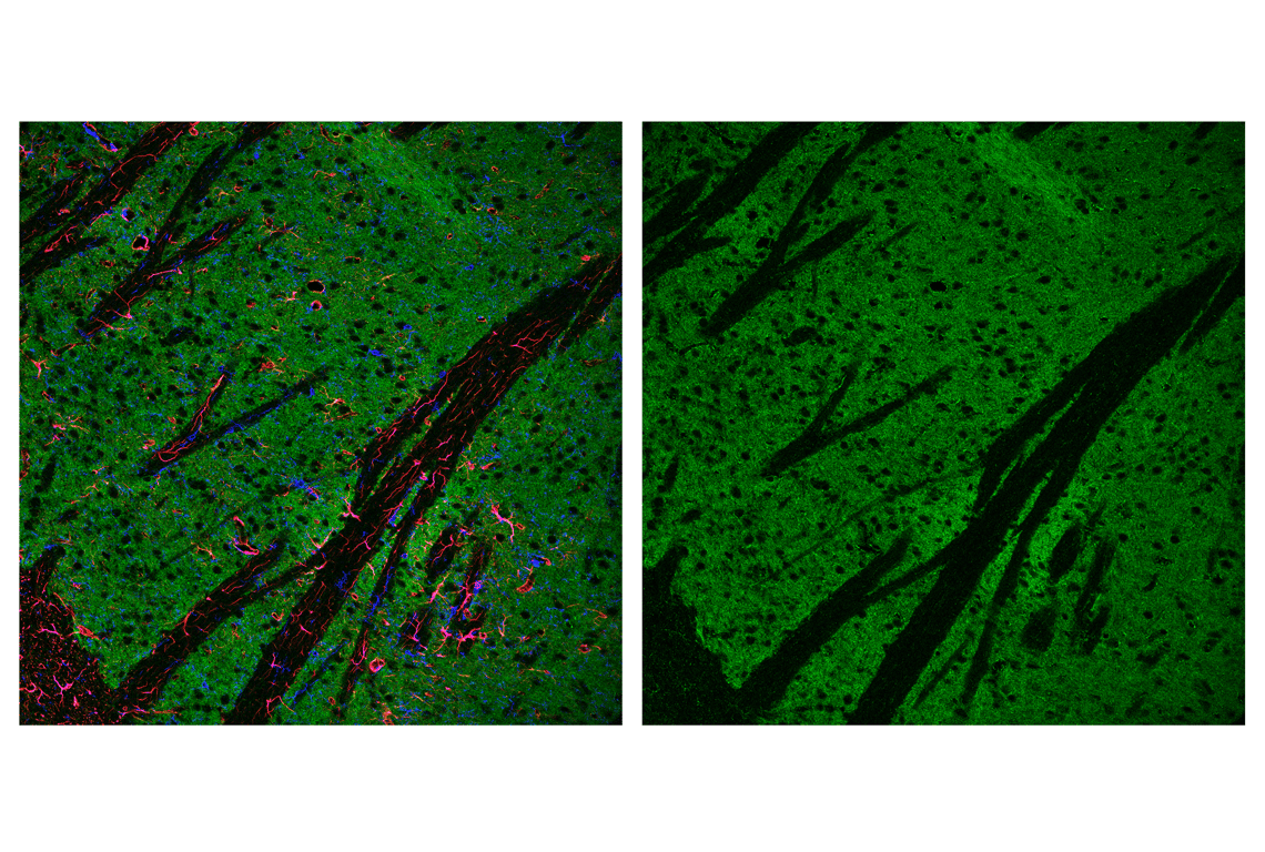
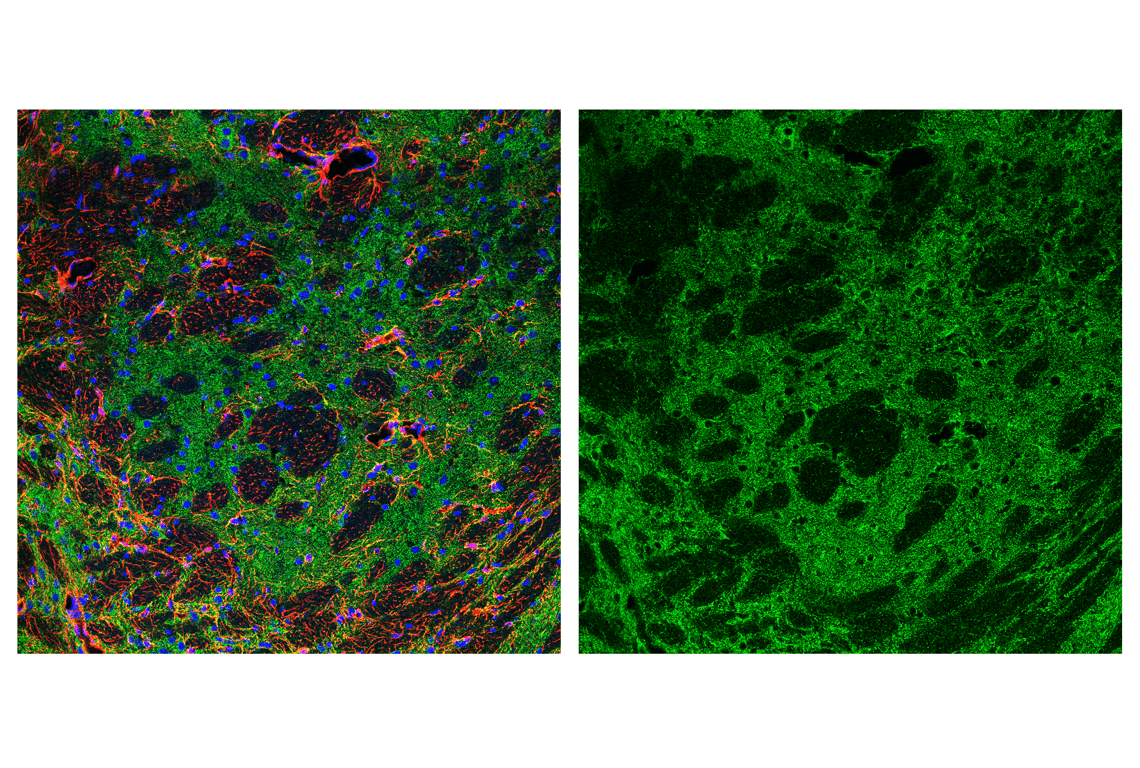
Confocal immunofluorescent analysis of mouse brainstem using AIF (D39D2) XP® Rabbit mAb #5318 (green) and GFAP (GA5) Mouse mAb #3670 (red). Sections were mounted in ProLong® Gold Antifade Reagent with DAPI #8961 (blue).
Orders: 877-616-CELL (2355) • [email protected] • Support: 877-678-TECH (8324) • [email protected] •
Web:
cellsignal.com For Research Use Only. Not for Use in Diagnostic Procedures.
Revision 8
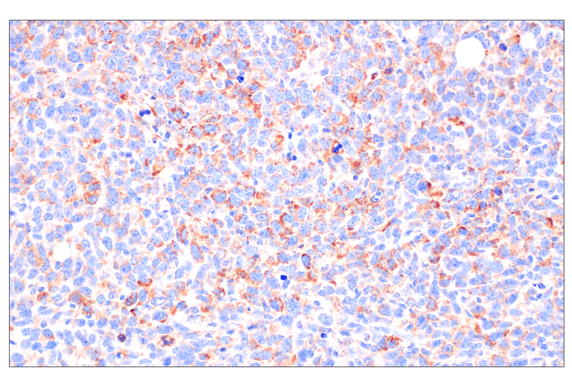
Immunohistochemical analysis of paraffin-embedded human hepatocellular carcinoma using AIF (D39D2) XP® Rabbit mAb.
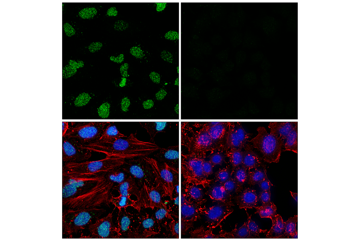
Confocal immunofluorescent analysis of U-87 MG cells (left, positive) or SNB-19 cells (right, negative) using MIF (E8S8H) Rabbit mAb (green). Actin filaments were labeled with DyLight™ 554 Phalloidin #13054 (red). Samples were mounted in ProLong® Gold Antifade Reagent with DAPI #8961 (blue).
Confocal immunofluorescent analysis of HeLa cells, either treated with hydrogen peroxide (500 mM, 5 min; left) or treated with hydrogen peroxide (500 mM, 5 min), then treated post fixation with recombinant human PARG (right), using Poly/Mono-ADP Ribose (D9P7Z) Rabbit mAb (green), β-Actin (8H10D10) Mouse mAb #3700 (red), and DAPI #4083 (blue).
Orders: 877-616-CELL (2355) • [email protected] • Support: 877-678-TECH (8324) • [email protected] •
Web:
cellsignal.com For Research Use Only. Not for Use in Diagnostic Procedures.
Revision 8
Confocal immunofluorescent analysis of mouse cerebellum using AIF (D39D2) XP® Rabbit mAb #5318 (green), GFAP (GA5) Mouse mAb #3670 (red), and F4/80 (BM8.1) Rat mAb #71299 (blue).
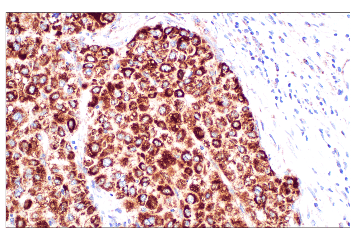
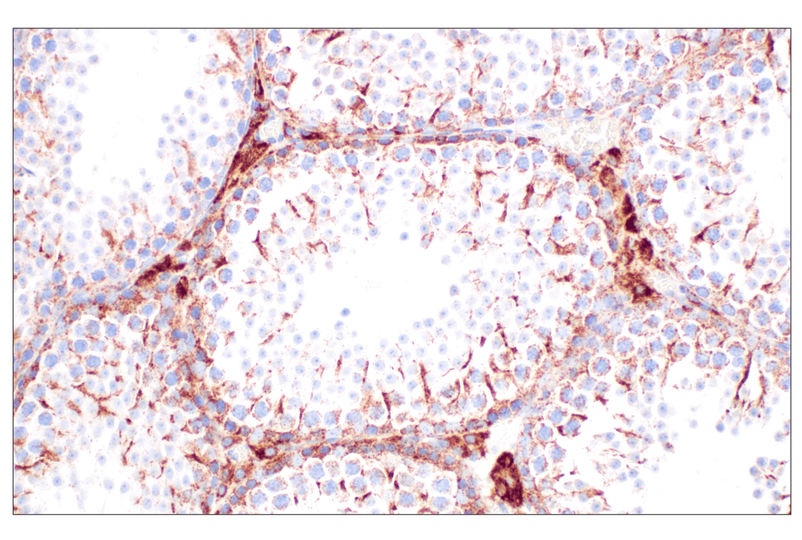
Immunohistochemical analysis of paraffin-embedded human renal cell carcinoma using AIF (D39D2) XP® Rabbit mAb.
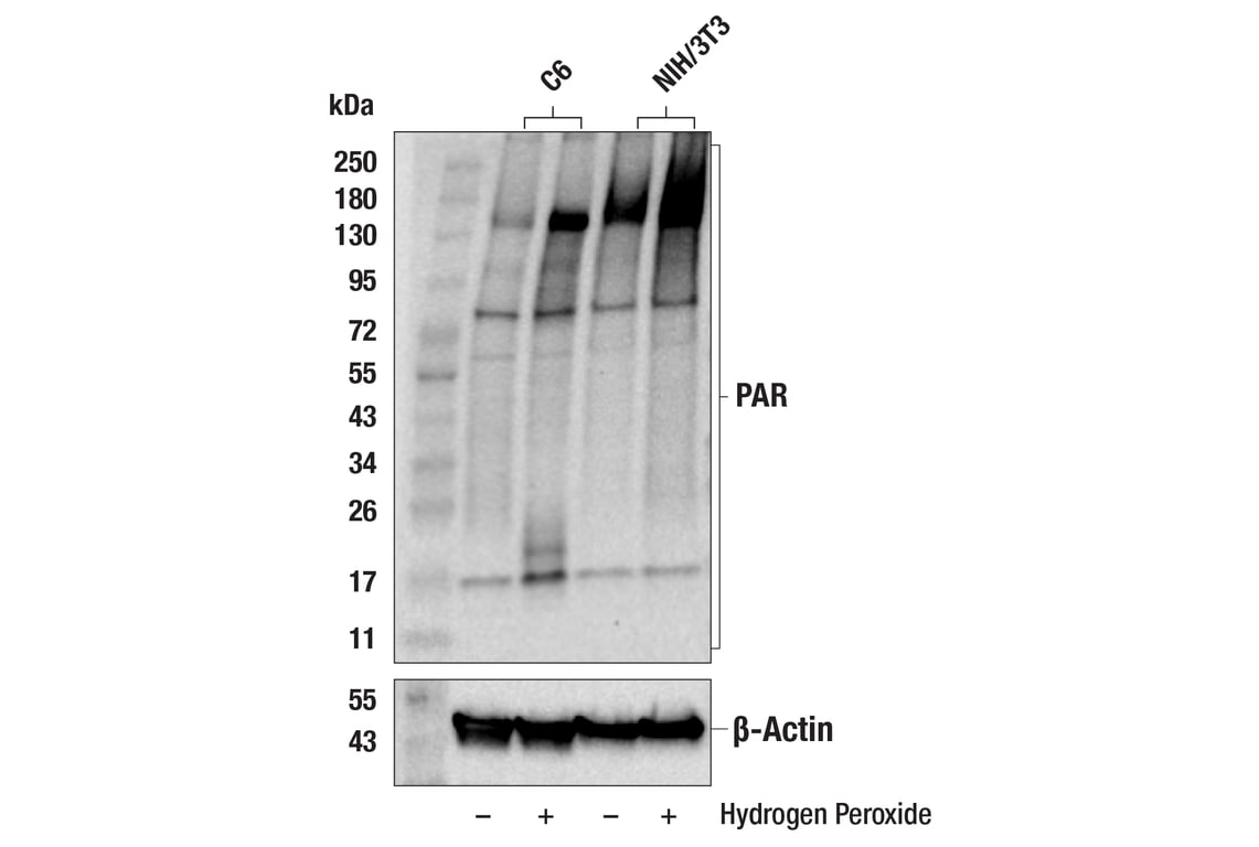
Western blot analysis of extracts from C6 and NIH/3T3 cells that were serum starved overnight, untreated (-) or treated (+) with hydrogen peroxide (1,000 μM, 5 min), using Poly/Mono-ADP Ribose (D9P7Z) Rabbit mAb (upper) or B-Actin (13E5) Rabbit mAb # 4970.
Orders: 877-616-CELL (2355) • [email protected] • Support: 877-678-TECH (8324) • [email protected] •
Web:
cellsignal.com For Research Use Only. Not for Use in Diagnostic Procedures.
Revision 8
Confocal immunofluorescent analysis of mouse striatum using AIF (D39D2) XP® Rabbit mAb #5318 (green), GFAP (GA5) Mouse mAb #3670 (red), and F4/80 (BM8.1) Rat mAb #71299 (blue).
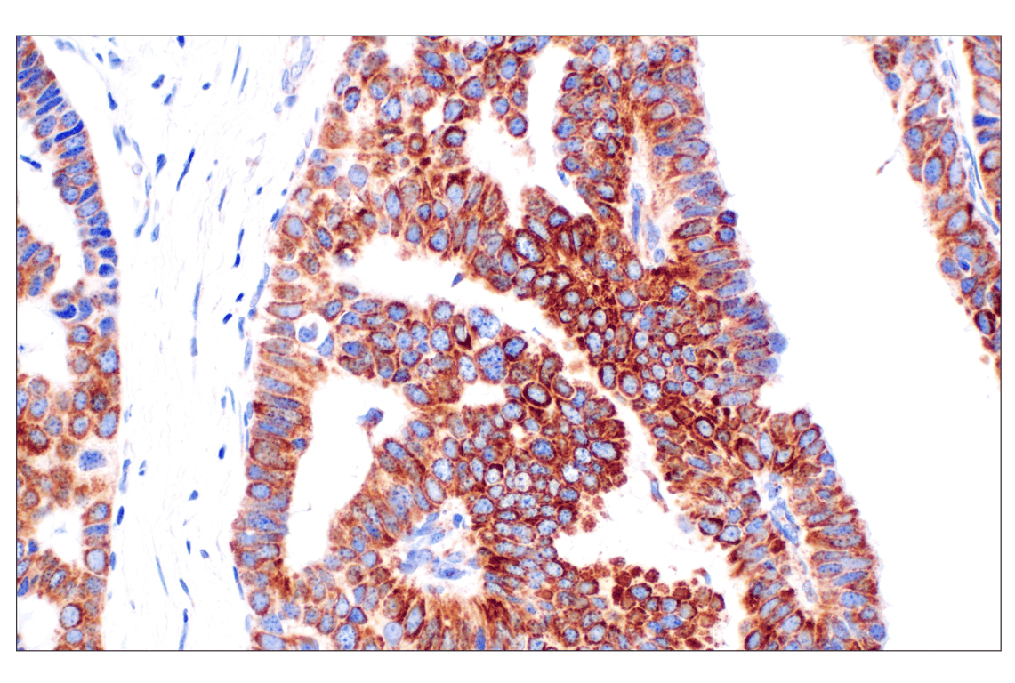
Immunohistochemical analysis of paraffin-embedded human colon adenocarcinoma using AIF (D39D2) XP® Rabbit mAb.
Confocal immunofluorescent analysis of mouse thalamus using AIF (D39D2) XP® Rabbit mAb #5318 (green), GFAP (GA5) Mouse mAb #3670 (red). Sections were mounted in ProLong® Gold Antifade Reagent with DAPI #8961 (blue).
Orders: 877-616-CELL (2355) • [email protected] • Support: 877-678-TECH (8324) • [email protected] •
Web:
cellsignal.com For Research Use Only. Not for Use in Diagnostic Procedures.
Revision 8
Immunohistochemical analysis of paraffin-embedded human papillary carcinoma of the breast using AIF (D39D2) XP® Rabbit mAb.
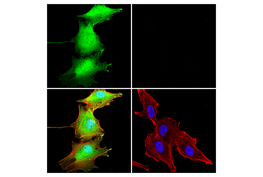
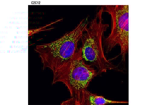
Confocal immunofluorescent analysis of C2C12 cells using AIF (D39D2) XP® Rabbit mAb #5318 (green). Actin filaments were labeled with DY-554 phalloidin (red). Blue pseudocolor = DRAQ5® #4084 (fluorescent DNA dye).
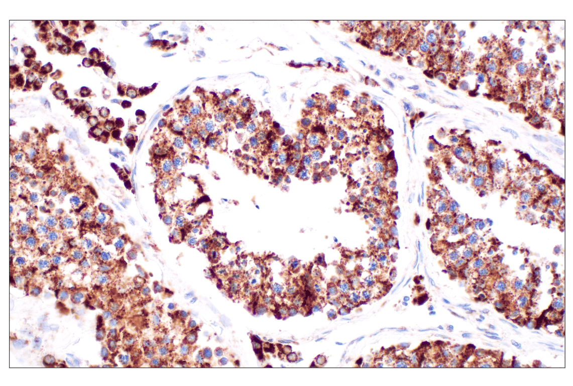
Immunohistochemical analysis of paraffin-embedded normal human testis using AIF (D39D2) XP® Rabbit mAb.
Orders: 877-616-CELL (2355) • [email protected] • Support: 877-678-TECH (8324) • [email protected] •
Web:
cellsignal.com For Research Use Only. Not for Use in Diagnostic Procedures.
Revision 8
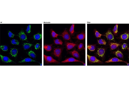
Confocal immunofluorescent analysis of HeLa cells using AIF (D39D2) XP ™ Rabbit mAb (green), showing colocalization with mitochondria that have been labeled with MitoTracker® Red CMXRos (red). Blue pseudocolor = DRAQ5® #4084 (fluorescent DNA dye).
Immunohistochemical analysis of paraffin-embedded normal human spleen using AIF (D39D2) XP® Rabbit mAb.
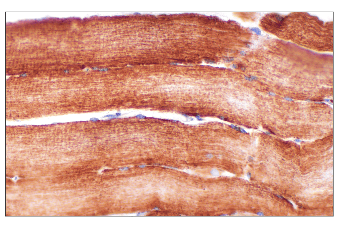
Immunohistochemical analysis of paraffin-embedded normal human skeletal muscle using AIF (D39D2) XP® Rabbit mAb.
Orders: 877-616-CELL (2355) • [email protected] • Support: 877-678-TECH (8324) • [email protected] •
Web:
cellsignal.com For Research Use Only. Not for Use in Diagnostic Procedures.
Revision 8
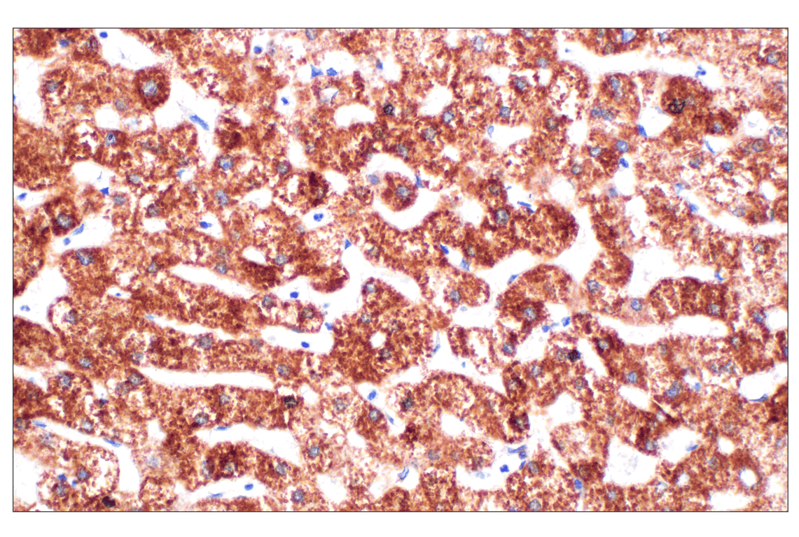
Immunohistochemical analysis of paraffin-embedded normal human liver using AIF (D39D2) XP® Rabbit mAb.
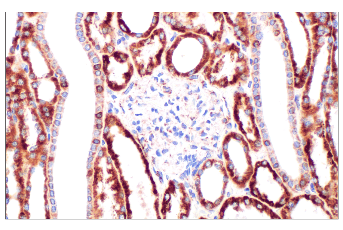
Immunohistochemical analysis of paraffin-embedded normal human kidney using AIF (D39D2) XP® Rabbit mAb.
Immunohistochemical analysis of paraffin-embedded normal human breast using AIF (D39D2) XP® Rabbit mAb.
Orders: 877-616-CELL (2355) • [email protected] • Support: 877-678-TECH (8324) • [email protected] •
Web:
cellsignal.com For Research Use Only. Not for Use in Diagnostic Procedures.
Revision 8
Immunohistochemical analysis of paraffin-embedded A20 syngeneic tumor using AIF (D39D2) XP® Rabbit mAb.
Immunohistochemical analysis of paraffin-embedded LL/2 syngeneic tumor using AIF (D39D2) XP® Rabbit mAb.
Immunohistochemical analysis of paraffin-embedded mouse testis using AIF (D39D2) XP® Rabbit mAb.
Orders: 877-616-CELL (2355) • [email protected] • Support: 877-678-TECH (8324) • [email protected] •
Web:
cellsignal.com For Research Use Only. Not for Use in Diagnostic Procedures.
Revision 8
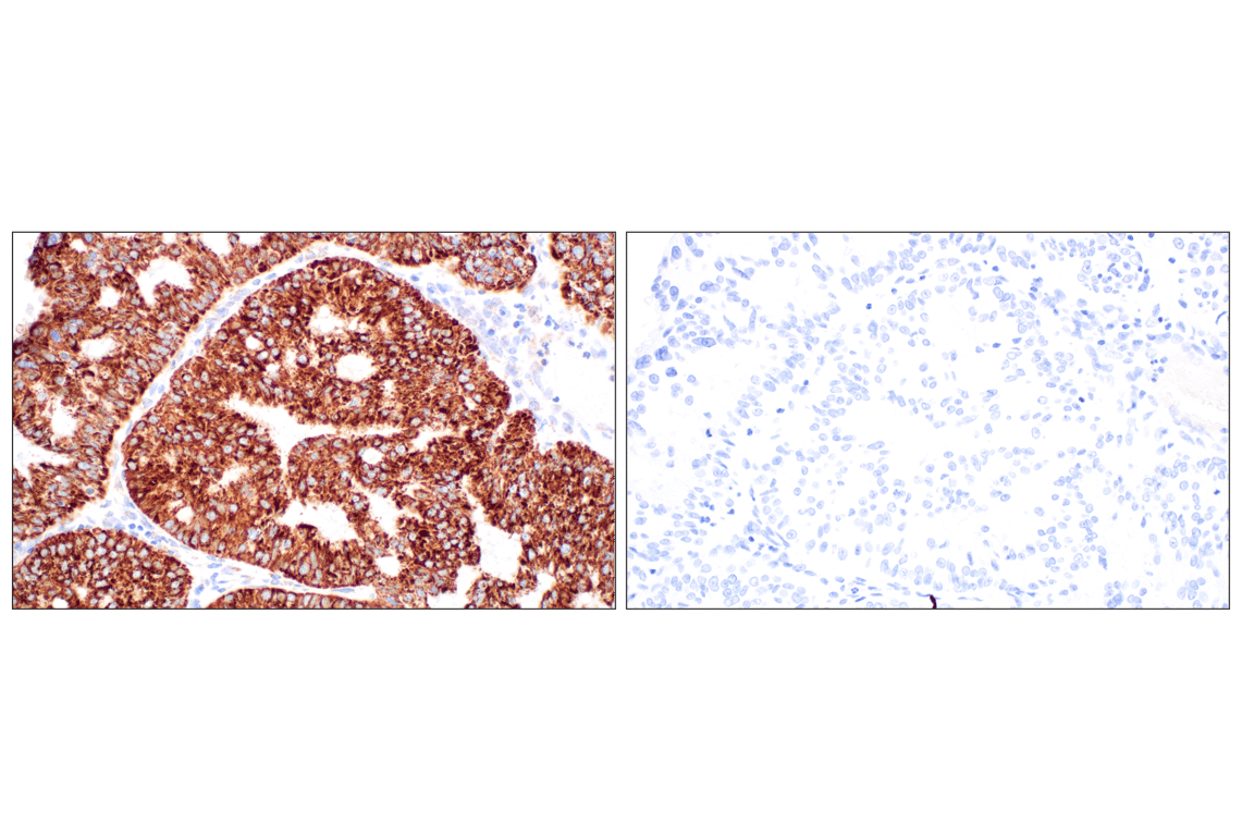
Immunohistochemical analysis of paraffin-embedded human endometrioid adenocarcinoma using AIF (D39D2) XP® Rabbit mAb (left) compared to concentration-matched Rabbit (DA1E) mAb IgG XP® Isotype Control #3900 (right).
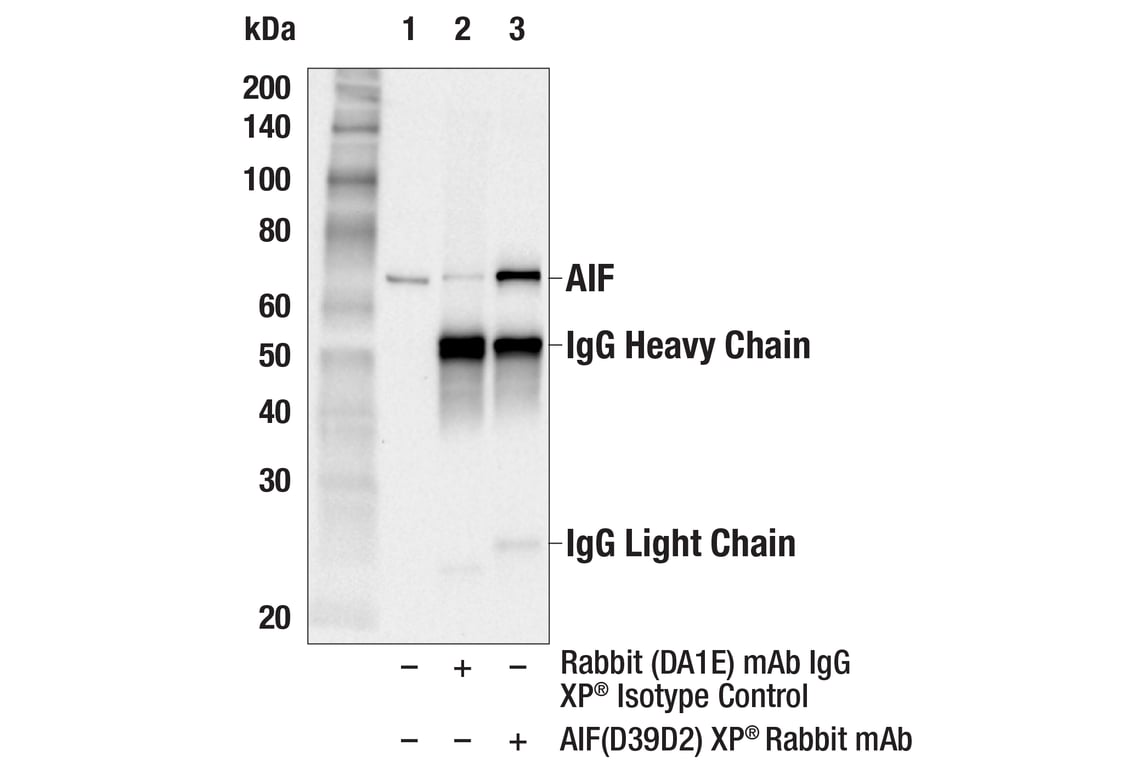
Immunoprecipitation of AIF protein from Jurkat cell extracts. Lane 1 is 10% input, lane 2 is Rabbit (DA1E) mAb IgG XP® Isotype Control #3900, and lane 3 is AIF (D39D2) XP® Rabbit mAb. Western blot analysis was performed using AIF (D39D2) XP® Rabbit mAb. Anti-rabbit IgG, HRP-linked Antibody #7074 was used as a secondary antibody.
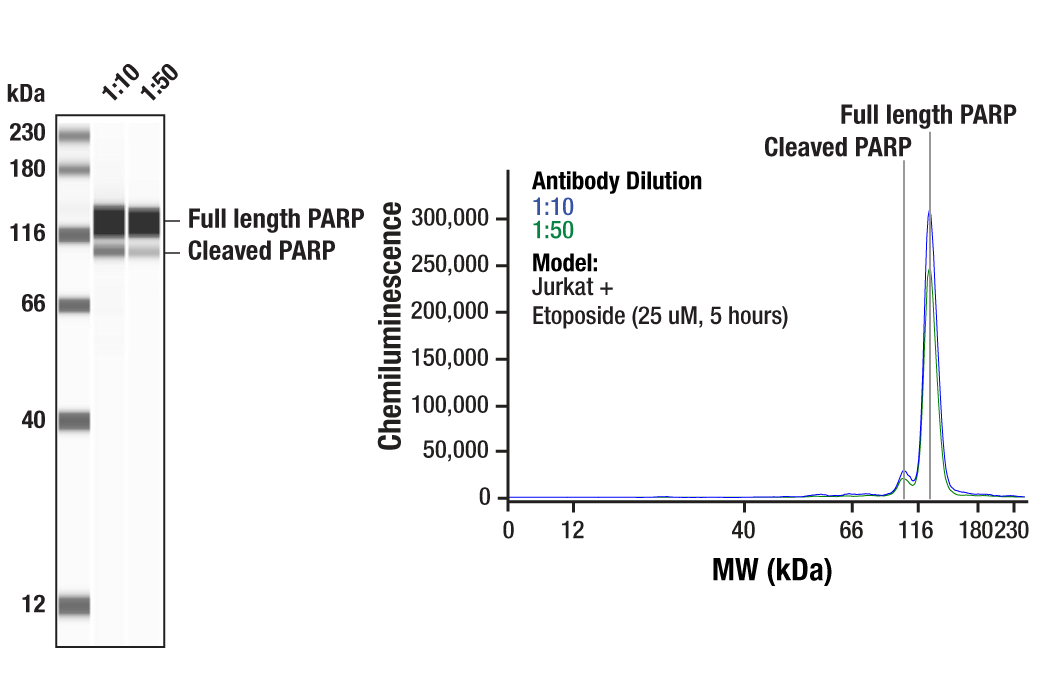
Simple Western™ analysis of lysates (1 mg/mL) from Jurkat cells treated with Etoposide (25 uM, 5 hours) using PARP (46D11) Rabbit mAb #9532. The virtual lane view (left) shows the target bands (as indicated) at 1:10 and 1:50 dilutions of primary antibody. The corresponding electropherogram view (right) plots chemiluminescence by molecular weight along the capillary at 1:10 (blue line) and 1:50 (green line) dilutions of primary antibody. This experiment was performed under reducing conditions on the Jess™ Simple Western instrument from ProteinSimple, a BioTechne brand, using the 12-230 kDa separation module.
Orders: 877-616-CELL (2355) • [email protected] • Support: 877-678-TECH (8324) • [email protected] •
Web:
cellsignal.com For Research Use Only. Not for Use in Diagnostic Procedures.
Revision 8
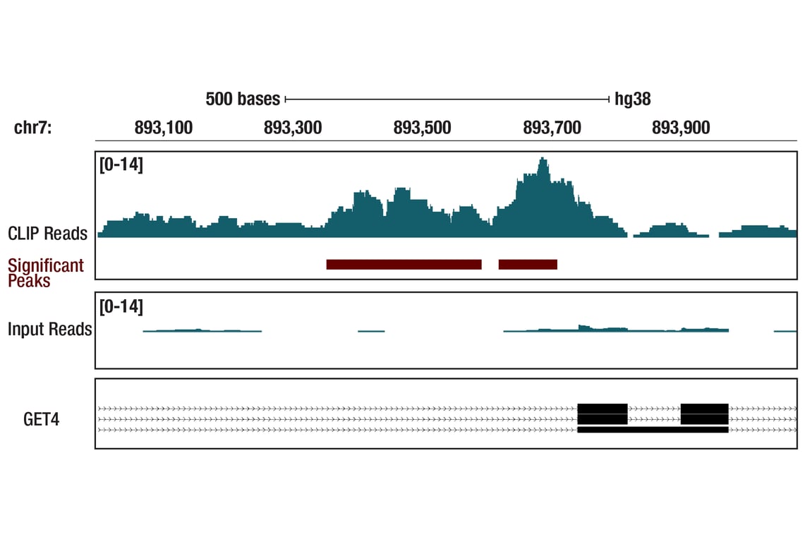
Enhanced cross-linking and immunoprecipitation (eCLIP) was performed with RNA from K-562 cells and PARP (46D11) Rabbit mAb using a protocol based on the RBP-eCLIP method from Eclipsebio. The figure shows binding across the GET4 transcript. Data is kindly provided by the laboratory of Dr. Gene Yeo and used with permission.
Orders: 877-616-CELL (2355) • [email protected] • Support: 877-678-TECH (8324) • [email protected] •
Web:
cellsignal.com For Research Use Only. Not for Use in Diagnostic Procedures.