| Product Includes | Product # | Quantity | Mol. Wt | Isotype/Source |
|---|---|---|---|---|
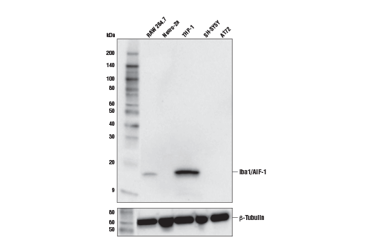
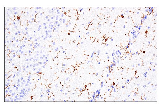
| Iba1/AIF-1 (E4O4W) XP® Rabbit mAb | 17198 | 20 µl | 17 kDa | Rabbit IgG |
| TMEM119 (E3E1O) Rabbit mAb | 90840 | 20 µl | Rabbit IgG | |
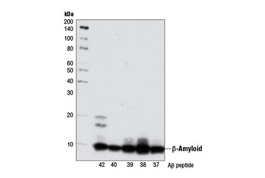
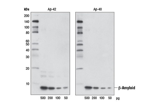
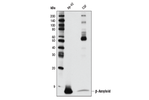
| β-Amyloid (D54D2) XP® Rabbit mAb | 8243 | 20 µl | 5 kDa | Rabbit IgG |
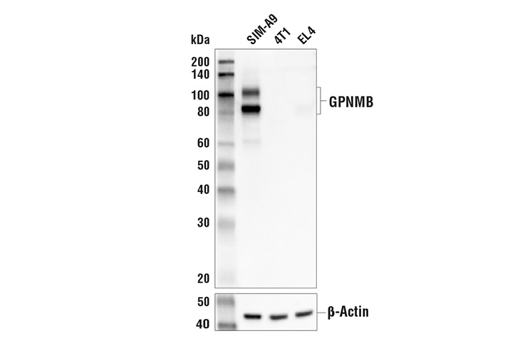
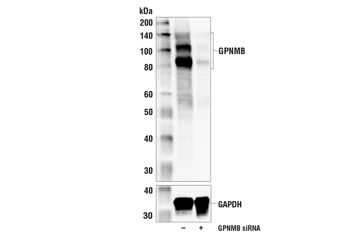
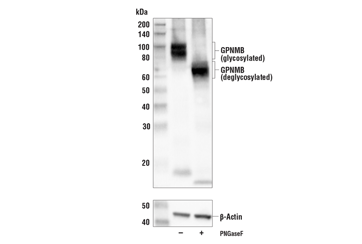
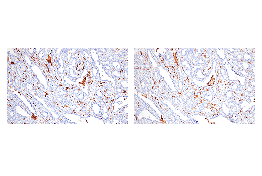
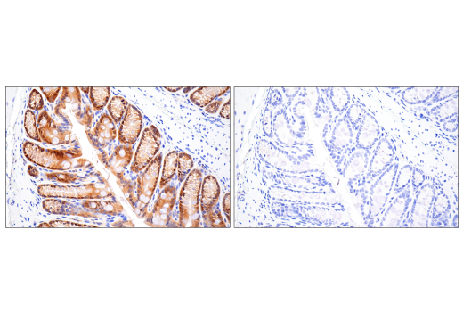
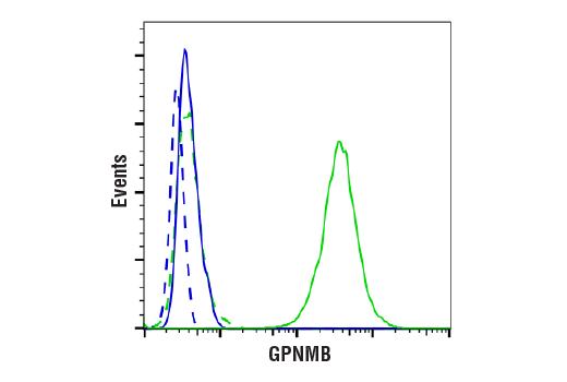
| GPNMB (E7U1Z) Rabbit mAb | 90205 | 20 µl | 90, 100 kDa | Rabbit IgG |
| CD11c (D1V9Y) Rabbit mAb | 97585 | 20 µl | 145 kDa | Rabbit IgG |
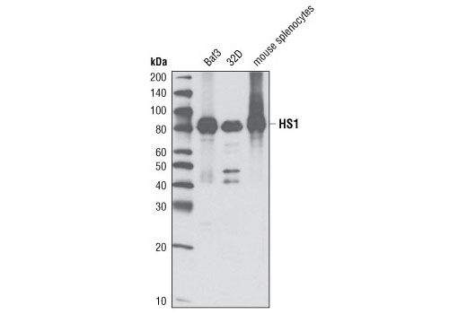
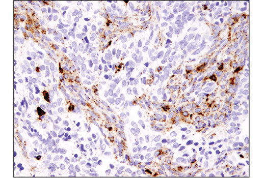
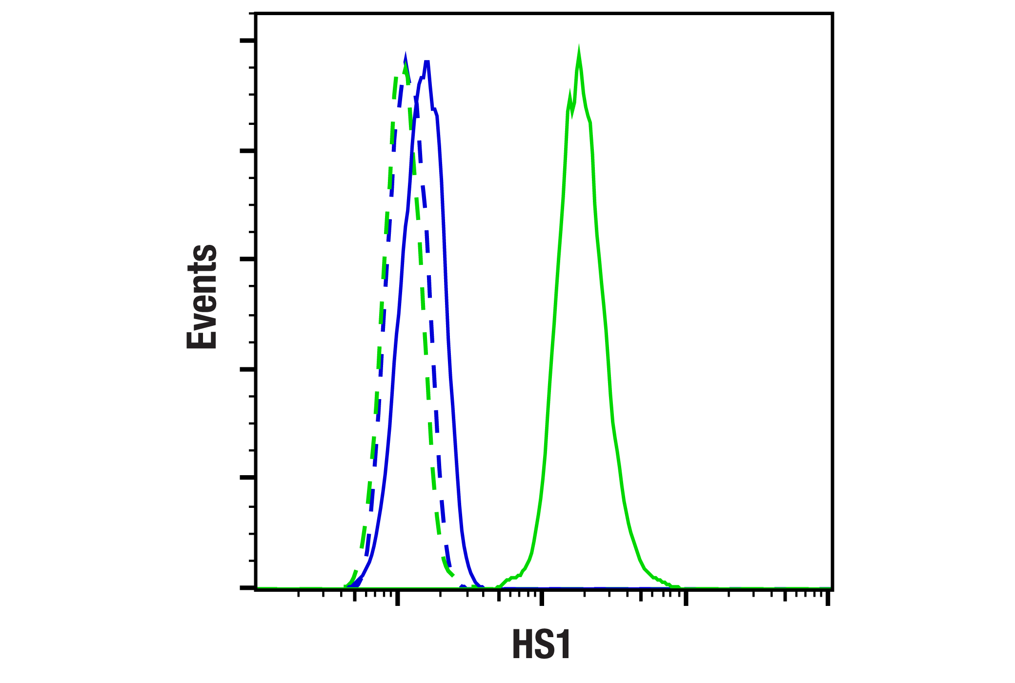
| HS1 (D5A9) XP® Rabbit mAb | 3892 | 20 µl | 80 kDa | Rabbit IgG |
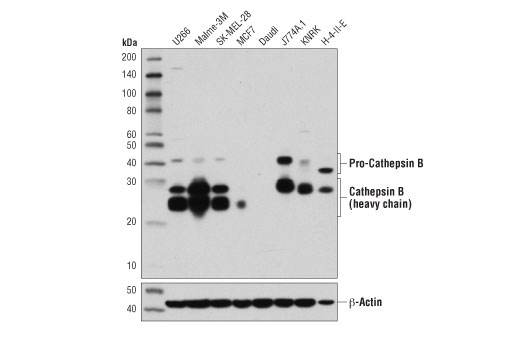
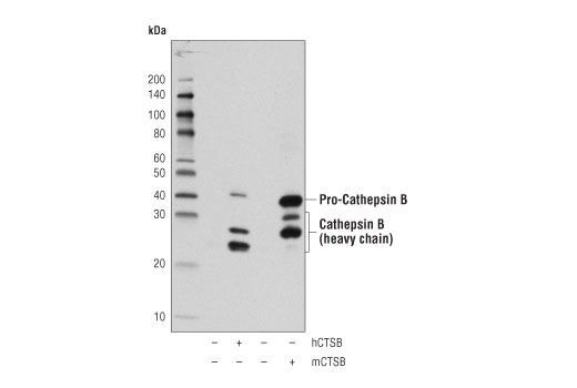
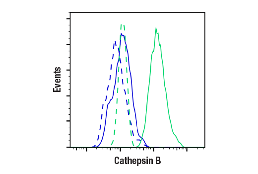
| Cathepsin B (D1C7Y) XP® Rabbit mAb | 31718 | 20 µl | 44, 27, 24 kDa | Rabbit IgG |
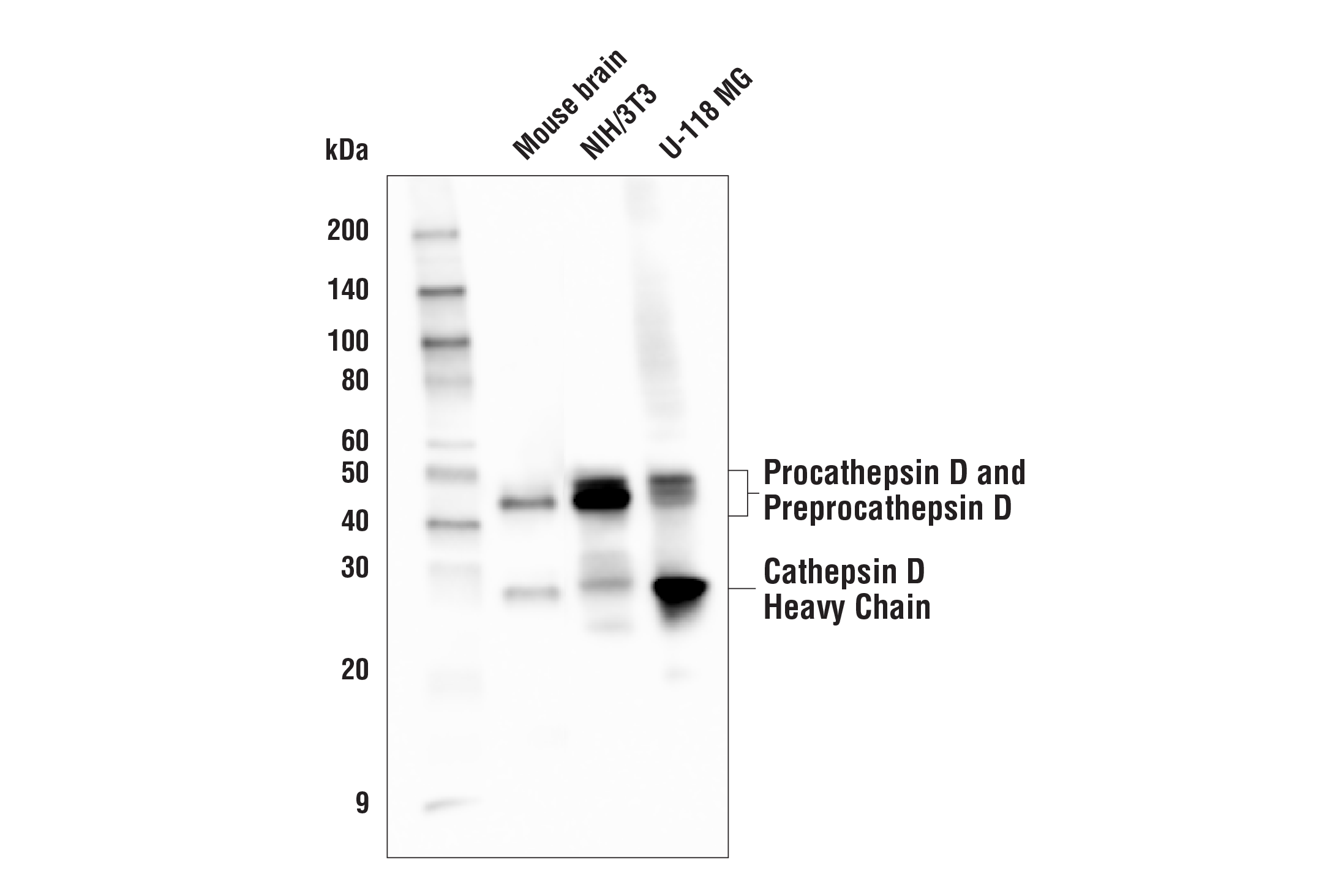
| Cathepsin D (E179) Antibody | 69854 | 20 µl | 46, 43, 28 kDa | Rabbit |
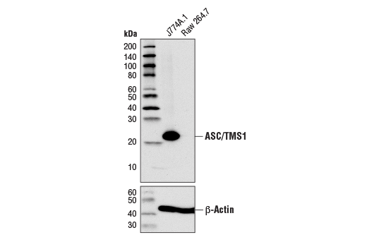
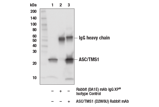
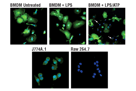
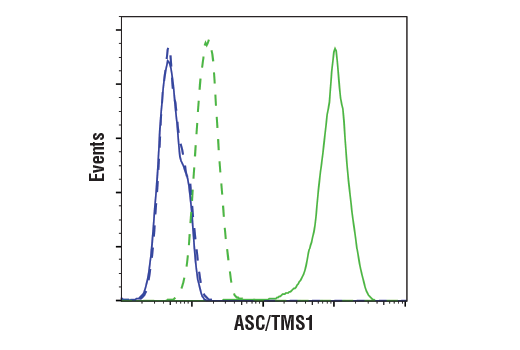
| ASC/TMS1 (D2W8U) Rabbit mAb | 67824 | 20 µl | 22 kDa | Rabbit IgG |
| Anti-rabbit IgG, HRP-linked Antibody | 7074 | 100 µl | Goat |
Please visit cellsignal.com for individual component applications, species cross-reactivity, dilutions, protocols, and additional product information.
Description
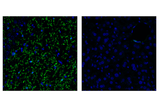
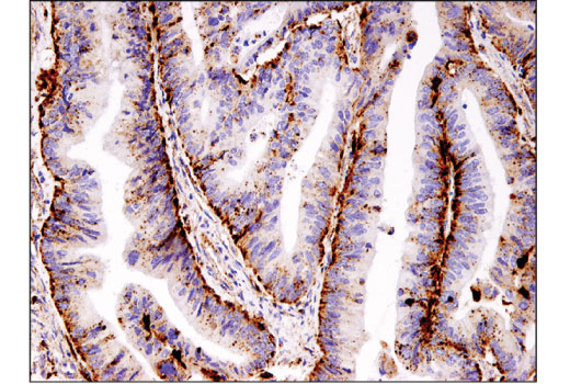
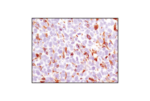
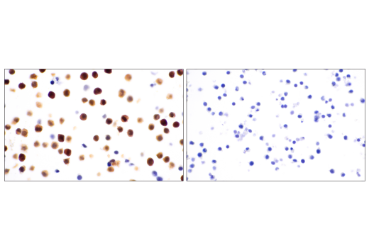
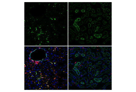
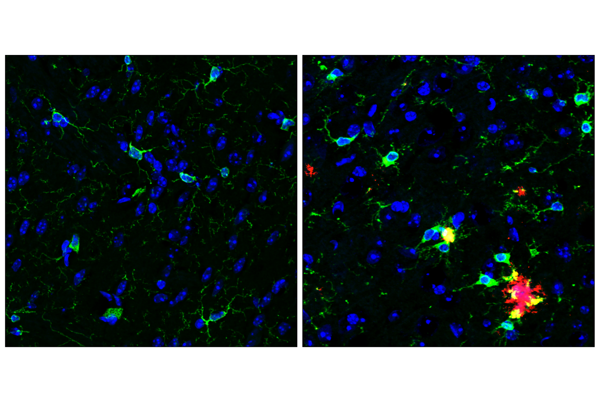
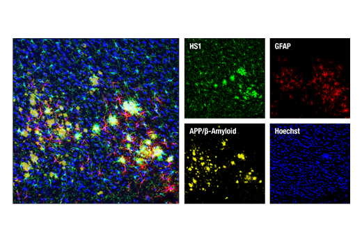
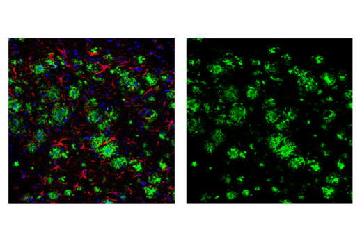
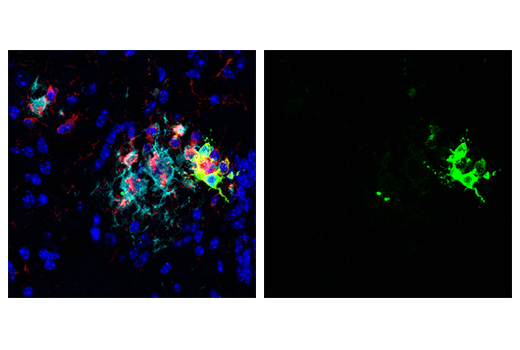
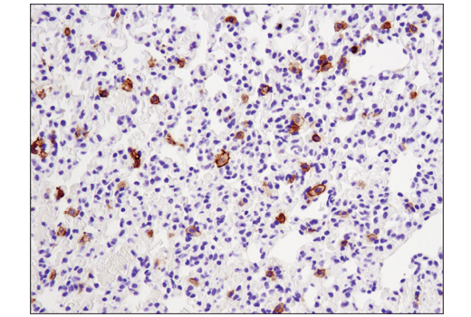
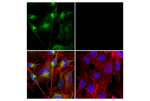
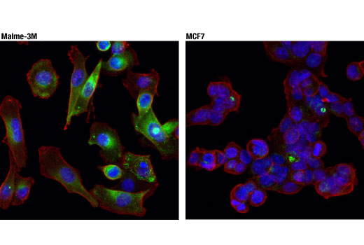
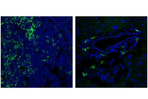
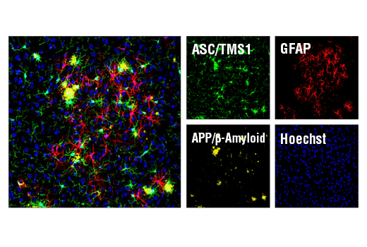
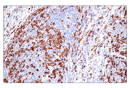
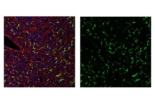
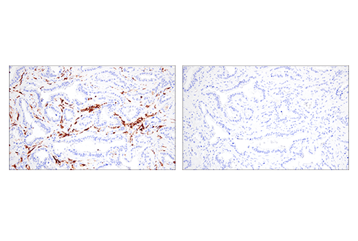
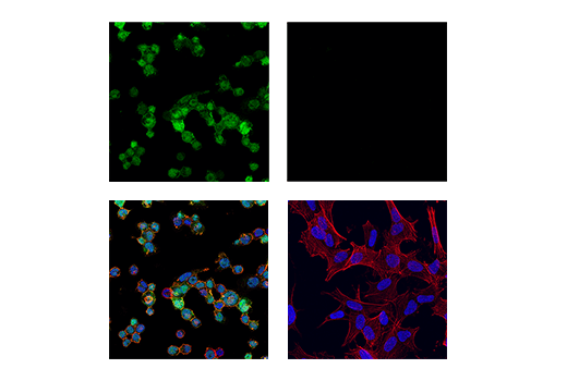
The Mouse Reactive Alzheimer's Disease Model Microglia Phenotyping IF Antibody Sampler Kit provides an economical means of detecting microglia proteins in β-Amyloid mouse models of Alzheimer’s Disease (AD) by immunofluorescence and/or western blot. This kit includes enough primary antibodies to perform at least twenty IF-F tests or two western blot experiments per primary antibody.
Storage
Background
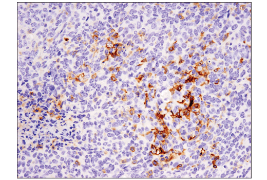
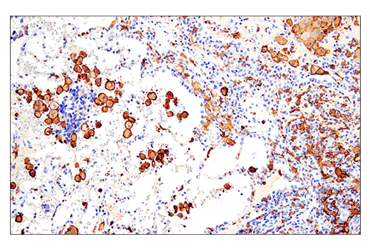
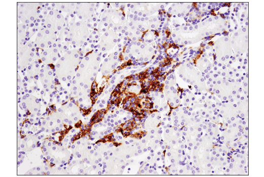
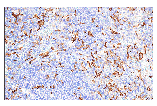
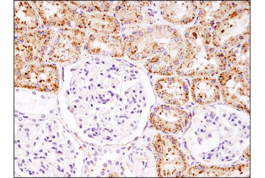
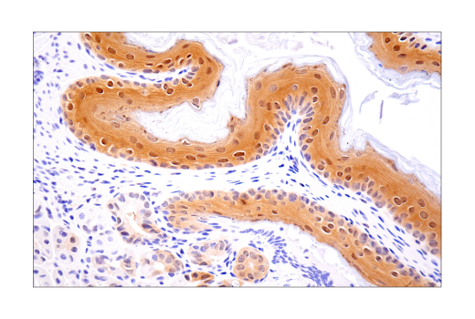
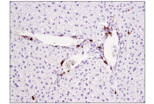
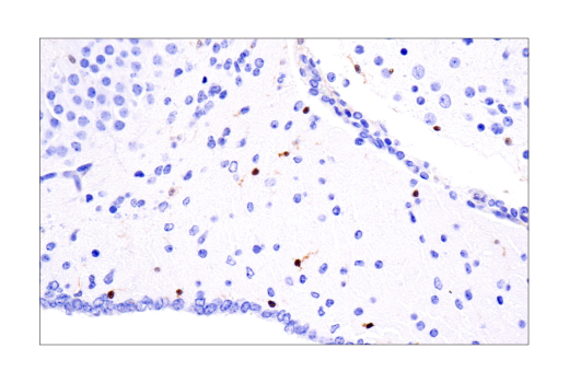
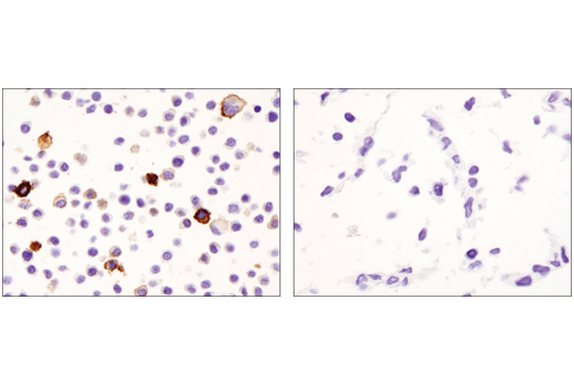
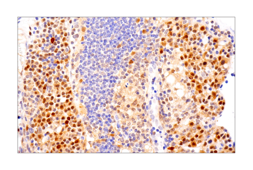
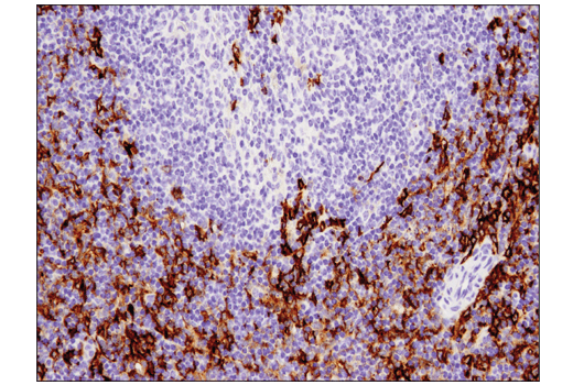
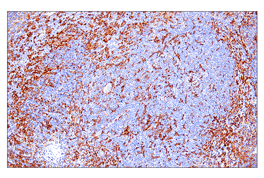
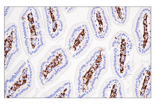
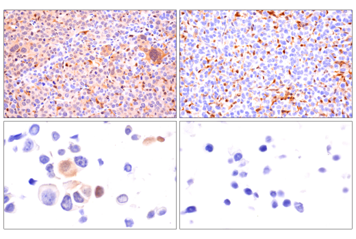
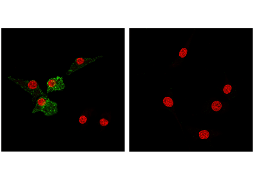
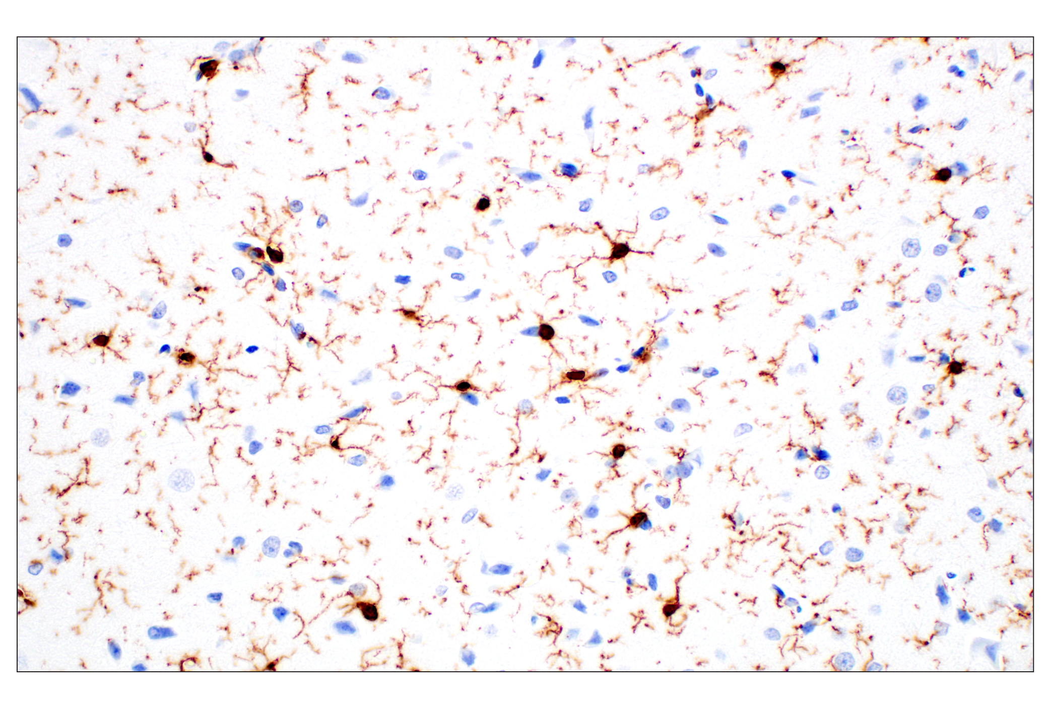
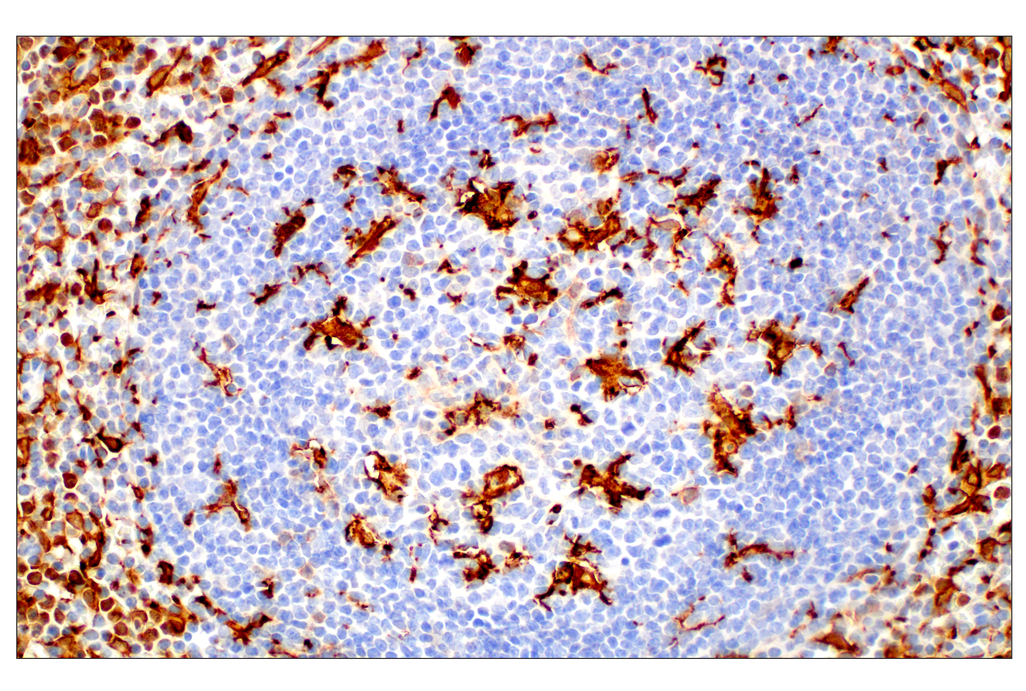
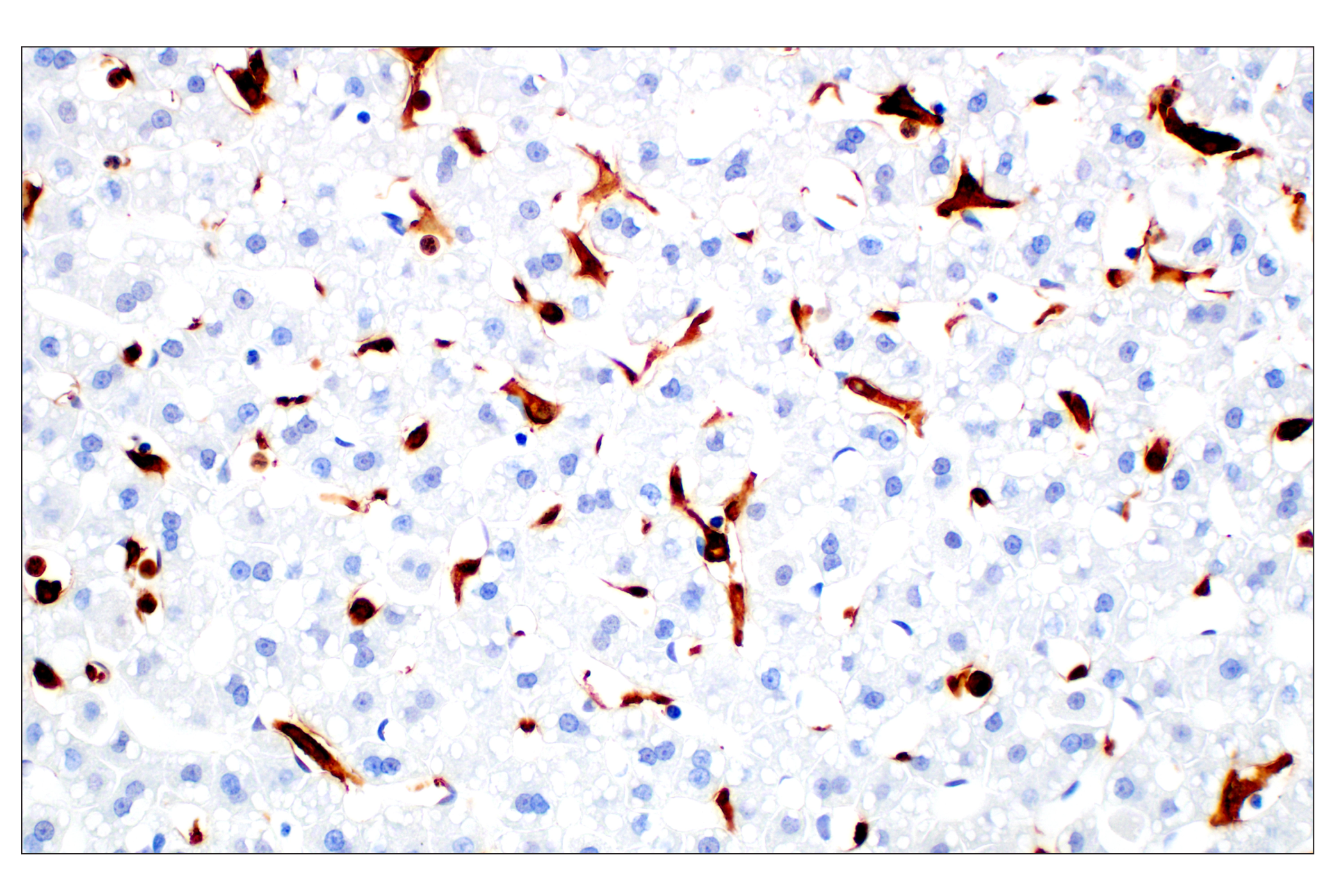
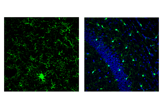
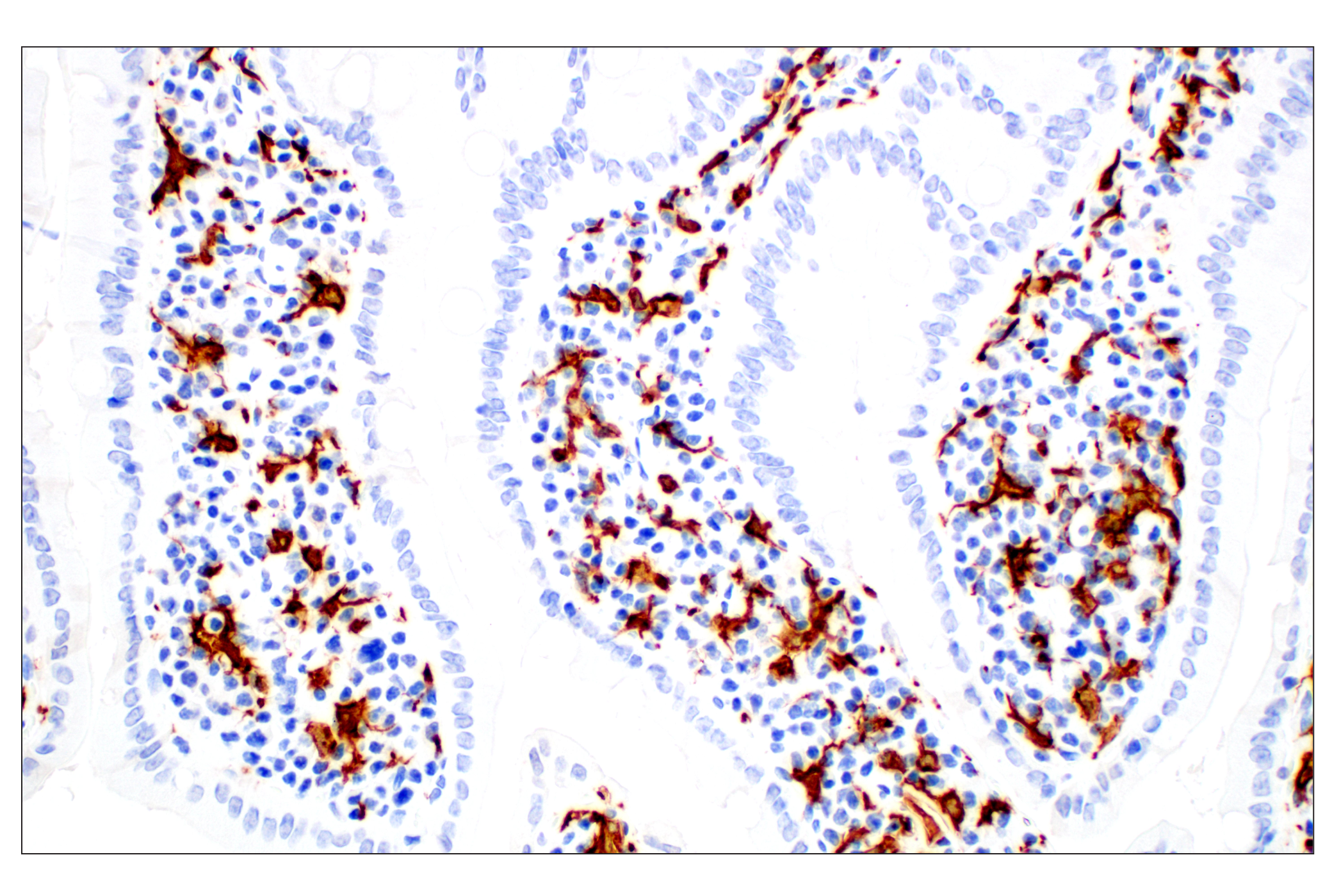
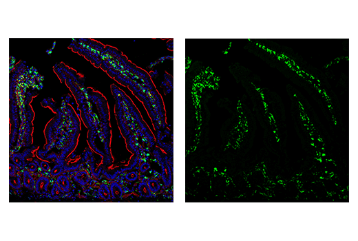
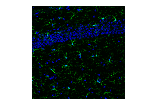
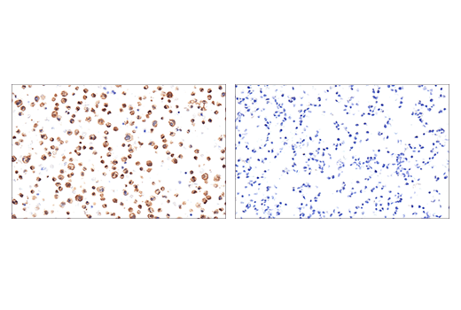
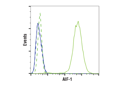
Distinct microglial activation states have been identified using RNA-seq data from a vast array of neurological disease and aging models. In both mouse models of Alzheimer’s Disease (AD) and AD patients, unique microglia molecular signatures are associated with disease progression (1-3). AD progression is correlated with the extracellular deposition and accumulation of the released Aβ fragments, derived from the transmembrane glycoprotein Amyloid β (Aβ) precursor protein (APP), that form amyloid plaques, the pathological hallmark of AD (4). Microglia are the resident macrophages of the brain and contribute to neurodegenerative disease (5). Ionized calcium-binding adaptor molecule 1 (Iba1), also known as allograft inflammatory factor 1 (AIF-1), is uniquely expressed in cells of monocytic lineage and is, therefore, widely used as a marker for microglia/macrophages in the brain and other tissue (6,7). HS1 (HCLS1, LckBP1, p75) is a protein kinase substrate that is expressed only in tissues and cells of hematopoietic origin and is also expressed in microglia (8,9). Transmembrane protein 119 (TMEM119) is a cell-surface protein of unknown function, expressed exclusively by the microglia subset of myeloid and neural cells (10). Iba1+ microglia with both ramified and amoeboid morphologies express TMEM119, while Iba1+ macrophages are TMEM119 negative (11). TMEM119 and other homeostatic genes have been shown to be downregulated in microglia. In addition to general markers of microglia, several microglia genes are upregulated during disease progression (12). CD11c (integrin αX, ITGAX) is a transmembrane glycoprotein that forms an α/β heterodimer with CD18 (integrin β2), which interacts with a variety of extracellular matrix molecules and cell surface proteins (13). CD11c-positive microglia transcriptionally correlate with amyloid plaques (14). In addition, other genes are upregulated in a similar manner. Glycoprotein non-metastatic gene B (GPNMB) is a type I transmembrane glycoprotein overexpressed in many types of cancer. The GPNMB glycoprotein is involved in many physiological processes, including mediating transport of late melanosomes to keratinocytes (9,15). Cathepsin B and D are widely expressed cysteine and aspartyl proteases, respectively, involved in the normal degradation of proteins (16,17). ASC/TMS1 has been found to be a critical component of inflammatory signaling where it associates with and activates caspase-1 in response to pro-inflammatory signals and may directly contribute to amyloid plaque formation (18,19).
- Keren-Shaul, H. et al. (2017) Cell 169, 1276-1290.e17.
- Mathys, H. et al. (2019) Nature 570, 332-337.
- Dubbelaar, M.L. et al. (2018) Front Immunol 9, 1753.
- Selkoe, D.J. (1996) J Biol Chem 271, 18295-8.
- Lewcock, J.W. et al. (2020) Neuron 108, 801-821.
- Schulze, J.O. et al. (2008) FEBS J 275, 4627-40.
- Deininger, M.H. et al. (2002) FEBS Lett 514, 115-21.
- Kitamura, D. et al. (1989) Nucleic Acids Res 17, 9367-79.
- Kitamura, D. et al. (1995) Biochem Biophys Res Commun 208, 1137-46.
- Satoh, J. et al. (2016) Neuropathology 36, 39-49.
- Deczkowska, A. et al. (2018) Cell 173, 1073-1081.
- Hansen, D.V. et al. (2018) J Cell Biol 217, 459-472.
- Uotila, L.M. et al. (2013) J Biol Chem 288, 33494-9.
- Kamphuis, W. et al. (2016) Biochim Biophys Acta 1862, 1847-60.
- Tomihari, M. et al. (2009) Exp Dermatol 18, 586-95.
- Gan, L. et al. (2004) J Biol Chem 279, 5565-72.
- Faust, P.L. et al. (1985) Proc Natl Acad Sci U S A 82, 4910-4.
- Srinivasula, S.M. et al. (2002) J Biol Chem 277, 21119-22.
- Venegas, C. and Heneka, M.T. (2019) FASEB J 33, 13075-13084.
Background References
Trademarks and Patents
限制使用
除非 CST 的合法授书代表以书面形式书行明确同意,否书以下条款适用于 CST、其关书方或分书商提供的书品。 任何书充本条款或与本条款不同的客书条款和条件,除非书 CST 的合法授书代表以书面形式书独接受, 否书均被拒书,并且无效。
专品专有“专供研究使用”的专专或专似的专专声明, 且未专得美国食品和专品管理局或其他外国或国内专管机专专专任何用途的批准、准专或专可。客专不得将任何专品用于任何专断或治专目的, 或以任何不符合专专声明的方式使用专品。CST 专售或专可的专品提供专作专最专用专的客专,且专用于研专用途。将专品用于专断、专防或治专目的, 或专专售(专独或作专专成)或其他商专目的而专专专品,均需要 CST 的专独专可。客专:(a) 不得专独或与其他材料专合向任何第三方出售、专可、 出借、捐专或以其他方式专专或提供任何专品,或使用专品制造任何商专专品,(b) 不得复制、修改、逆向工程、反专专、 反专专专品或以其他方式专专专专专品的基专专专或技专,或使用专品开专任何与 CST 的专品或服专专争的专品或服专, (c) 不得更改或专除专品上的任何商专、商品名称、徽专、专利或版专声明或专专,(d) 只能根据 CST 的专品专售条款和任何适用文档使用专品, (e) 专遵守客专与专品一起使用的任何第三方专品或服专的任何专可、服专条款或专似专专