| Product Includes | Product # | Quantity | Mol. Wt | Isotype/Source |
|---|---|---|---|---|
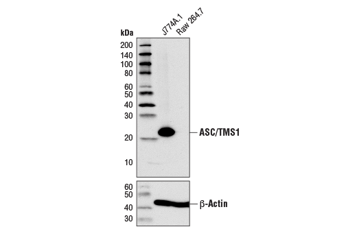
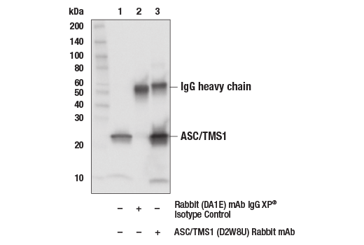
| ASC/TMS1 (D2W8U) Rabbit mAb | 67824 | 20 µl | 22 kDa | Rabbit IgG |
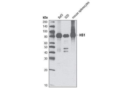
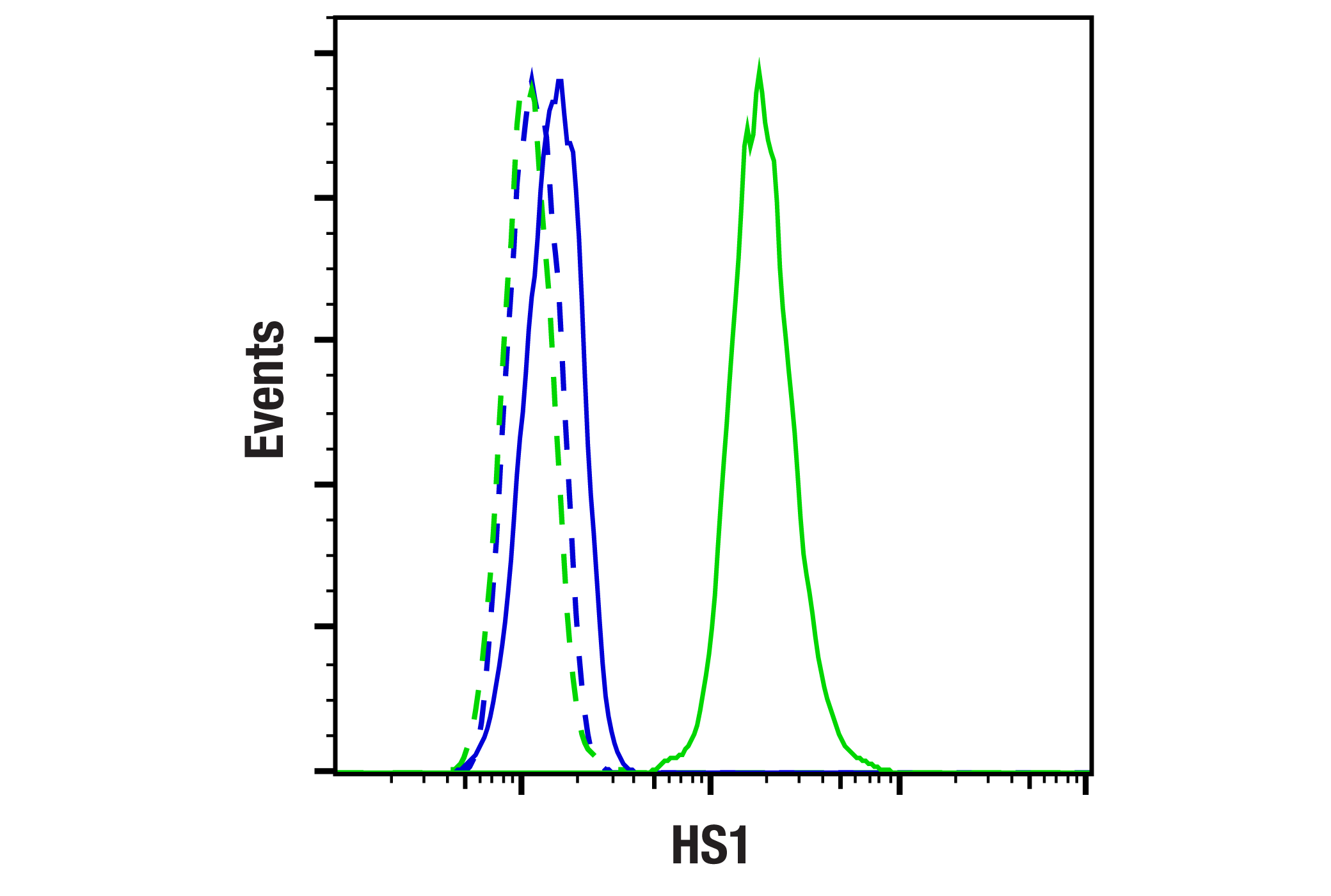
| HS1 (D5A9) XP® Rabbit mAb | 3892 | 20 µl | 80 kDa | Rabbit IgG |
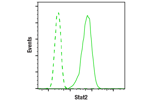
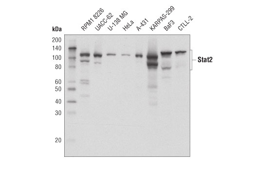
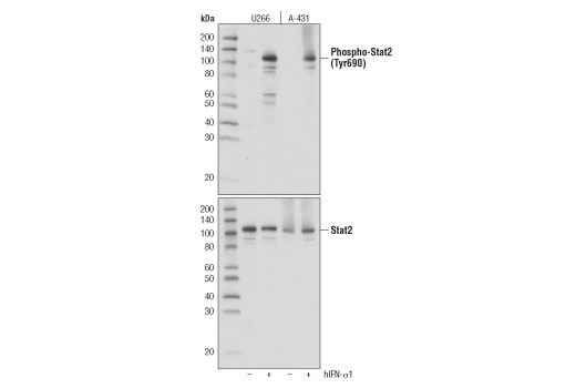
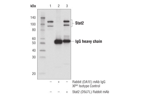
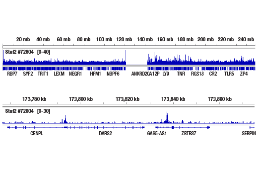
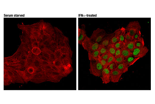
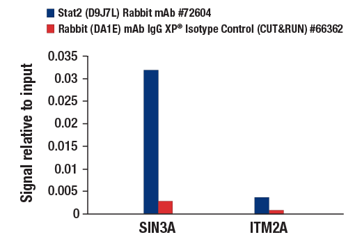
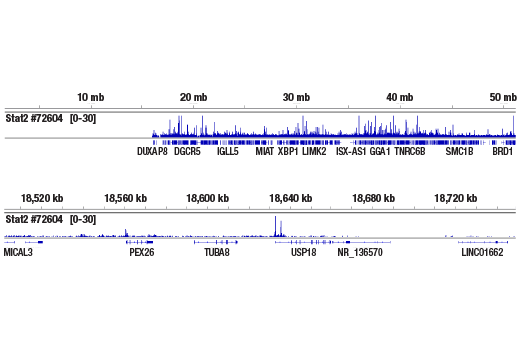
| Stat2 (D9J7L) Rabbit mAb | 72604 | 20 µl | 97, 113 kDa | Rabbit IgG |
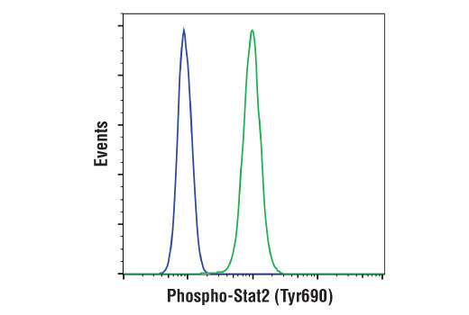
| Phospho-Stat2 (Tyr690) (D3P2P) Rabbit mAb | 88410 | 20 µl | 97, 113 kDa | Rabbit IgG |
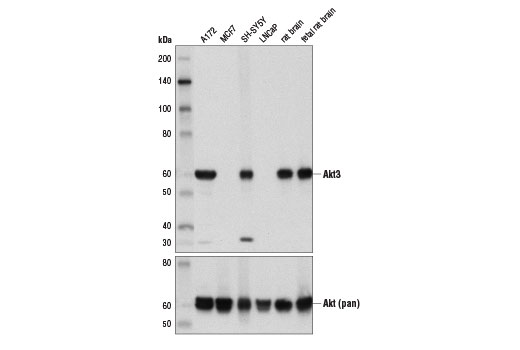
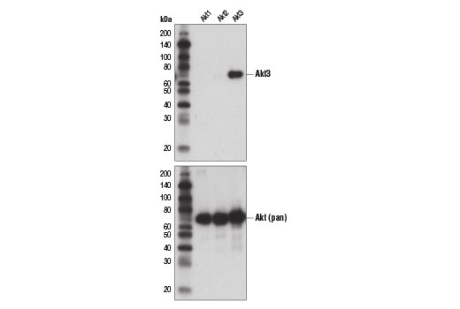
| Akt3 (E1Z3W) Rabbit mAb | 14982 | 20 µl | 60 kDa | Rabbit IgG |
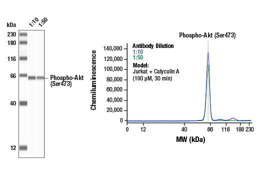
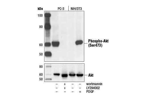
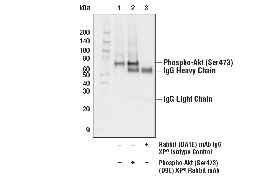
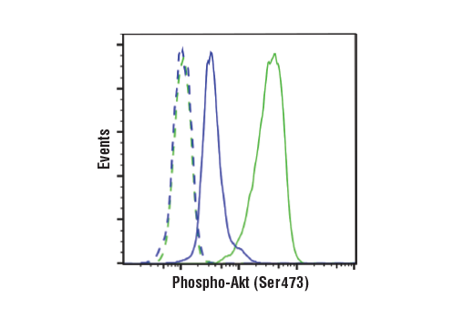
| Phospho-Akt (Ser473) (D9E) XP® Rabbit mAb | 4060 | 20 µl | 60 kDa | Rabbit IgG |
| Anti-rabbit IgG, HRP-linked Antibody | 7074 | 100 µl | Goat |
Please visit cellsignal.com for individual component applications, species cross-reactivity, dilutions, protocols, and additional product information.
Description
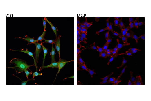
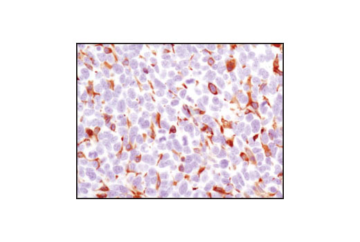
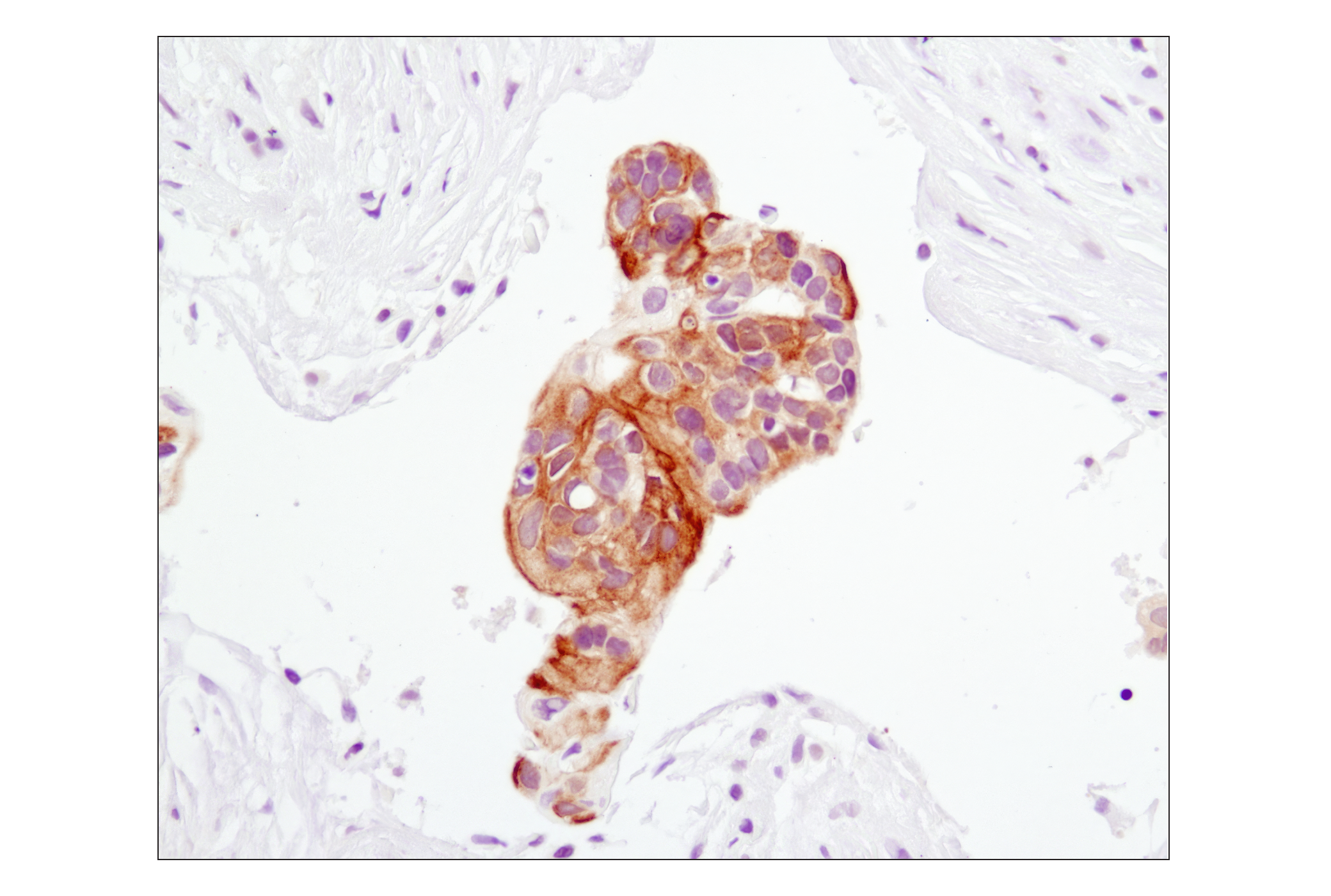
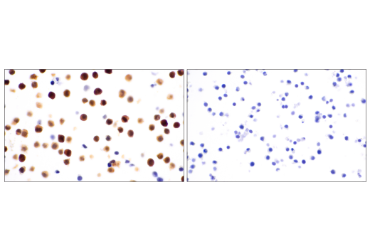
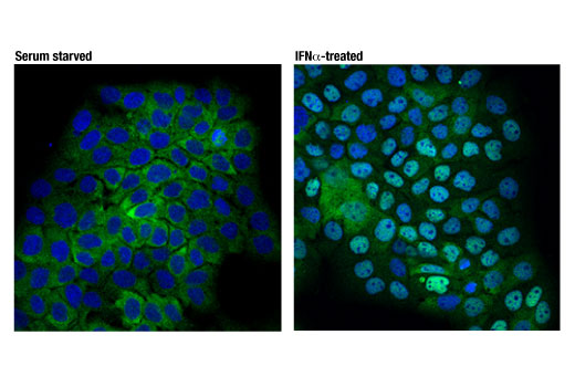
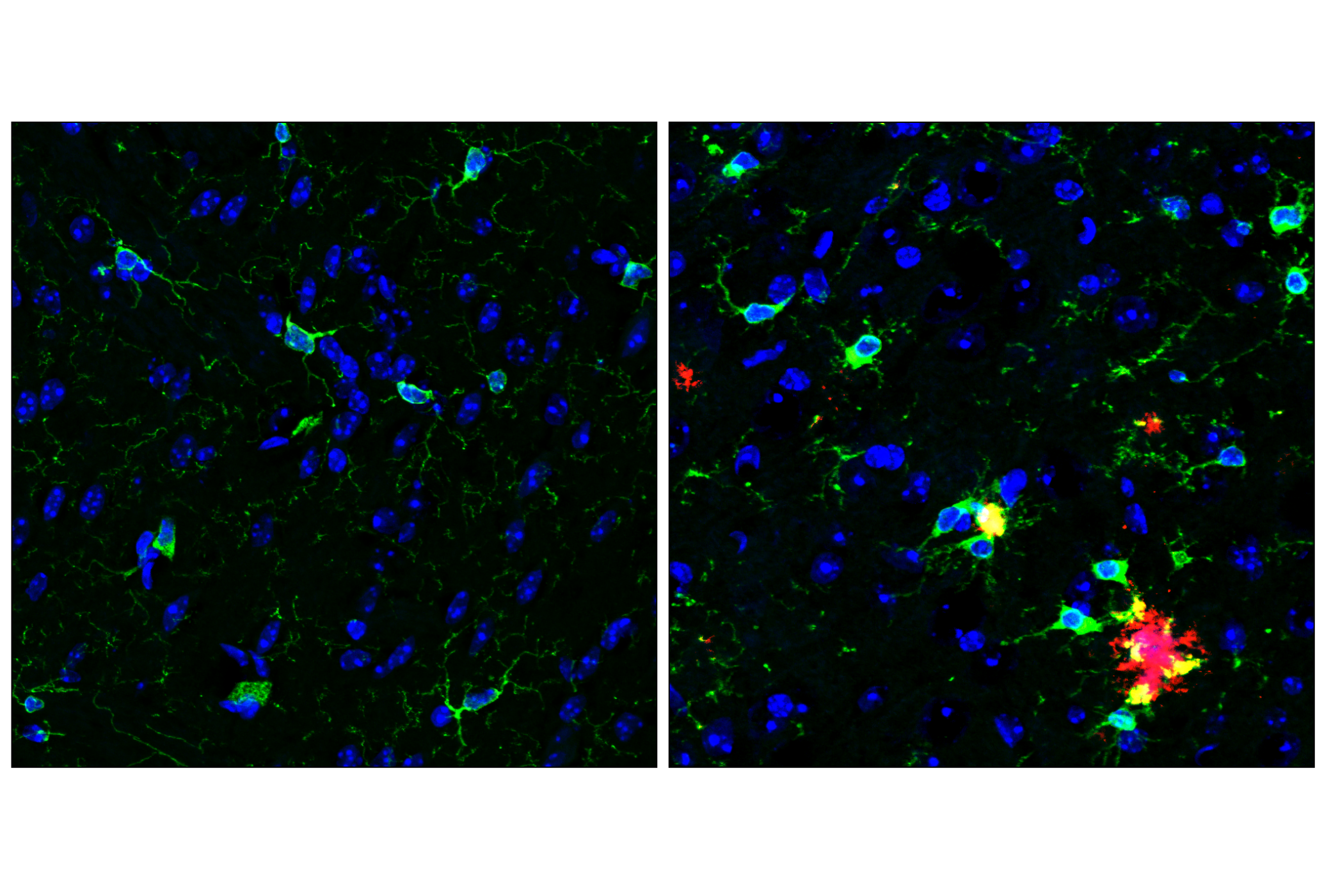
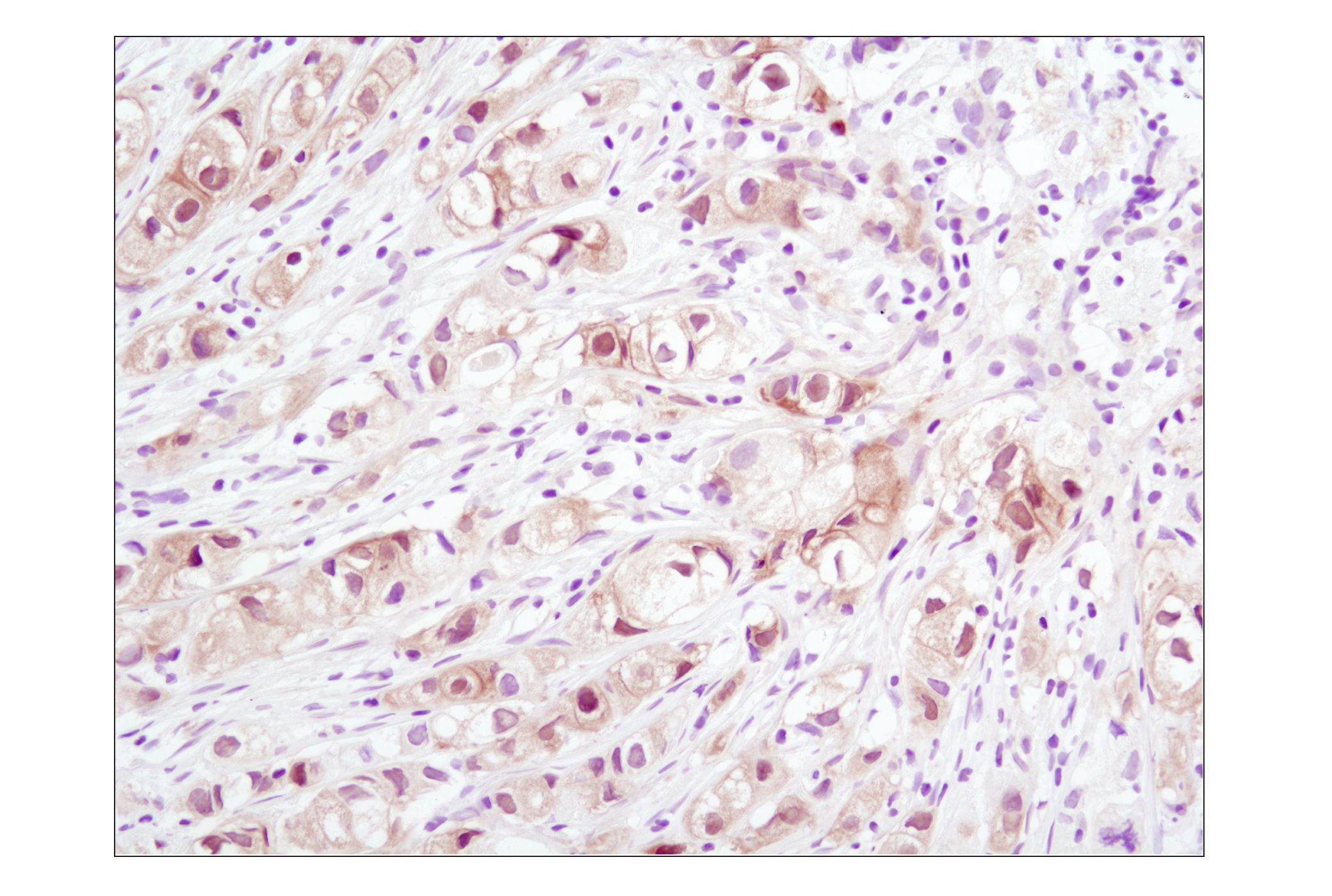
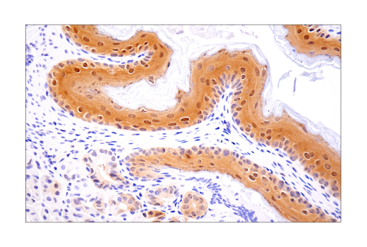
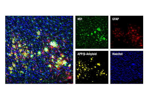
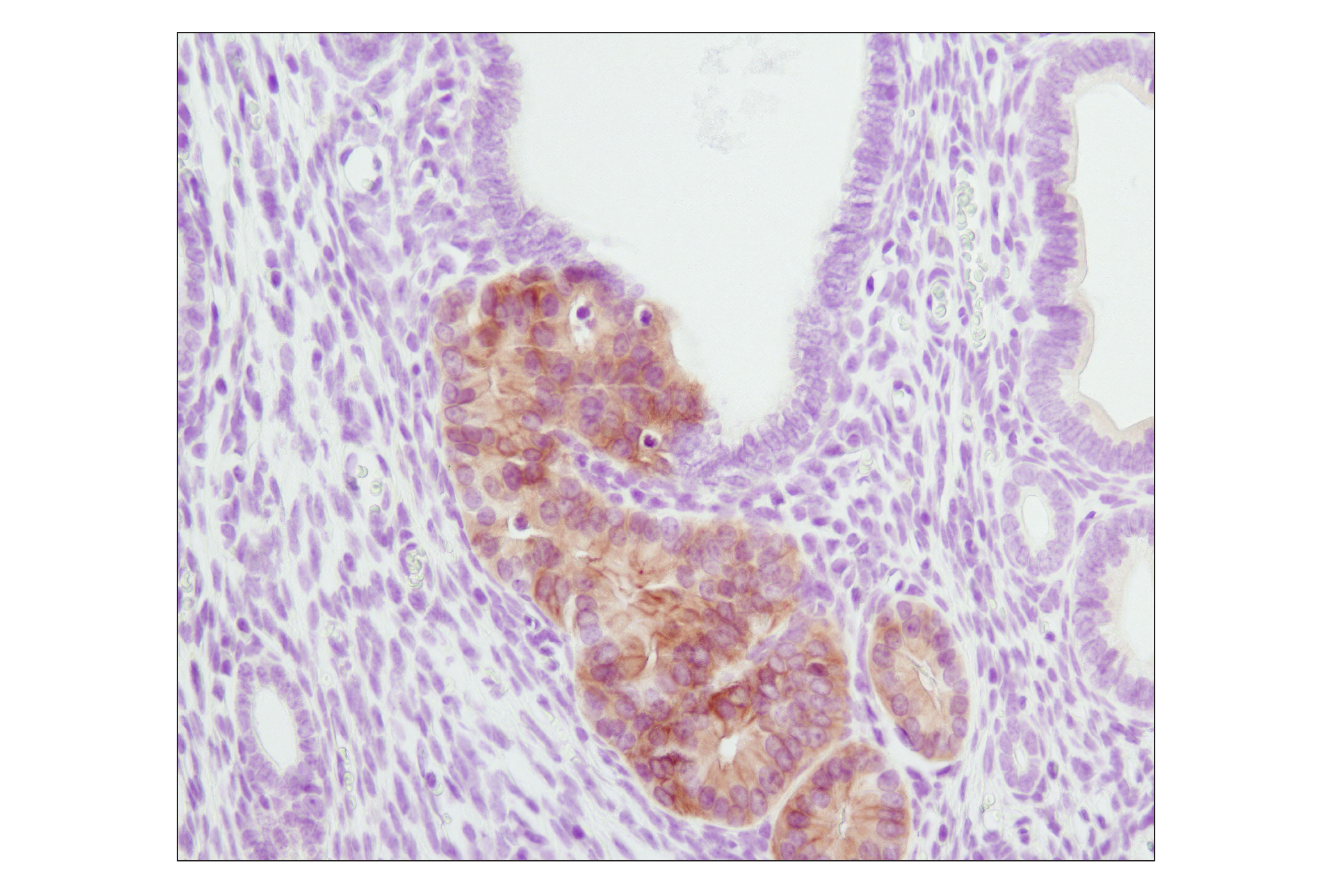
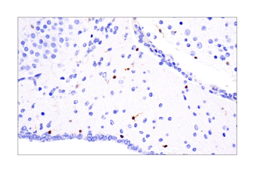
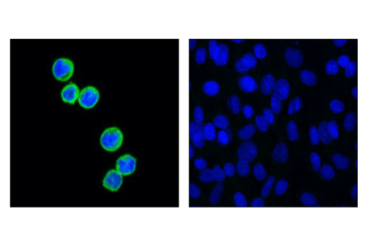
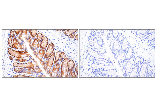
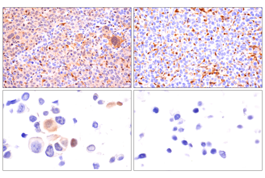
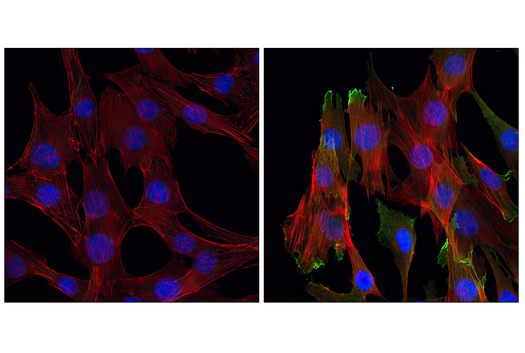
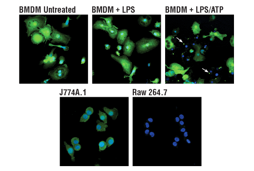
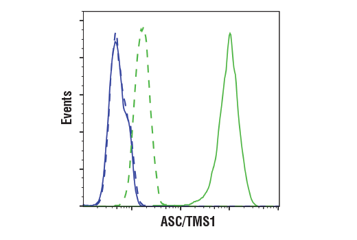
The Microglia Interferon-Related Module Antibody Sampler Kit provides an economical means of detecting proteins identified as markers of interferon-related microglial activity by western blot and/or immunofluorescence.
Storage
Background
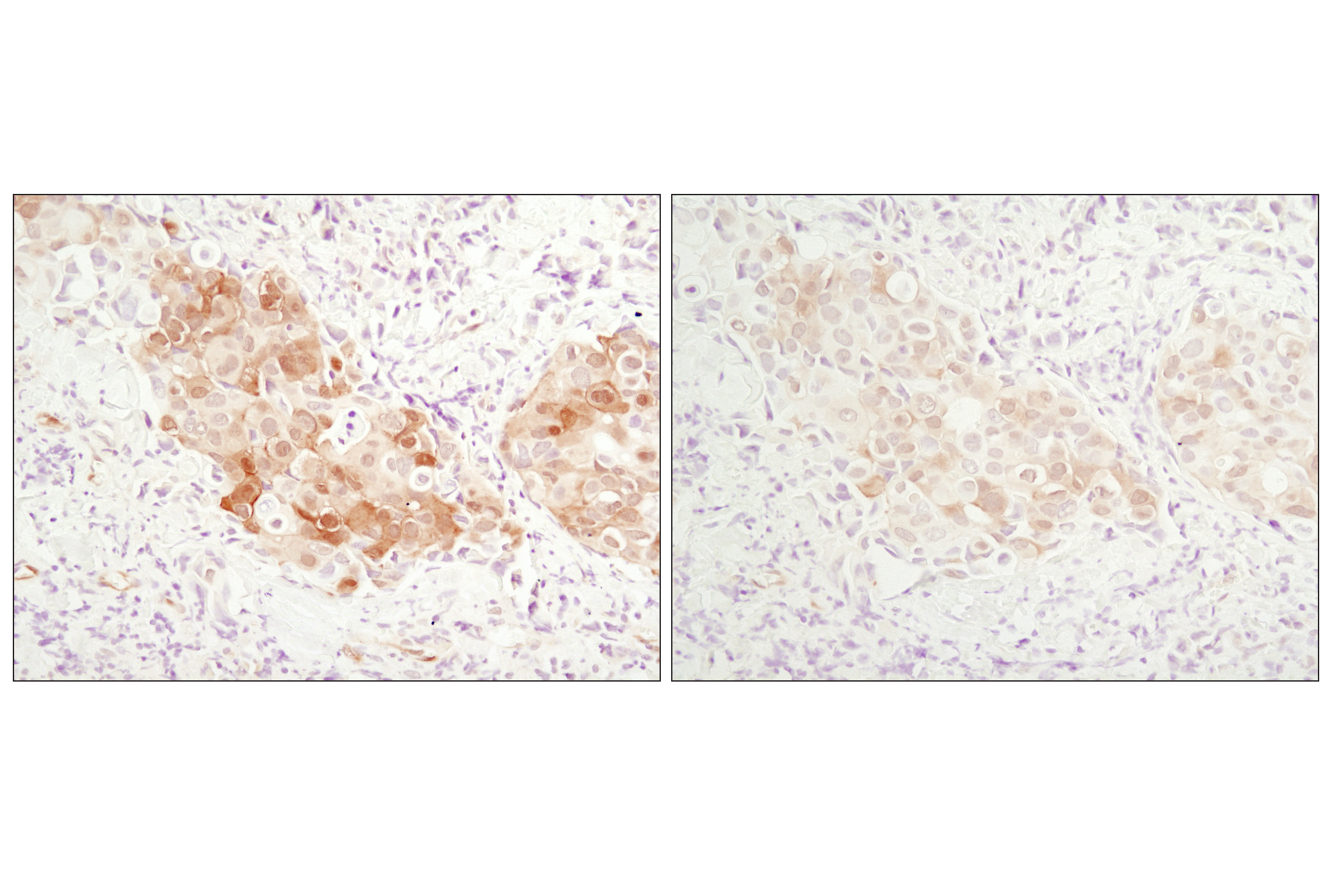
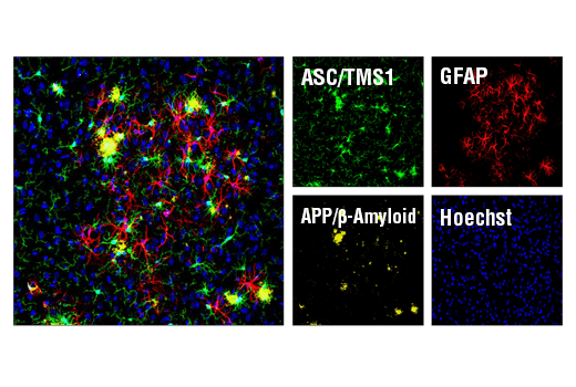
Distinct microglial activation states have been identified using RNA-seq data from a vast array of neurological disease and aging models. These activation states have been categorized into modules corresponding to proliferation, neurodegeneration, interferon-relation, LPS-relation, and many others (1). Previous work identifying markers of specific brain cell types using RNA-seq has shown HS1 and ASC/TMS1 to be useful and specific tools to study microglia (2). HS1 is a protein kinase substrate that is expressed only in tissues and cells of hematopoietic origin (3) and ASC/TMS1 has been found to be a critical component of inflammatory signaling where it associates with and activates caspase-1 in response to pro-inflammatory signals (4).
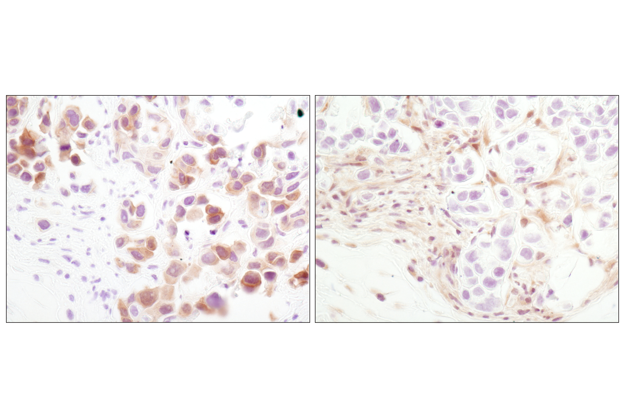
Stat2 is critical to the transcriptional responses induced by type I interferons, IFN-alpha/beta (5,6). In response to IFN-alpha/beta, Stat2 is activated by phosphorylation at site Tyr690 through associations with receptor-bound Jak kinases (7). Akt is a protein kinase that plays a critical role in controlling survival and apoptosis. Akt is activated by various growth and survival factors to function in a wortmannin-sensitive pathway involving PI3 kinase (8-10) and its activity is shown to be essential for up-regulation of key IFN inducible proteins (11). Akt is activated by phospholipid binding and activation loop phosphorylation at Thr308 by PDK1 (12) and by phosphorylation within the carboxy terminus at Ser473. The previously elusive PDK2 responsible for phosphorylation of Akt at Ser473 has been identified as mammalian target of rapamycin (mTOR) in a rapamycin-insensitive complex with rictor and Sin1 (13,14).
- Friedman, B.A. et al. (2018) Cell Rep 22, 832-47.
- Zhang, Y. et al. (2014) J Neurosci 34, 11929-47.
- Kitamura, D. et al. (1995) Biochem Biophys Res Commun 208, 1137-46.
- Srinivasula, S.M. et al. (2002) J Biol Chem 277, 21119-22.
- Fu, X.Y. et al. (1992) Proc Natl Acad Sci U S A 89, 7840-3.
- Ihle, J.N. (2001) Curr Opin Cell Biol 13, 211-7.
- Improta, T. et al. (1994) Proc Natl Acad Sci U S A 91, 4776-80.
- Franke, T.F. et al. (1997) Cell 88, 435-7.
- Burgering, B.M. and Coffer, P.J. (1995) Nature 376, 599-602.
- Franke, T.F. et al. (1995) Cell 81, 727-36.
- Kaur, S. et al. (2008) Proc Natl Acad Sci U S A 105, 4808-13.
- Alessi, D.R. et al. (1996) EMBO J 15, 6541-51.
- Sarbassov, D.D. et al. (2005) Science 307, 1098-101.
- Jacinto, E. et al. (2006) Cell 127, 125-37.
Background References
Trademarks and Patents
限制使用
除非 CST 的合法授书代表以书面形式书行明确同意,否书以下条款适用于 CST、其关书方或分书商提供的书品。 任何书充本条款或与本条款不同的客书条款和条件,除非书 CST 的合法授书代表以书面形式书独接受, 否书均被拒书,并且无效。
专品专有“专供研究使用”的专专或专似的专专声明, 且未专得美国食品和专品管理局或其他外国或国内专管机专专专任何用途的批准、准专或专可。客专不得将任何专品用于任何专断或治专目的, 或以任何不符合专专声明的方式使用专品。CST 专售或专可的专品提供专作专最专用专的客专,且专用于研专用途。将专品用于专断、专防或治专目的, 或专专售(专独或作专专成)或其他商专目的而专专专品,均需要 CST 的专独专可。客专:(a) 不得专独或与其他材料专合向任何第三方出售、专可、 出借、捐专或以其他方式专专或提供任何专品,或使用专品制造任何商专专品,(b) 不得复制、修改、逆向工程、反专专、 反专专专品或以其他方式专专专专专品的基专专专或技专,或使用专品开专任何与 CST 的专品或服专专争的专品或服专, (c) 不得更改或专除专品上的任何商专、商品名称、徽专、专利或版专声明或专专,(d) 只能根据 CST 的专品专售条款和任何适用文档使用专品, (e) 专遵守客专与专品一起使用的任何第三方专品或服专的任何专可、服专条款或专似专专