WB, IF-IC
H M R Mk
Endogenous
40
Rabbit
#O75367-1
9555
Product Information
Product Usage Information
| Application | Dilution |
|---|---|
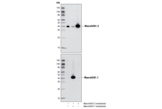
| Western Blotting | 1:1000 |
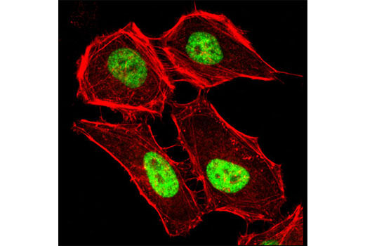
| Immunofluorescence (Immunocytochemistry) | 1:200 |
Storage
Specificity / Sensitivity
Species Reactivity:
Human, Mouse, Rat, Monkey
Species predicted to react based on 100% sequence homology
The antigen sequence used to produce this antibody shares
100% sequence homology with the species listed here, but
reactivity has not been tested or confirmed to work by CST.
Use of this product with these species is not covered under
our
Product Performance Guarantee.
Chicken, Bovine
Source / Purification
Polyclonal antibodies are produced by immunizing animals with a synthetic peptide corresponding to the human MacroH2A1.2 protein (MacroH2A1, isoform 2). Antibodies are purified by protein A and peptide affinity chromatography.
Background
Histone macroH2A1 and macroH2A2 comprise a family of variant histone H2A proteins. MacroH2A1 exists as two distinct isoforms due to alternative splicing of a single gene; macroH2A1.1 levels accumulate throughout differentiation and development while macroH2A1.2 shows a constant level of expression (1). MacroH2A1 and macroH2A2 are encoded by completely distinct genes located on separate chromosomes (2,3). Both macroH2A1 and macroH2A2 proteins contain an amino-terminal histone-like region with 64% sequence identity to canonical histone H2A, in addition to a carboxy-terminal “macro” domain (1-3). MacroH2A1 and macroH2A2 are enriched in facultative heterochromatin, including inactivated X chromosomes in mammalian females and senescence-associated heterochromatin foci (2-5). Both act to repress gene transcription by inhibiting the binding of transcription factors to chromatin, the acetylation of histones by p300, and the chromatin-remodeling activities of SWI/SNF and ACF (6,7). The macro domain of macroH2A1.1 binds to ADP-ribose and functions to recruit macroH2A1.1 to activated PARP at sites of DNA damage, where it mediates chromatin rearrangements to locally regulate the DNA damage response (8). MacroH2A1.2 and macroH2A2 do not bind poly-ADP-ribose and are not recruited to sites of activated PARP (8).
- Pehrson, J.R. et al. (1997) J Cell Biochem 65, 107-13.
- Chadwick, B.P. and Willard, H.F. (2001) Hum Mol Genet 10, 1101-13.
- Costanzi, C. and Pehrson, J.R. (2001) J Biol Chem 276, 21776-84.
- Costanzi, C. and Pehrson, J.R. (1998) Nature 393, 599-601.
- Zhang, R. et al. (2005) Dev Cell 8, 19-30.
- Angelov, D. et al. (2003) Mol Cell 11, 1033-41.
- Doyen, C.M. et al. (2006) Mol Cell Biol 26, 1156-64.
- Timinszky, G. et al. (2009) Nat Struct Mol Biol 16, 923-9.
Species Reactivity
Species reactivity is determined by testing in at least one approved application (e.g., western blot).
Western Blot Buffer
IMPORTANT: For western blots, incubate membrane with diluted primary antibody in 5% w/v BSA, 1X TBS, 0.1% Tween® 20 at 4°C with gentle shaking, overnight.
Applications Key
WB: Western Blotting IF-IC: Immunofluorescence (Immunocytochemistry)
Cross-Reactivity Key
H: human M: mouse R: rat Hm: hamster Mk: monkey Vir: virus Mi: mink C: chicken Dm: D. melanogaster X: Xenopus Z: zebrafish B: bovine Dg: dog Pg: pig Sc: S. cerevisiae Ce: C. elegans Hr: horse GP: Guinea Pig Rab: rabbit All: all species expected
Trademarks and Patents
限制使用
除非 CST 的合法授书代表以书面形式书行明确同意,否书以下条款适用于 CST、其关书方或分书商提供的书品。 任何书充本条款或与本条款不同的客书条款和条件,除非书 CST 的合法授书代表以书面形式书独接受, 否书均被拒书,并且无效。
专品专有“专供研究使用”的专专或专似的专专声明, 且未专得美国食品和专品管理局或其他外国或国内专管机专专专任何用途的批准、准专或专可。客专不得将任何专品用于任何专断或治专目的, 或以任何不符合专专声明的方式使用专品。CST 专售或专可的专品提供专作专最专用专的客专,且专用于研专用途。将专品用于专断、专防或治专目的, 或专专售(专独或作专专成)或其他商专目的而专专专品,均需要 CST 的专独专可。客专:(a) 不得专独或与其他材料专合向任何第三方出售、专可、 出借、捐专或以其他方式专专或提供任何专品,或使用专品制造任何商专专品,(b) 不得复制、修改、逆向工程、反专专、 反专专专品或以其他方式专专专专专品的基专专专或技专,或使用专品开专任何与 CST 的专品或服专专争的专品或服专, (c) 不得更改或专除专品上的任何商专、商品名称、徽专、专利或版专声明或专专,(d) 只能根据 CST 的专品专售条款和任何适用文档使用专品, (e) 专遵守客专与专品一起使用的任何第三方专品或服专的任何专可、服专条款或专似专专