Revision 3
#36833
Store at -20C
Late-Onset Alzheimer's Disease Risk Gene Antibody Sampler Kit
1 Kit
(9 x 20 microliters)
877-616-CELL (2355)
877-678-TECH (8324)
3 Trask Lane | Danvers | Massachusetts | 01923 | USA
For Research Use Only. Not for Use in Diagnostic Procedures.
| Product Includes | Product # | Quantity | Mol. Wt | Isotype/Source |
|---|---|---|---|---|
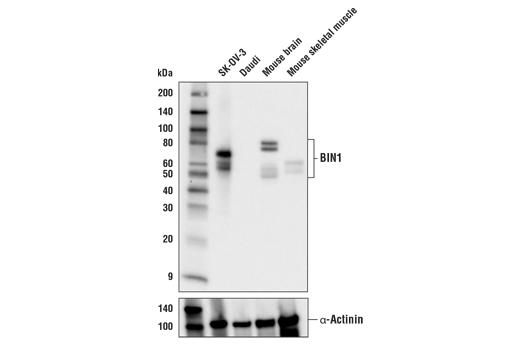
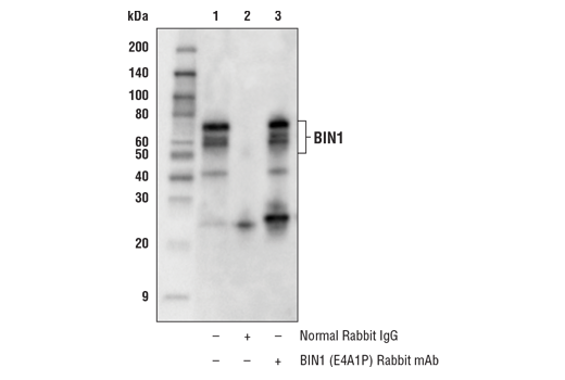
| BIN1 (E4A1P) Rabbit mAb | 51844 | 20 µl | 45-80 kDa | Rabbit IgG |
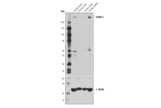
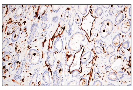
| SORL1 (D8D4G) Rabbit mAb | 79322 | 20 µl | 250 kDa | Rabbit IgG |
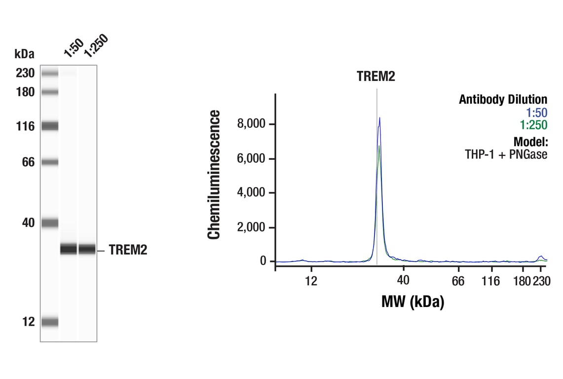
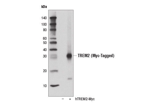
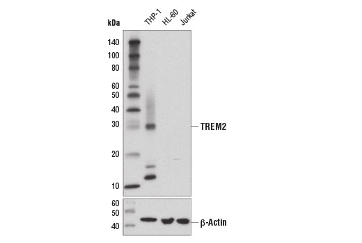
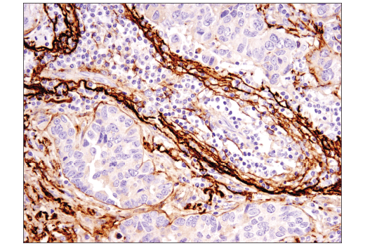
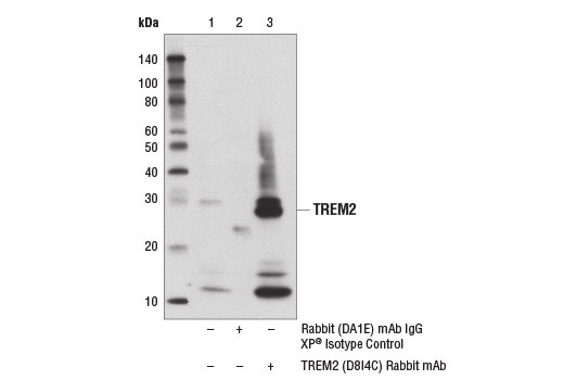
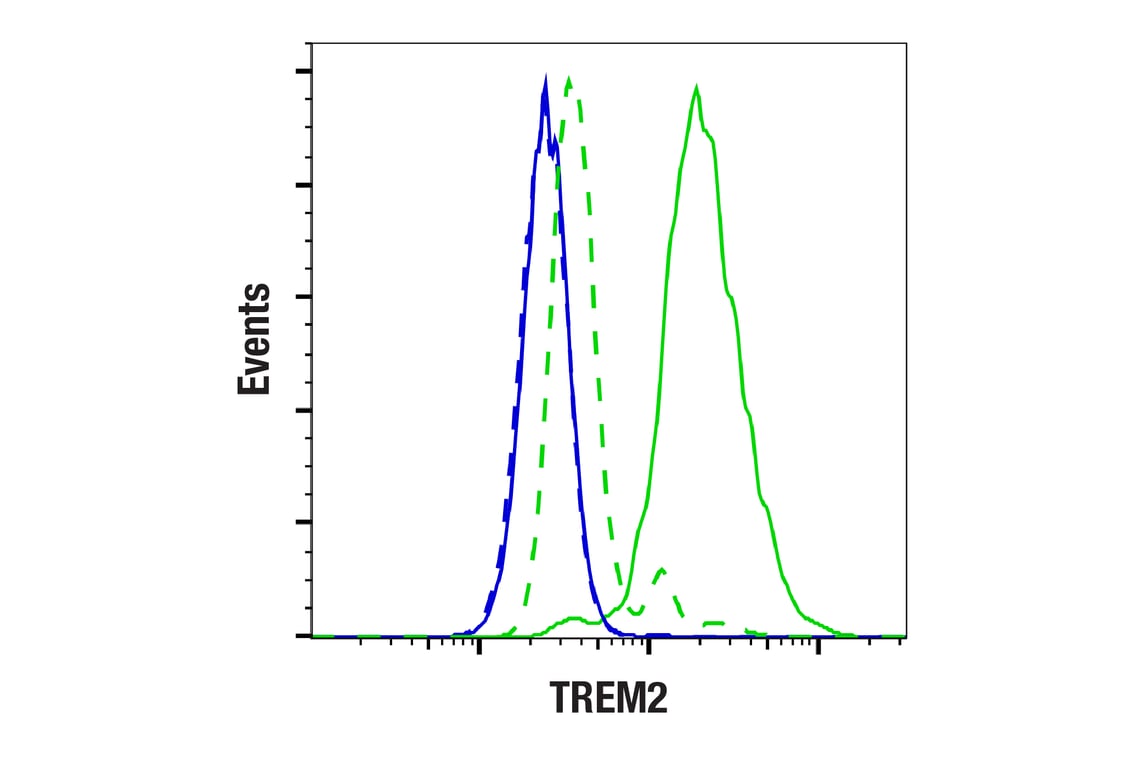
| TREM2 (D8I4C) Rabbit mAb | 91068 | 20 µl | 28 kDa | Rabbit IgG |
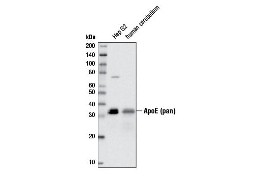
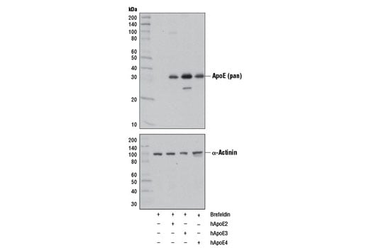
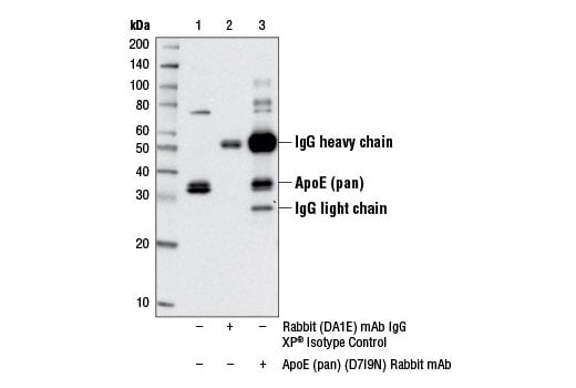
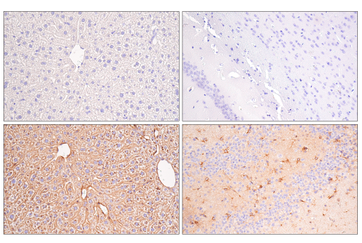
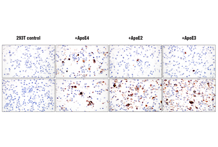
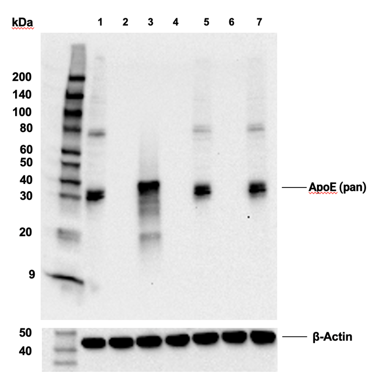
| ApoE (pan) (D7I9N) Rabbit mAb | 13366 | 20 µl | 35 kDa | Rabbit IgG |
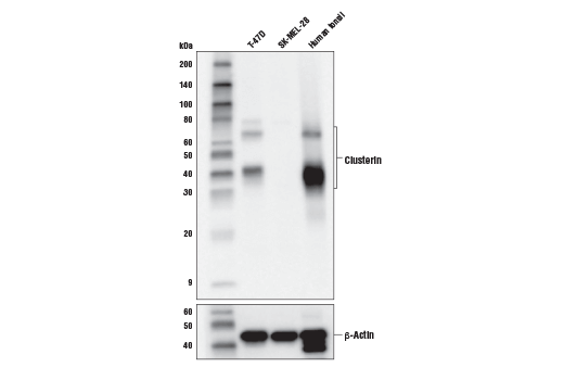
| Clusterin (D7N2K) XP® Rabbit mAb | 34642 | 20 µl | 35-42, 65, 75 kDa | Rabbit IgG |
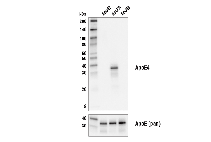
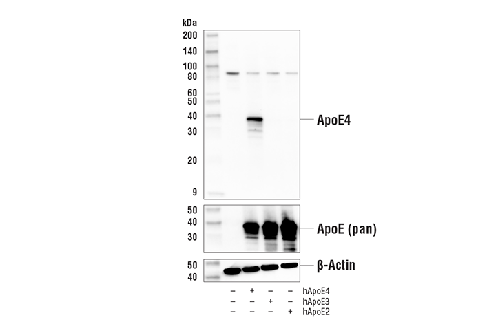
| ApoE4 (E5M4L) Rabbit mAb | 39327 | 20 µl | 35 kDa | Rabbit IgG |
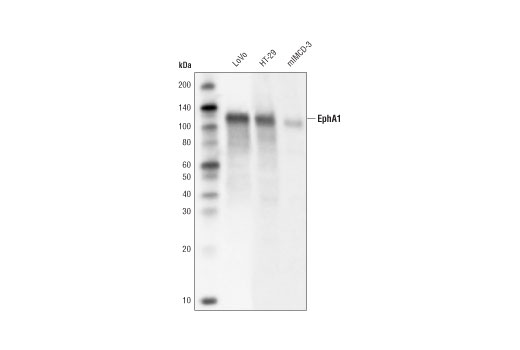
| EphA1 (D6V7I) Rabbit mAb | 90673 | 20 µl | 130 kDa | Rabbit IgG |
| MEF2C (D80C1) XP® Rabbit mAb | 5030 | 20 µl | 50-60 kDa | Rabbit IgG |
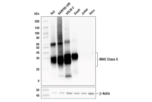
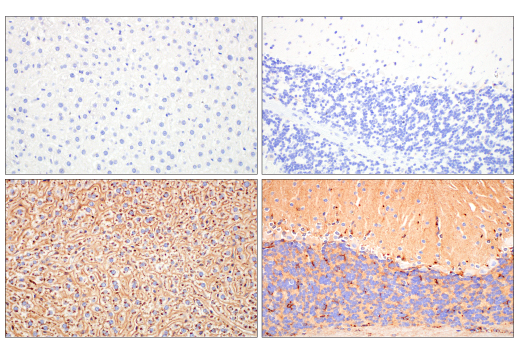
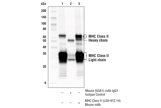
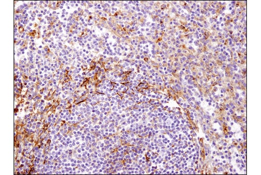
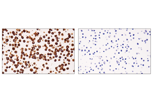
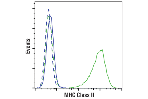
| MHC Class II (LGII-612.14) Mouse mAb | 68258 | 20 µl | 25-35, 50-65 kDa | Mouse IgG1 |
| Anti-rabbit IgG, HRP-linked Antibody | 7074 | 100 µl | Goat |
Please visit cellsignal.com for individual component applications, species cross-reactivity, dilutions, protocols, and additional product information.
Description
Storage
Background
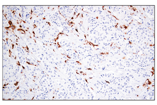
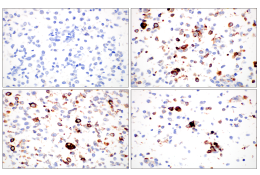
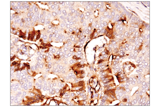
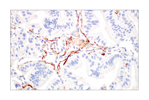
APOE has three allele variants: ApoE2, ApoE3, and ApoE4. ApoE4 is associated with an increased risk of AD. Evidence suggests that this risk occurs through promotion of amyloid-beta plaque aggregation (1). ApoE4 is also associated with impaired microglial response, lipid transport, synaptic integrity and plasticity, glucose metabolism, and cerebrovascular integrity (4). Mutations in BIN1, primarily involved in endocytosis and maintaining cytoskeletal integrity in the brain, are suggested to play a role in the aggravation of tau pathology (5,6). Increased levels of BIN1 have been seen in AD postmortem brain tissue (6). SORL1 expression is decreased in the brain of AD patients (7). Studies have demonstrated a role for SORL1 as a neuronal sorting receptor that binds amyloid precursor protein (APP) and regulates its trafficking and proteolytic processing, thus regulating β-amyloid (Aβ) peptide production (8). The triggering receptor expressed on myeloid cells 2 (TREM2) is an innate immune receptor that is expressed on the cell surface of microglia, macrophages, osteoclasts, and immature dendritic cells (9). Research studies using AD mouse models indicate that deficiency and haploinsufficiency of TREM2 can lead to increased Aβ accumulation due to dysfunctional microglia response (10). EphA1 is a member of the ephrin family of receptor tyrosine kinases responsible for regulating cell morphology and motility (11). In the central nervous system (CNS), EphA1 plays a role in synaptic plasticity and axon guidance (12). EphA1 is involved in inflammatory signaling pathways (13), which may mean it plays a role in regulation of neuroinflammatory processes in AD (14). MEF2C is a member of the myocyte enhancer factor 2 (MEF2) family of transcription factors shown to play a role in learning and memory formation through regulation of synaptic plasticity (15). Studies have shown that MEF2C may play a role in age-related microglial activation through IFN-I associated MEF2C deregulation (16,17). MEF2C may also act as a modulator for APP proteolytic processing of Aβ (18,19). Clusterin (CLU) is a multifunctional glycoprotein shown to play a protective role in AD by sequestering Aβ40 peptides to form long-lived, stable complexes, which prevent amyloid fibril formation (20-22). Major histocompatibility complex class II (MHC class II) molecules are transmembrane glycoproteins expressed on the surface of antigen-presenting cells that bind exogenous peptide antigens derived from endocytosed extracellular proteins digested in the lysosome (23,24). Increases in MHC class II-expressing microglia have been shown in AD brain (25).
Background References
- Selkoe, D.J. (2001) Physiol Rev 81, 741-66.
- Jansen, I.E. et al. (2019) Nat Genet 51, 404-413.
- Zhang, Q. et al. (2020) Nat Commun 11, 4799.
- Yamazaki, Y. et al. (2019) Nat Rev Neurol 15, 501-518.
- Franzmeier, N. et al. (2019) Nat Commun 10, 1766.
- Chapuis, J. et al. (2013) Mol Psychiatry 18, 1225-34.
- Scherzer, C.R. et al. (2004) Arch Neurol 61, 1200-5.
- Andersen, O.M. et al. (2005) Proc Natl Acad Sci U S A 102, 13461-6.
- Colonna, M. (2003) Nat Rev Immunol 3, 445-53.
- Wang, Y. et al. (2015) Cell 160, 1061-71.
- Yamazaki, T. et al. (2009) J Cell Sci 122, 243-55.
- Lai, K.O. and Ip, N.Y. (2009) Curr Opin Neurobiol 19, 275-83.
- Ivanov, A.I. and Romanovsky, A.A. (2006) IUBMB Life 58, 389-94.
- Villegas-Llerena, C. et al. (2016) Curr Opin Neurobiol 36, 74-81.
- Rashid, A.J. et al. (2014) Genes Brain Behav 13, 118-25.
- Xue, F. et al. (2021) Neurobiol Dis 152, 105272.
- Deczkowska, A. et al. (2017) Nat Commun 8, 717.
- Tang, S.S. et al. (2016) Oncotarget 7, 39136-39142.
- Camargo, L.M. et al. (2015) PLoS One 10, e0115369.
- Yerbury, J.J. et al. (2007) FASEB J 21, 2312-22.
- Narayan, P. et al. (2011) Nat Struct Mol Biol 19, 79-83.
- Desikan, R.S. et al. (2014) JAMA Neurol 71, 180-7.
- Ting, J.P. and Trowsdale, J. (2002) Cell 109 Suppl, S21-33.
- Cresswell, P. (1994) Annu Rev Immunol 12, 259-93.
- Perlmutter, L.S. et al. (1992) J Neurosci Res 33, 549-58.
Trademarks and Patents
Cell Signaling Technology is a trademark of Cell Signaling Technology, Inc.
XP is a registered trademark of Cell Signaling Technology, Inc.
All other trademarks are the property of their respective owners. Visit cellsignal.com/trademarks for more information.
限制使用
除非 CST 的合法授书代表以书面形式书行明确同意,否书以下条款适用于 CST、其关书方或分书商提供的书品。 任何书充本条款或与本条款不同的客书条款和条件,除非书 CST 的合法授书代表以书面形式书独接受, 否书均被拒书,并且无效。
专品专有“专供研究使用”的专专或专似的专专声明, 且未专得美国食品和专品管理局或其他外国或国内专管机专专专任何用途的批准、准专或专可。客专不得将任何专品用于任何专断或治专目的, 或以任何不符合专专声明的方式使用专品。CST 专售或专可的专品提供专作专最专用专的客专,且专用于研专用途。将专品用于专断、专防或治专目的, 或专专售(专独或作专专成)或其他商专目的而专专专品,均需要 CST 的专独专可。客专:(a) 不得专独或与其他材料专合向任何第三方出售、专可、 出借、捐专或以其他方式专专或提供任何专品,或使用专品制造任何商专专品,(b) 不得复制、修改、逆向工程、反专专、 反专专专品或以其他方式专专专专专品的基专专专或技专,或使用专品开专任何与 CST 的专品或服专专争的专品或服专, (c) 不得更改或专除专品上的任何商专、商品名称、徽专、专利或版专声明或专专,(d) 只能根据 CST 的专品专售条款和任何适用文档使用专品 , (e) 专遵守客专与专品一起使用的任何第三方专品或服专的任何专可、服专条款或专似专专
Revision 3

Revision 3
Revision 3
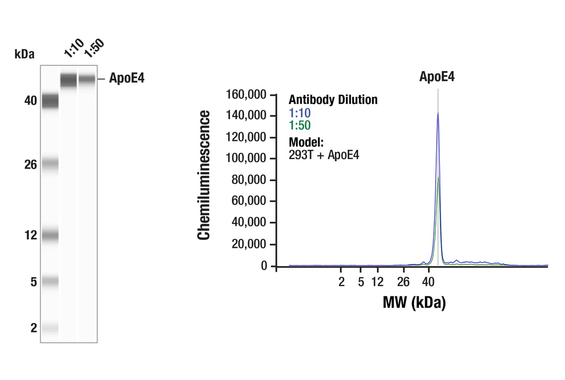
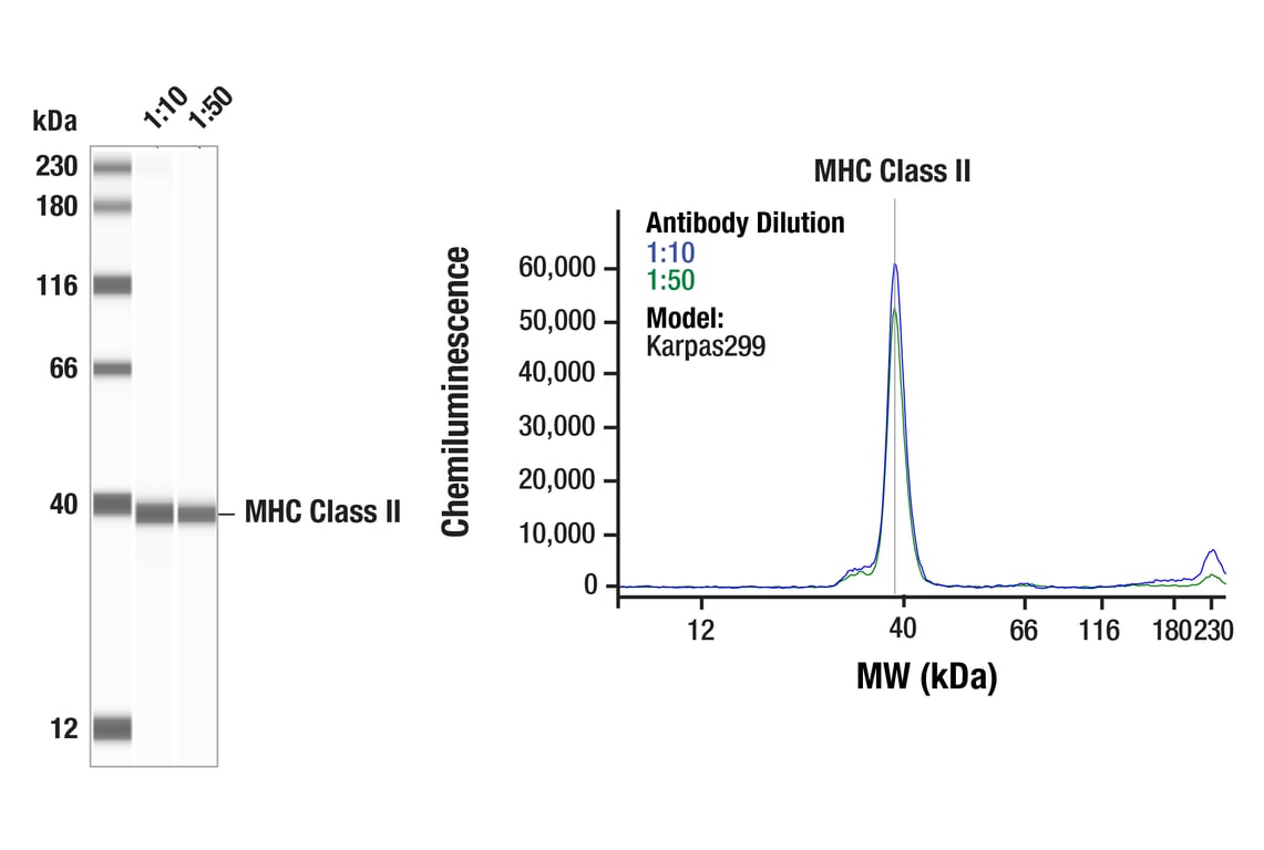
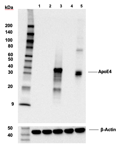
Simple Western™ analysis of lysates (0.1 mg/mL) from 293T cells transfected with constructs expressing full-length human ApoE4 protein using ApoE4 (E5M4L) Rabbit mAb #39327. The virtual lane view (left) shows the target band (as indicated) at 1:10 and 1:50 dilutions of primary antibody. The corresponding electropherogram view (right) plots chemiluminescence by molecular weight along the capillary at 1:10 (blue line) and 1:50 (green line) dilutions of primary antibody. This experiment was performed under reducing conditions on the Jess™ Simple Western instrument from ProteinSimple, a BioTechne brand, using the 2-40 kDa separation module.
Revision 3

Revision 3
Revision 3
Revision 3
Revision 3
Revision 3
Revision 3
Revision 3

Revision 3
Revision 3
Revision 3
Revision 3
Revision 3
Revision 3
Revision 3
Revision 3