| Product Includes | Product # | Quantity | Mol. Wt | Isotype/Source |
|---|---|---|---|---|
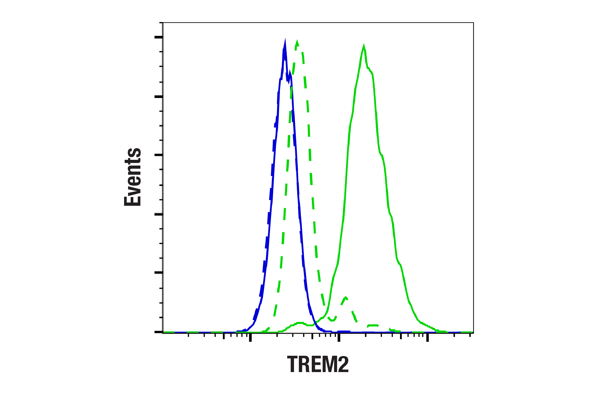
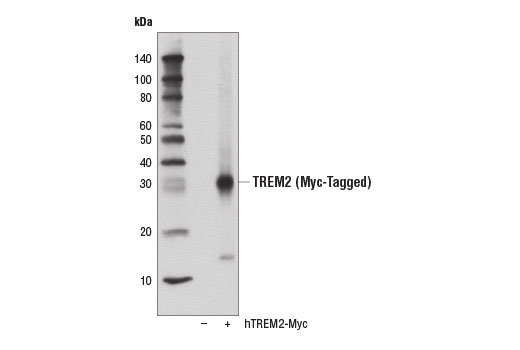
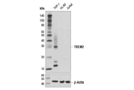
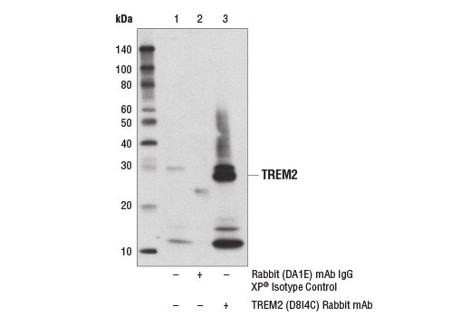
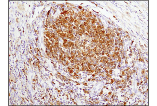
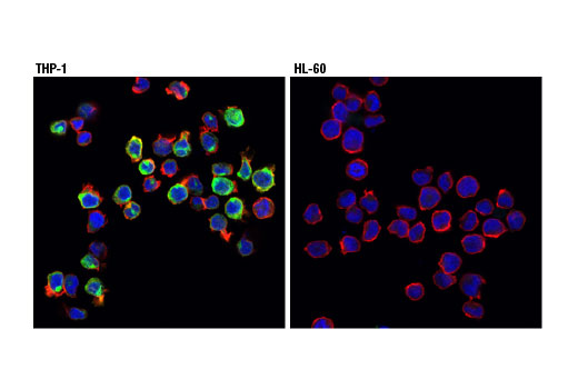
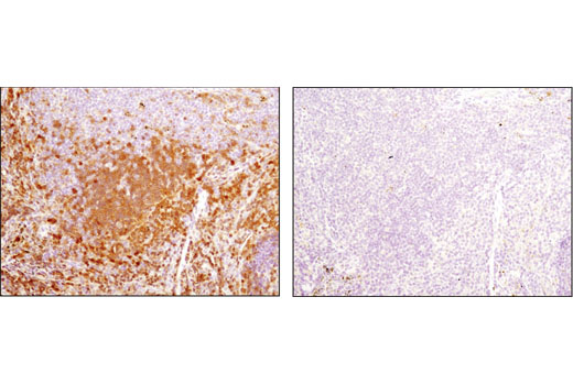
| TREM2 (D8I4C) Rabbit mAb | 91068 | 20 µl | 28 kDa | Rabbit IgG |
| TREM2 (E9U8L) Rabbit mAb (Amino-terminal Antigen) | 70551 | 20 µl | 28 kDa | Rabbit IgG |
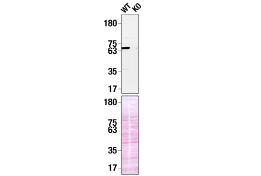
| CD33 Antibody | 77576 | 20 µl | 70-80 kDa | Rabbit |
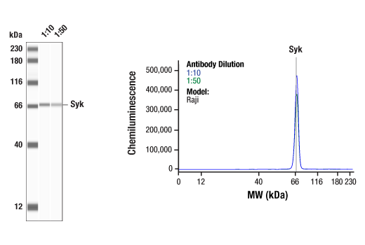
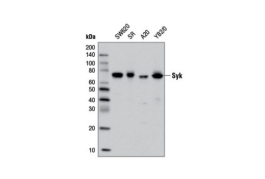
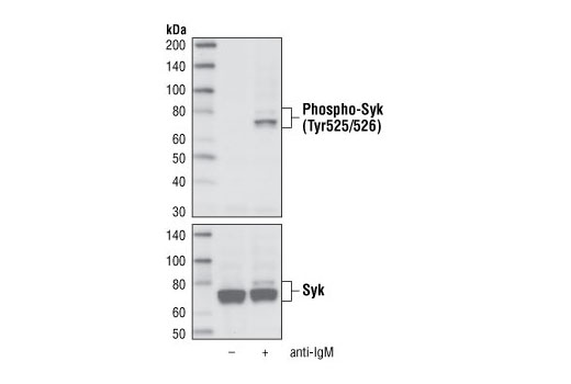
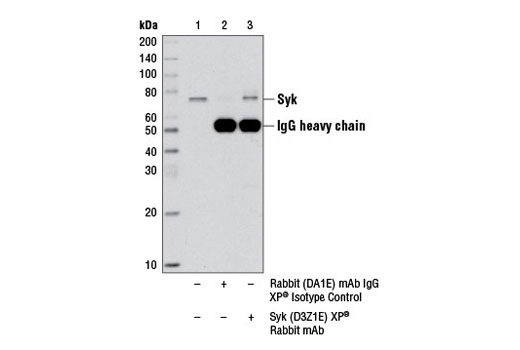
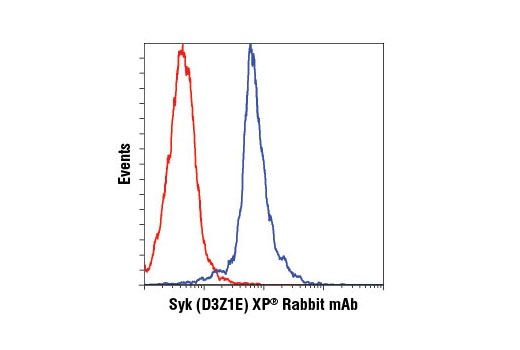
| Syk (D3Z1E) XP® Rabbit mAb | 13198 | 20 µl | 72 kDa | Rabbit IgG |
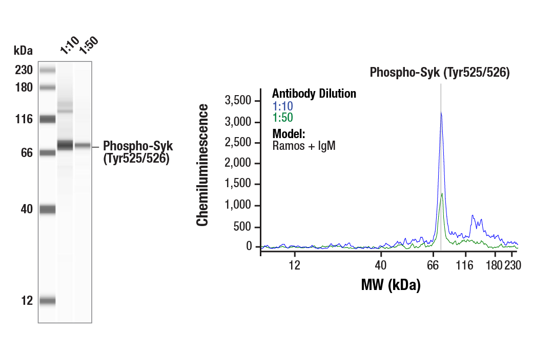
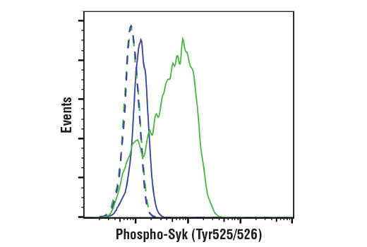
| Phospho-Syk (Tyr525/526) (C87C1) Rabbit mAb | 2710 | 20 µl | 72 kDa | Rabbit IgG |
| DAP12 (E7U7T) Rabbit mAb | 97415 | 20 µl | 10, 12 kDa | Rabbit IgG |
| Anti-rabbit IgG, HRP-linked Antibody | 7074 | 100 µl | Goat |
Please visit cellsignal.com for individual component applications, species cross-reactivity, dilutions, protocols, and additional product information.
Description
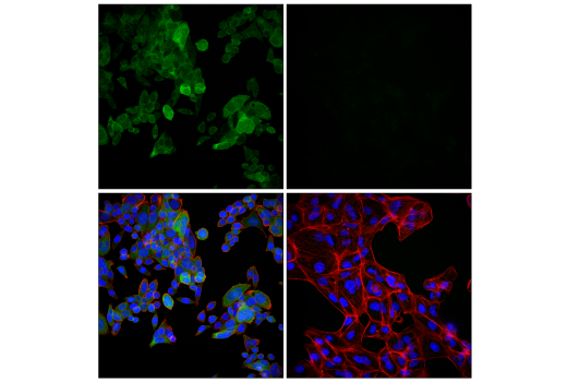
The Human TREM2 Activity Antibody Sampler Kit provides an economical means of evaluating key members of the human TREM2 signaling pathway using phospho-specific and control antibodies. The kit includes enough antibodies to perform two western blot experiments with each primary antibody.
Storage
Background
Alzheimer's Disease (AD) is one of the most common neurodegenerative diseases worldwide. Clinically, it is characterized by the presence of extracellular amyloid plaques and intracellular neurofibrillary tangles, resulting in neuronal dysfunction and cell death. Triggering receptor expressed on myeloid cells 2 (TREM2), a protein localized at the membrane of innate immune cells, including microglia in the brain, has been genetically linked to AD, with specific variants increasing disease risk by as much as threefold (1,2). The TREM2 receptor is a single-pass type I membrane glycoprotein that consists of an extracellular immunoglobulin-like domain, a transmembrane domain, and a cytoplasmic tail. Upon activation, TREM2 interacts with the tyrosine kinase-binding protein DNAX-activating protein 12 (DAP12, TYROBP) to form a receptor-signaling complex. The DAP12 protein structure consists of a short extracellular domain, a transmembrane domain, and a cytoplasmic immunoreceptor tyrosine-based activation motif (ITAM) (2-9). ITAMs function as a binding site for tyrosine kinases, including spleen tyrosine kinase (Syk). Syk is comprised of two tandem amino-terminal Src homology (SH) 2 domains separated by an SH2-kinase linker, and a C-terminal tyrosine kinase domain, separated from the SH2 domains by an inter-domain linker. When Syk binds to an ITAM, it changes conformation, allowing for residues within the inter-domain linker region, including Tyr352, to become phosphorylated. Residues within the activation loop subsequently become phosphorylated, leading to full Syk activation. Tyr525 and Tyr526 are located in the activation loop of the Syk kinase domain and phosphorylation at these residues (equivalent to Tyr519/520 of mouse Syk) is essential for Syk function (10-12). This activation can lead to the mediation of a variety of cellular responses, including proliferation, differentiation, inflammation, and phagocytosis. Evidence suggests that TREM2 and DAP12 may act in a Syk-dependent manner to drive microglial cellular responses in AD (2,4-8,13).
There is also evidence that these processes may be regulated via crosstalk between TREM2 and the cell surface receptor CD33, a sialic acid-binding Ig-like lectin (Siglec-3) type I transmembrane protein. Much like TREM2, CD33 has been identified as a risk gene in AD. CD33 binds preferentially to alpha-2, 6-linked sialic acid, which can be found in sialylated gangliosides in the brain. Activation of CD33 has been shown to be inhibitory to a variety of cellular processes. Evidence suggests that TREM2 may act downstream of CD33 and that TREM2-dependent microglial signaling in AD may be directly inhibited by CD33 activation (14-17).
- Nguyen, A.T. et al. (2020) Acta Neuropathol 140, 477-493.
- Gratuze, M. et al. (2018) Mol Neurodegener 13, 66.
- Jonsson, T. et al. (2013) N Engl J Med 368, 107-16.
- Jay, T.R. et al. (2017) Mol Neurodegener 12, 56.
- McQuade, A. et al. (2020) Nat Commun 11, 5370.
- Schlepckow, K. et al. (2020) EMBO Mol Med 12, e11227.
- Zhao, Y. et al. (2018) Neuron 97, 1023-1031.e7.
- Colonna, M. (2003) Nat Rev Immunol 3, 445-53.
- Lanier, L.L. et al. (1998) Nature 391, 703-7.
- Zhang, J. et al. (2000) J Biol Chem 275, 35442-7.
- Mansueto, M.S. et al. (2019) J Biol Chem 294, 7658-7668.
- Grädler, U. et al. (2013) J Mol Biol 425, 309-33.
- Turner, M. et al. (2000) Immunol Today 21, 148-54.
- Karch, C.M. et al. (2012) PLoS One 7, e50976.
- Griciuc, A. et al. (2013) Neuron 78, 631-43.
- Griciuc, A. et al. (2019) Neuron 103, 820-835.e7.
- Salminen, A. et al. (2021) Neurochem Int 150, 105186.
Background References
Trademarks and Patents
限制使用
除非 CST 的合法授书代表以书面形式书行明确同意,否书以下条款适用于 CST、其关书方或分书商提供的书品。 任何书充本条款或与本条款不同的客书条款和条件,除非书 CST 的合法授书代表以书面形式书独接受, 否书均被拒书,并且无效。
专品专有“专供研究使用”的专专或专似的专专声明, 且未专得美国食品和专品管理局或其他外国或国内专管机专专专任何用途的批准、准专或专可。客专不得将任何专品用于任何专断或治专目的, 或以任何不符合专专声明的方式使用专品。CST 专售或专可的专品提供专作专最专用专的客专,且专用于研专用途。将专品用于专断、专防或治专目的, 或专专售(专独或作专专成)或其他商专目的而专专专品,均需要 CST 的专独专可。客专:(a) 不得专独或与其他材料专合向任何第三方出售、专可、 出借、捐专或以其他方式专专或提供任何专品,或使用专品制造任何商专专品,(b) 不得复制、修改、逆向工程、反专专、 反专专专品或以其他方式专专专专专品的基专专专或技专,或使用专品开专任何与 CST 的专品或服专专争的专品或服专, (c) 不得更改或专除专品上的任何商专、商品名称、徽专、专利或版专声明或专专,(d) 只能根据 CST 的专品专售条款和任何适用文档使用专品, (e) 专遵守客专与专品一起使用的任何第三方专品或服专的任何专可、服专条款或专似专专