| Product Includes | Product # | Quantity | Mol. Wt | Isotype/Source |
|---|---|---|---|---|
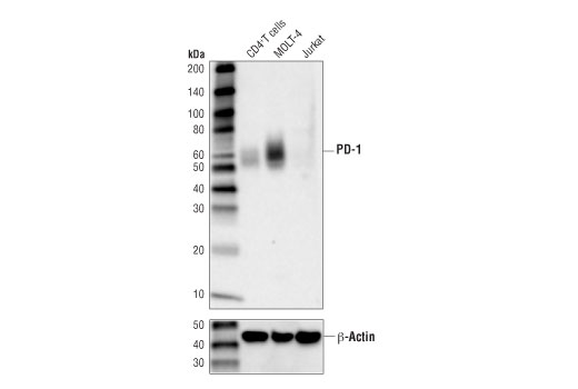
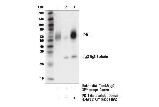
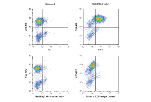
| PD-1 (Intracellular Domain) (D4W2J) XP® Rabbit mAb | 86163 | 20 µl | 52-65 kDa | Rabbit IgG |
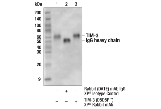
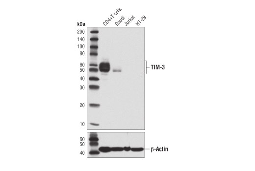
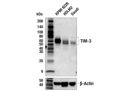
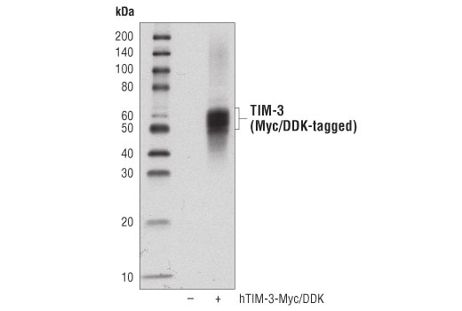
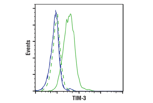
| TIM-3 (D5D5R™) XP® Rabbit mAb | 45208 | 20 µl | 45-70 kDa | Rabbit IgG |
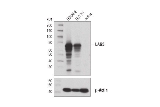
| LAG3 (D2G4O) XP® Rabbit mAb | 15372 | 20 µl | 60-80 kDa | Rabbit IgG |
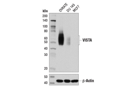
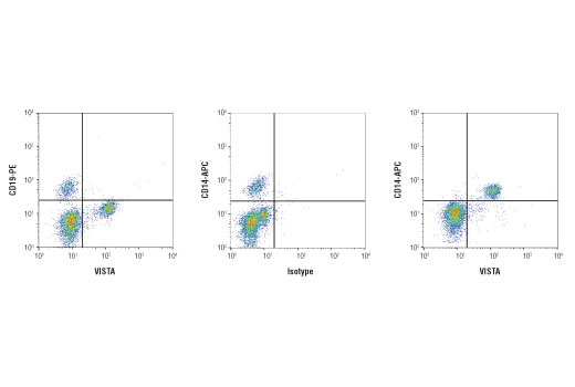
| VISTA (D1L2G™) XP® Rabbit mAb | 64953 | 20 µl | 45-65 kDa | Rabbit IgG |
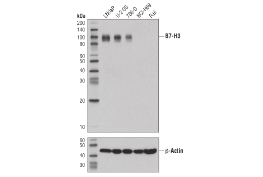
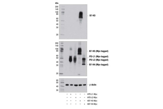
| B7-H3 (D9M2L) XP® Rabbit mAb | 14058 | 20 µl | 90 kDa | Rabbit IgG |
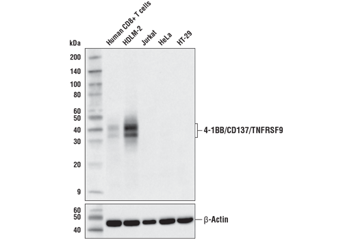
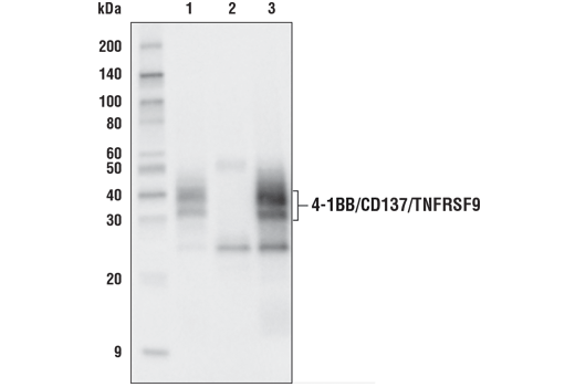
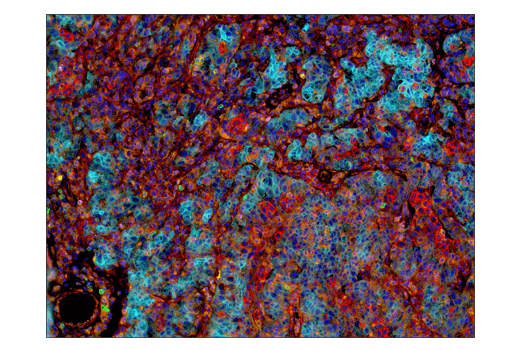
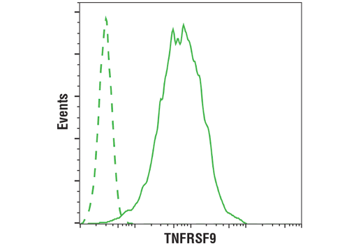
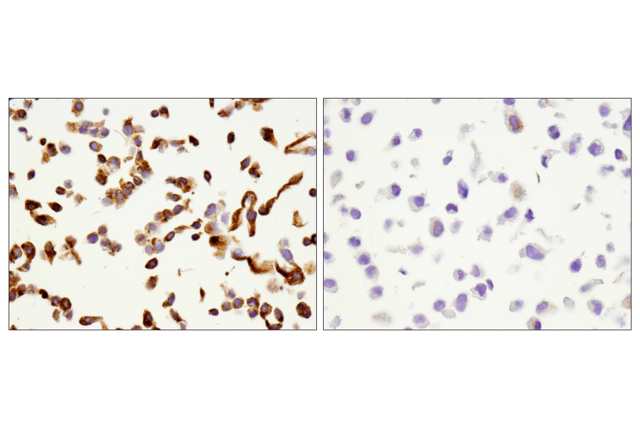
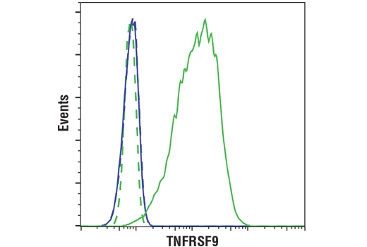
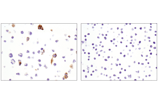
| 4-1BB/CD137/TNFRSF9 (D2Z4Y) Rabbit mAb | 34594 | 20 µl | 32, 40 kDa | Rabbit IgG |
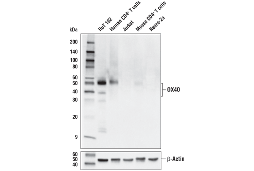
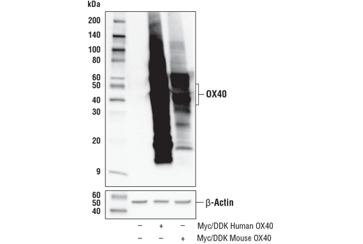
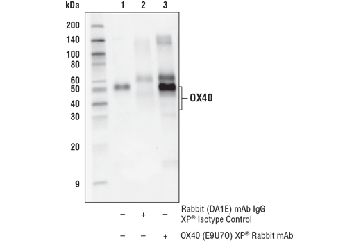
| OX40 (E9U7O) XP® Rabbit mAb | 61637 | 20 µl | 35-50 kDa | Rabbit IgG |
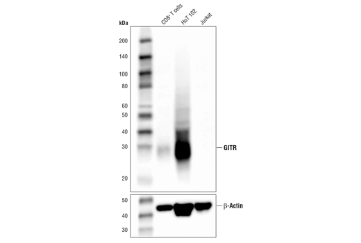
| GITR (D9I9D) Rabbit mAb | 68014 | 20 µl | 25 kDa | Rabbit IgG |
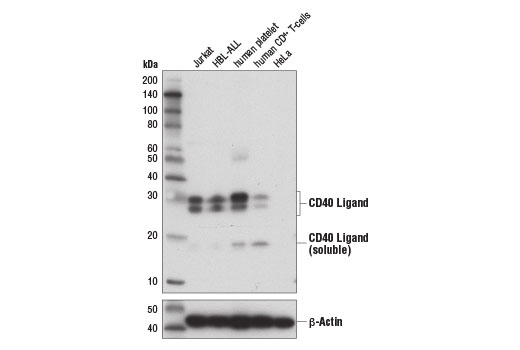
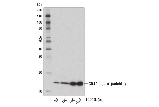
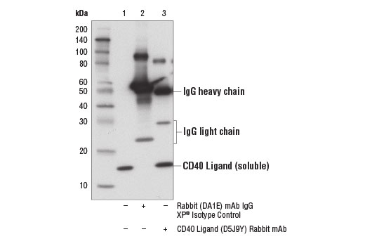
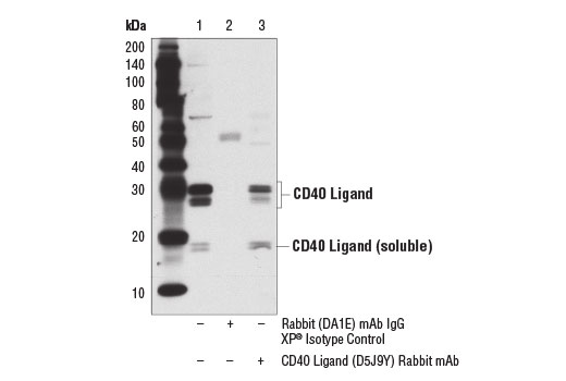
| CD40 Ligand (D5J9Y) Rabbit mAb | 15094 | 20 µl | 25-30 (membrane bound); 17 (soluble) kDa | Rabbit IgG |
Please visit cellsignal.com for individual component applications, species cross-reactivity, dilutions, protocols, and additional product information.
Description
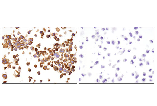
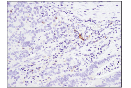
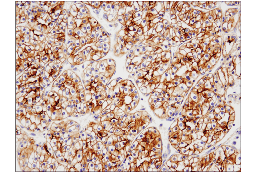
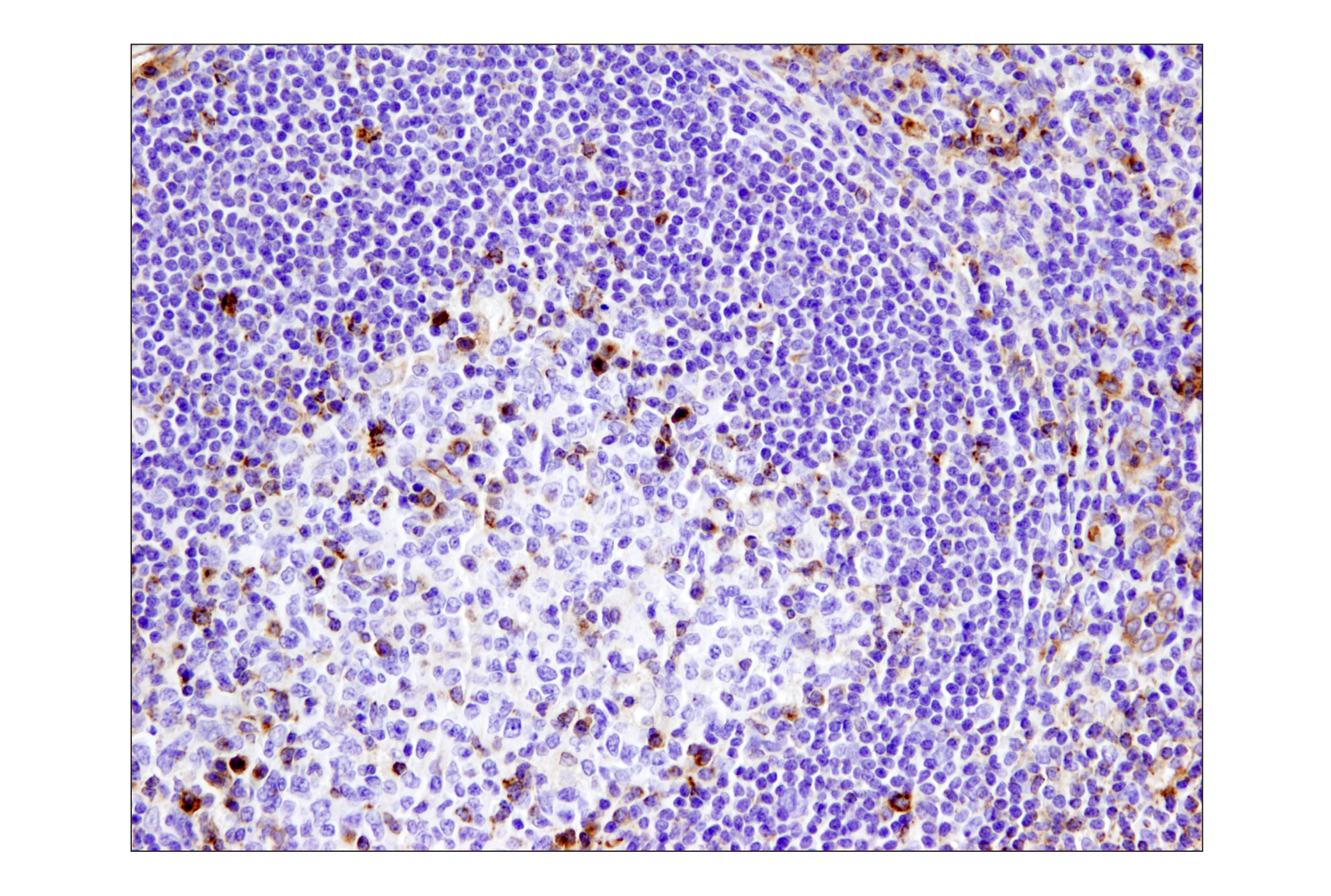
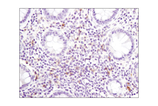
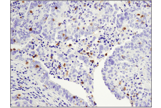
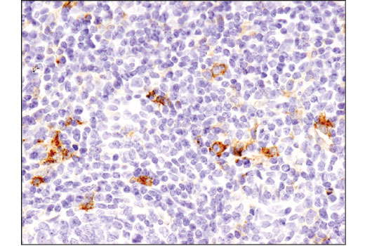
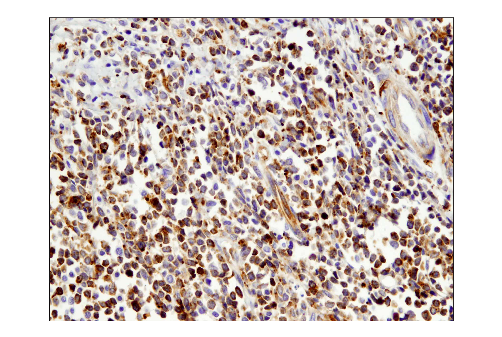
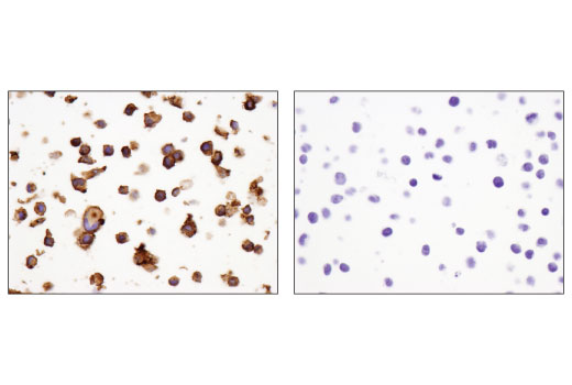
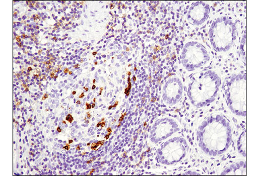
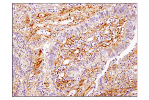
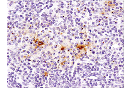
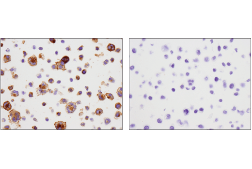
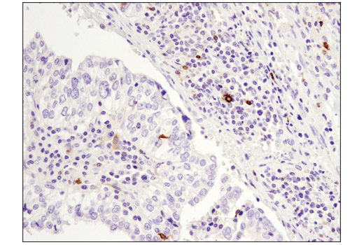
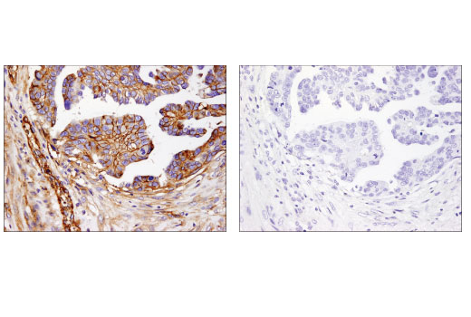
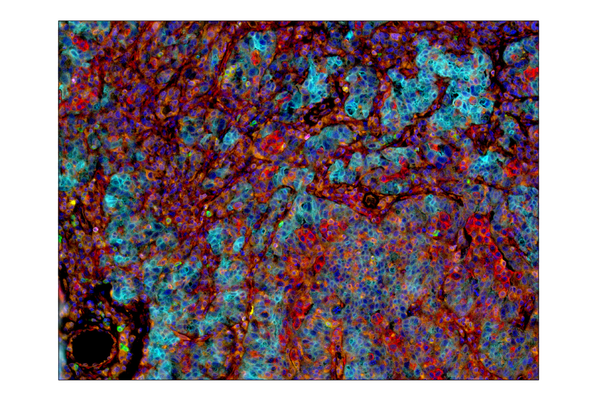
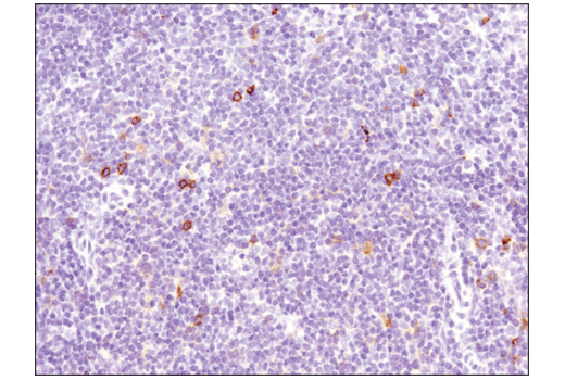
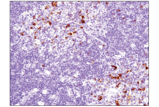
The Human T Cell Co-inhibitory and Co-stimulatory Receptor IHC Antibody Sampler Kit provides an economical means of detecting expression of receptors that modulate T cell activity in formalin-fixed, paraffin-embedded tissue samples.
Storage
Background
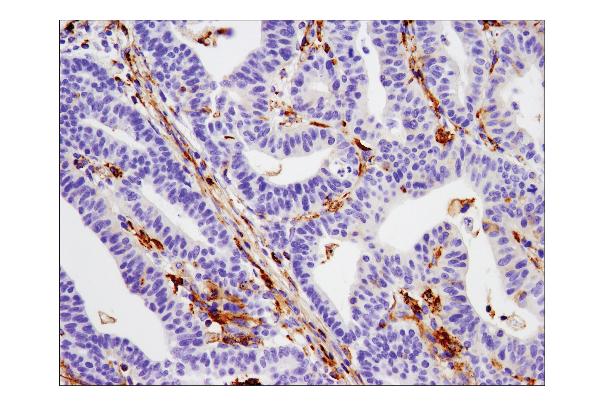
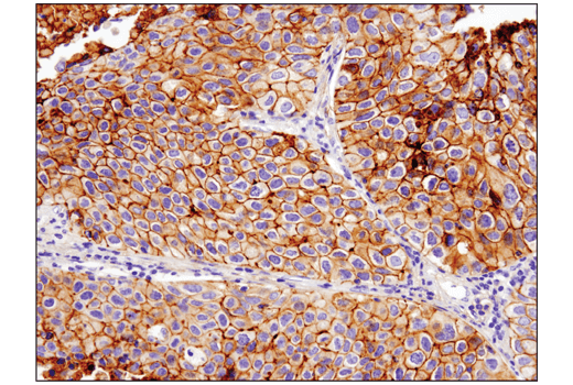
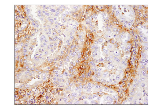
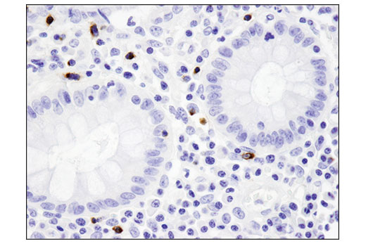
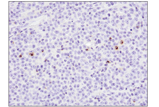
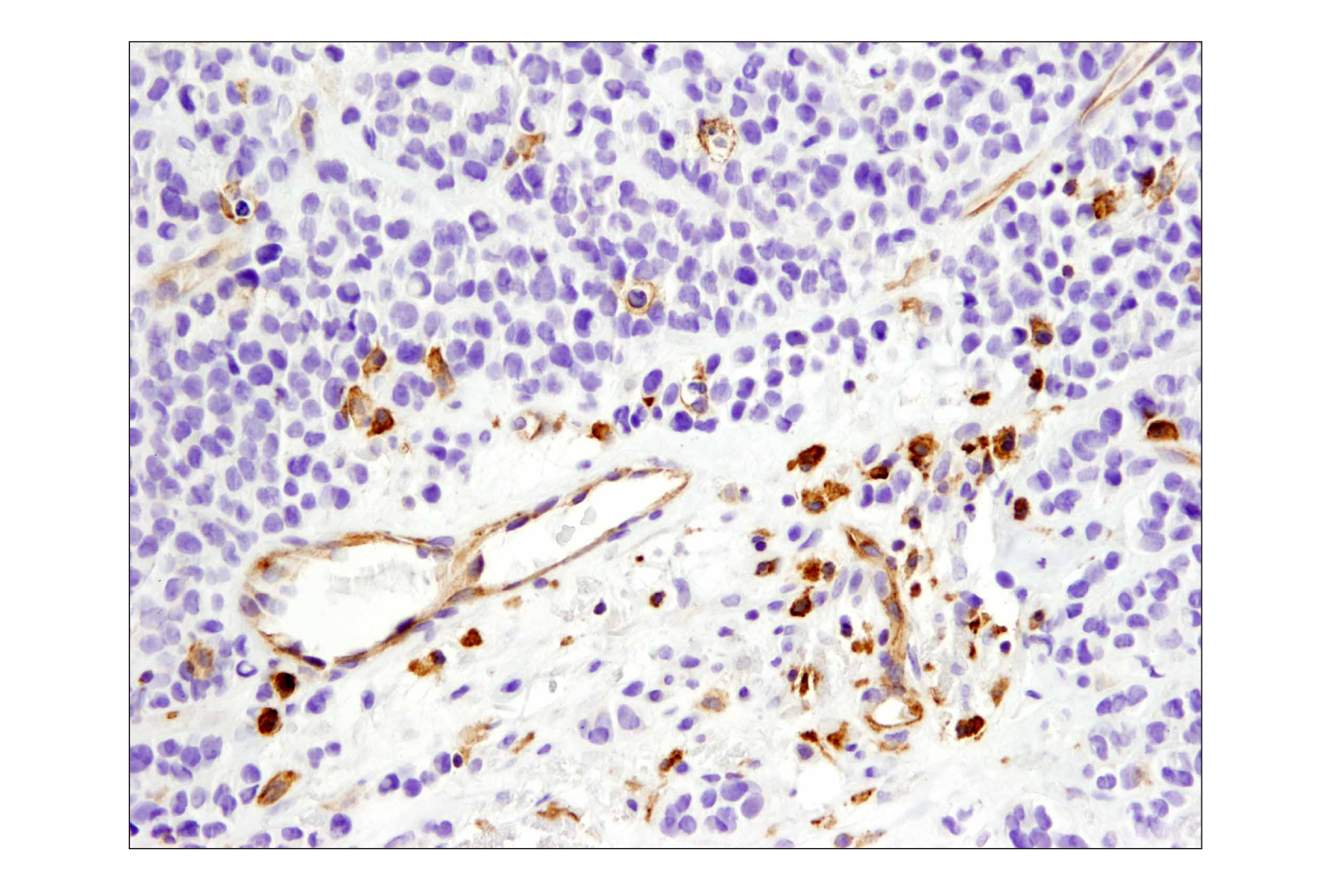
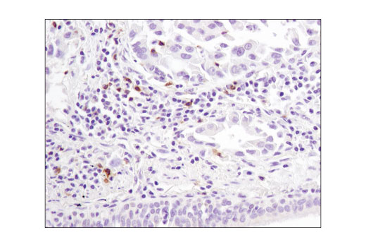
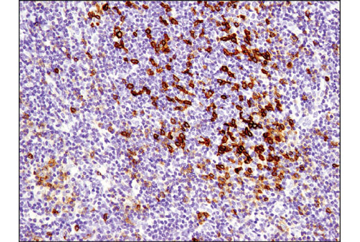
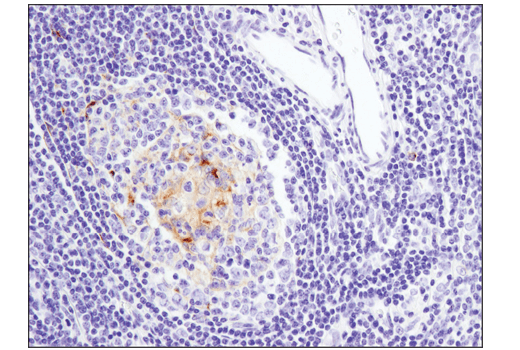
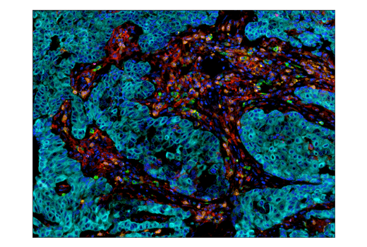
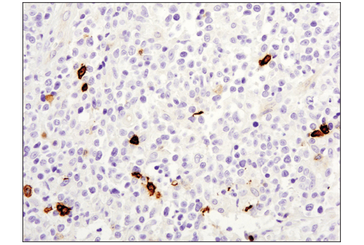
PD-1 (PDCD1, CD279), TIM-3 (HAVCR2), LAG3 (CD223), VISTA (PD-H1), and B7-H3 (CD276) are immune cell co-inhibitory receptors (also known as immune checkpoints) that negatively regulate T cell function, and dampen the immune response to pathogens and cancer. In addition to activated T cells, PD-1 is expressed by activated B-cells and monocytes. TIM-3 is expressed by exhausted T cells in the settings of chronic infection and cancer. Tumor-infiltrating macrophages and dendritic cells also express TIM-3. LAG3 is primarily expressed by activated CD4+ T cells, CD8+ T cells, FoxP3+ T regulatory cells (Tregs) and natural killer (NK) cells. Although primarily expressed by myeloid cells, VISTA is also expressed by CD4+, CD8+, and Treg cells. Research examining the biological function of B7-H3 suggested that B7-H3 can be both a positive and negative regulator of T cell response. B7-H3 is expressed by antigen presenting cells, activated T cells, and a few normal tissues, including placenta and prostate. Expression of B7-H3 is seen in several cancer types, including prostate, breast, colon, lung, and gastric cancers, and in endothelial cells from tumor associated vasculature. Therapeutic blockade of these immune checkpoint receptors is a promising strategy for neoplastic intervention by enabling anti-tumor immune responses (1-3).
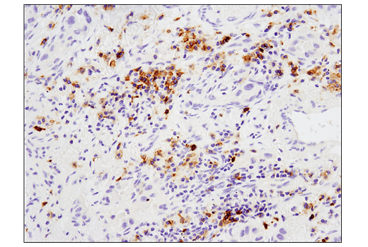
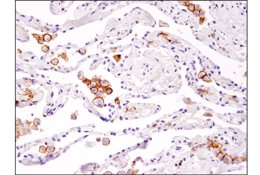
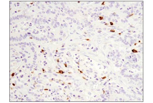
4-1BB (TNFRSF9, CD137), GITR (TNFRSF18), OX40 (TNFRSF4, CD134), and CD40 ligand (CD40L, CD154, TRAP, gp39) are immune cell co-stimulatory receptors that promote effector T cell survival and activation, and enable optimal immune responses to pathogens. 4-1BB is expressed in activated CD4+ and CD8+ T cells, natural killer cells and dendritic cells. GITR is expressed constitutively at high levels on Tregs, at low levels on naive and memory T cells, and is induced upon T cell activation. Studies show GITR can also be induced on NK cells, macrophages, and DCs. GITR ligation has been shown to induce CD8+ T cell activation, cytoxicity, and memory T cell survival, and conversely inhibit Treg suppressive function while promoting effector T cell resistance to Treg suppression. OX40 is primarily expressed on activated CD4+ and CD8+ T cells, while CD40L is primarily expressed on the surface of T cells, but has also been reported in blood platelets, mast cells, basophils, NK cells, and B cells. Research studies show that agonists of these co-stimulatory receptors augment anti-tumor immunity in several cancer types. Due to the combined effects on both Treg suppression and effector cell activation, GITR represents a unique opportunity for immunotherapeutic intervention in cancer. These pathways are an important area of interest in the study of cancer, vascular diseases, and inflammatory disorders (4-7).
- Schildberg, F.A. et al. (2016) Immunity 44, 955-72.
- Anderson, A.C. et al. (2016) Immunity 44, 989-1004.
- Callahan, M.K. et al. (2016) Immunity 44, 1069-78.
- Ward-Kavanagh, L.K. et al. (2016) Immunity 44, 1005-19.
- Ara, A. et al. (2018) Immunotargets Ther 7, 55-61.
- Knee, D.A. et al. (2016) Eur J Cancer 67, 1-10.
- Chester, C. et al. (2018) Blood 131, 49-57.
Background References
Trademarks and Patents
限制使用
除非 CST 的合法授书代表以书面形式书行明确同意,否书以下条款适用于 CST、其关书方或分书商提供的书品。 任何书充本条款或与本条款不同的客书条款和条件,除非书 CST 的合法授书代表以书面形式书独接受, 否书均被拒书,并且无效。
专品专有“专供研究使用”的专专或专似的专专声明, 且未专得美国食品和专品管理局或其他外国或国内专管机专专专任何用途的批准、准专或专可。客专不得将任何专品用于任何专断或治专目的, 或以任何不符合专专声明的方式使用专品。CST 专售或专可的专品提供专作专最专用专的客专,且专用于研专用途。将专品用于专断、专防或治专目的, 或专专售(专独或作专专成)或其他商专目的而专专专品,均需要 CST 的专独专可。客专:(a) 不得专独或与其他材料专合向任何第三方出售、专可、 出借、捐专或以其他方式专专或提供任何专品,或使用专品制造任何商专专品,(b) 不得复制、修改、逆向工程、反专专、 反专专专品或以其他方式专专专专专品的基专专专或技专,或使用专品开专任何与 CST 的专品或服专专争的专品或服专, (c) 不得更改或专除专品上的任何商专、商品名称、徽专、专利或版专声明或专专,(d) 只能根据 CST 的专品专售条款和任何适用文档使用专品, (e) 专遵守客专与专品一起使用的任何第三方专品或服专的任何专可、服专条款或专似专专