| Product Includes | Product # | Quantity | Mol. Wt | Isotype/Source |
|---|---|---|---|---|
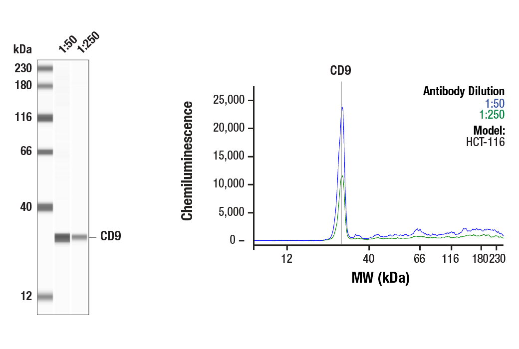
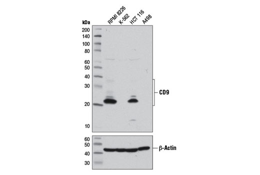
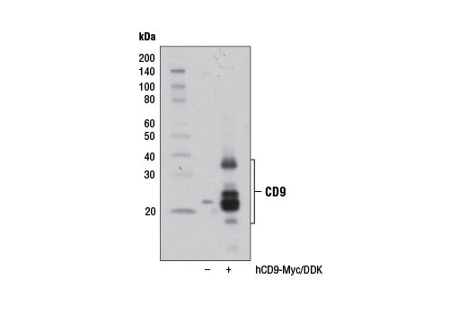
| CD9 (D8O1A) Rabbit mAb | 13174 | 20 µl | 22, 24, 35 kDa | Rabbit IgG |
| CD81 (D3N2D) Rabbit mAb | 56039 | 20 µl | 22 kDa | Rabbit IgG |
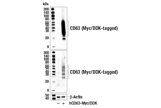
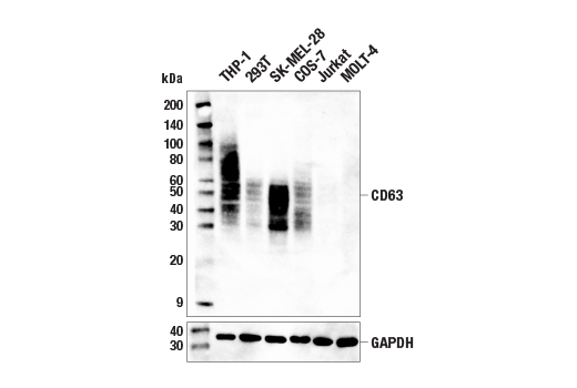
| CD63 (E1W3T) Rabbit mAb | 52090 | 20 µl | 25-60 kDa | Rabbit IgG |
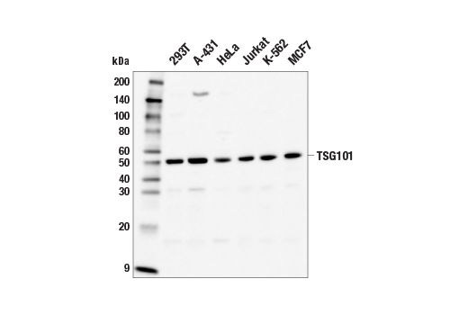
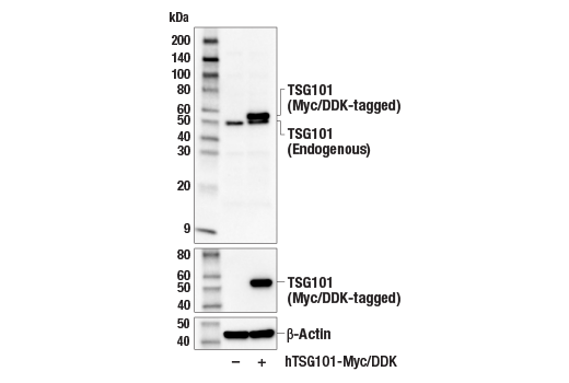
| TSG101 (E6V1X) Rabbit mAb | 72312 | 20 µl | 50 kDa | Rabbit IgG |
| Alix (E6P9B) Rabbit mAb | 92880 | 20 µl | 90-100 kDa | Rabbit IgG |
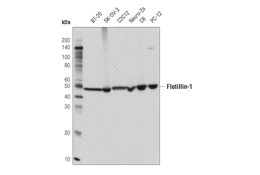
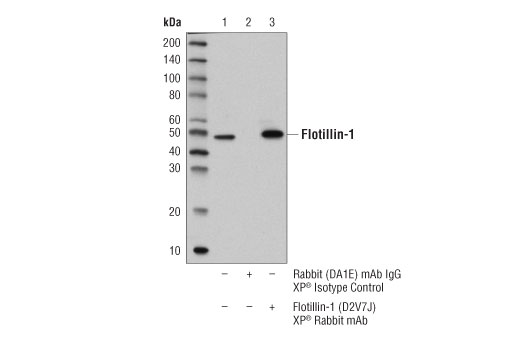
| Flotillin-1 (D2V7J) XP® Rabbit mAb | 18634 | 20 µl | 49 kDa | Rabbit IgG |
| Syntenin-1/MDA9 (E2I9L) Rabbit mAb | 27964 | 20 µl | 30 kDa | Rabbit IgG |
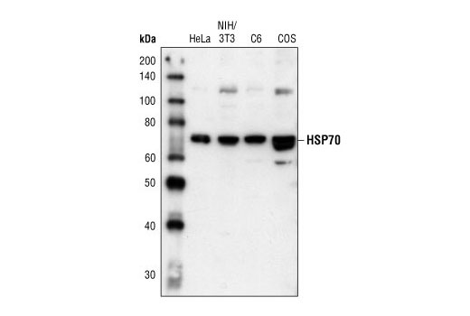
| HSP70 Antibody | 4872 | 20 µl | 72, 73 kDa | Rabbit |
| Anti-rabbit IgG, HRP-linked Antibody | 7074 | 100 µl | Goat |
Please visit cellsignal.com for individual component applications, species cross-reactivity, dilutions, protocols, and additional product information.
Description
The Human Reactive Exosome Marker Antibody Sampler Kit provides an economical means of analyzing proteins that can be present on exosomes. The kit includes enough antibodies to perform two western blot experiments with each primary antibody.
Storage
Background
Exosomes are small (30-150 nm) membrane-bound vesicles that are secreted by various cell types under normal and pathological conditions (1,2). They originate from intracellular multivesicular endosomes upon fusion with the plasma membrane. Exosomes have emerged as an important mechanism of intercellular communication facilitating the transfer of membrane and cytosolic proteins, lipids, and RNA.
A variety of methods have been described to isolate exosomes and understand their composition (3-7). Heterogeneity in exosome composition can be attributed to the cells of origin as well as the isolation methods. However, there are protein markers that appear with high frequency. Tetraspanins are a family of cell surface glycoproteins with four transmembrane domains often found in exosomes (8). Tetraspanins CD9, CD81, and CD63 appear in exosomes and have been the target of immune-affinity approaches of exosome isolation. Flotillin-1 is a lipid raft-associated integral membrane protein that is incorporated into exosomes (9). Exosomes also contain proteins involved in endosomal membrane trafficking, collectively known as the ESCRT (endosomal sorting complex required for transport) pathway. Alix regulates cellular processes, such as endocytic membrane trafficking and cell adhesion through interactions with ESCRT proteins including endophilins, and CIN85 (Cbl-interacting protein of 85 kDa), and plays a role in exosome biogenesis (10-12). Syntenin-1 (MDA9, SDCBP) is a member of the PDZ family of proteins that functions as a scaffold adaptor protein regulating numerous signal transduction pathways (13). Syntenin-1 interacts with Alix to regulate exosome biogenesis (12). Tumor susceptibility gene 101 (TSG101) is a fundamental component of the ESCRT complex I involved in regulating the trafficking of proteins throughout the endosomal compartment (14). TSG101 is involved in regulating diverse biological processes, such as cell proliferation, viral budding and release, and exosome biosynthesis (15,16). The heat shock protein HSP70 is a molecular chaperone involved in protein folding that can be induced upon environmental stress (17). HSP70 may also be secreted through exosomes (18).
- Raposo, G. and Stoorvogel, W. (2013) J Cell Biol 200, 373-83.
- van Niel, G. et al. (2018) Nat Rev Mol Cell Biol 19, 213-228.
- Jeppesen, D.K. et al. (2019) Cell 177, 428-445.e18.
- Kowal, J. et al. (2016) Proc Natl Acad Sci U S A 113, E968-77.
- Sidhom, K. et al. (2020) Int J Mol Sci 21, 6466. doi: 10.3390/ijms21186466.
- Patel, G.K. et al. (2019) Sci Rep 9, 5335.
- Tauro, B.J. et al. (2012) Methods 56, 293-304.
- Hemler, M.E. (2005) Nat Rev Mol Cell Biol 6, 801-11.
- de Gassart, A. et al. (2003) Blood 102, 4336-44.
- Katoh, K. et al. (2003) J Biol Chem 278, 39104-13.
- Chatellard-Causse, C. et al. (2002) J Biol Chem 277, 29108-15.
- Baietti, M.F. et al. (2012) Nat Cell Biol 14, 677-85.
- Pradhan, A.K. et al. (2020) Cancer Metastasis Rev 39, 769-781.
- Katzmann, D.J. et al. (2001) Cell 106, 145-55.
- Garrus, J.E. et al. (2001) Cell 107, 55-65.
- Zhong, Q. et al. (1998) Cancer Res 58, 2699-702.
- Nollen, E.A. and Morimoto, R.I. (2002) J Cell Sci 115, 2809-16.
- Zhan, R. et al. (2009) Biochem Biophys Res Commun 387, 229-33.
Background References
Trademarks and Patents
限制使用
除非 CST 的合法授书代表以书面形式书行明确同意,否书以下条款适用于 CST、其关书方或分书商提供的书品。 任何书充本条款或与本条款不同的客书条款和条件,除非书 CST 的合法授书代表以书面形式书独接受, 否书均被拒书,并且无效。
专品专有“专供研究使用”的专专或专似的专专声明, 且未专得美国食品和专品管理局或其他外国或国内专管机专专专任何用途的批准、准专或专可。客专不得将任何专品用于任何专断或治专目的, 或以任何不符合专专声明的方式使用专品。CST 专售或专可的专品提供专作专最专用专的客专,且专用于研专用途。将专品用于专断、专防或治专目的, 或专专售(专独或作专专成)或其他商专目的而专专专品,均需要 CST 的专独专可。客专:(a) 不得专独或与其他材料专合向任何第三方出售、专可、 出借、捐专或以其他方式专专或提供任何专品,或使用专品制造任何商专专品,(b) 不得复制、修改、逆向工程、反专专、 反专专专品或以其他方式专专专专专品的基专专专或技专,或使用专品开专任何与 CST 的专品或服专专争的专品或服专, (c) 不得更改或专除专品上的任何商专、商品名称、徽专、专利或版专声明或专专,(d) 只能根据 CST 的专品专售条款和任何适用文档使用专品, (e) 专遵守客专与专品一起使用的任何第三方专品或服专的任何专可、服专条款或专似专专