Revision 1
#42867
Store at -20C
Human Reactive Cell Death and Autophagy Antibody Sampler Kit
1 Kit
(9 x 20 microliters)
877-616-CELL (2355)
877-678-TECH (8324)
3 Trask Lane | Danvers | Massachusetts | 01923 | USA
For Research Use Only. Not for Use in Diagnostic Procedures.
| Product Includes | Product # | Quantity | Mol. Wt | Isotype/Source |
|---|---|---|---|---|
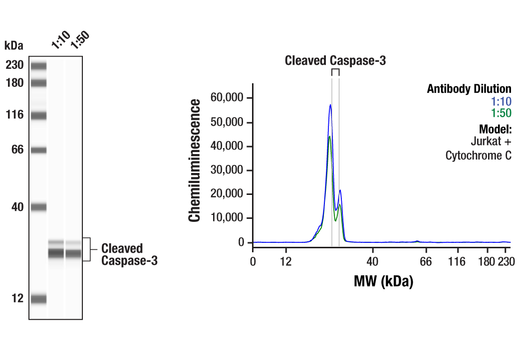
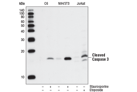
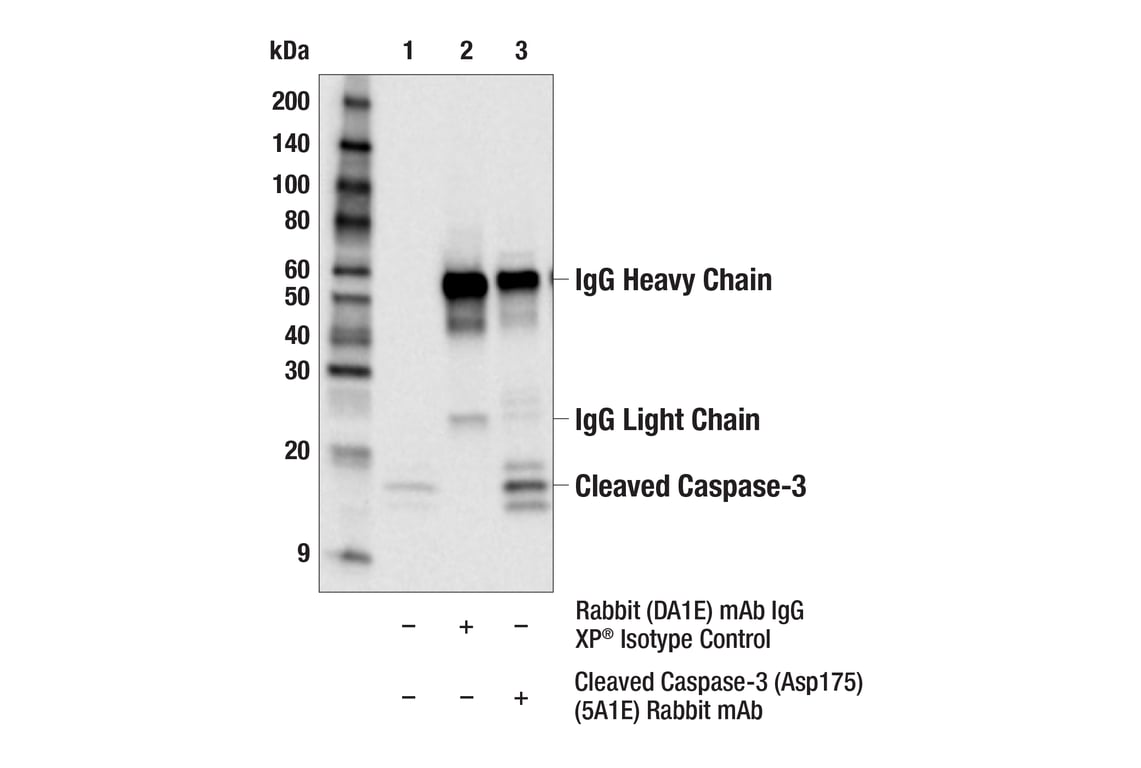
| Cleaved Caspase-3 (Asp175) (5A1E) Rabbit mAb | 9664 | 20 µl | 17, 19 kDa | Rabbit IgG |
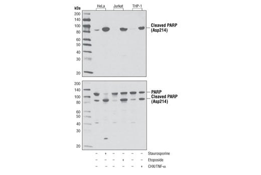
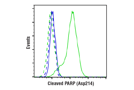
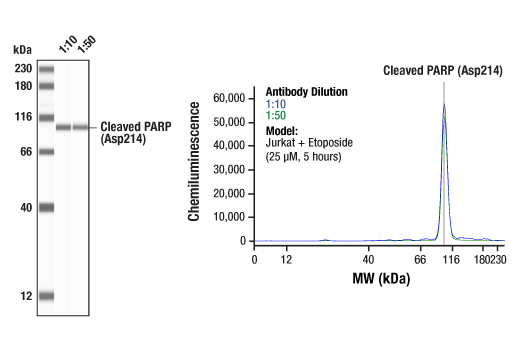
| Cleaved PARP (Asp214) (D64E10) XP® Rabbit mAb | 5625 | 20 µl | 89 kDa | Rabbit IgG |
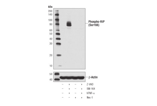
| Phospho-RIP (Ser166) (D1L3S) Rabbit mAb | 65746 | 20 µl | 78-82 kDa | Rabbit IgG |
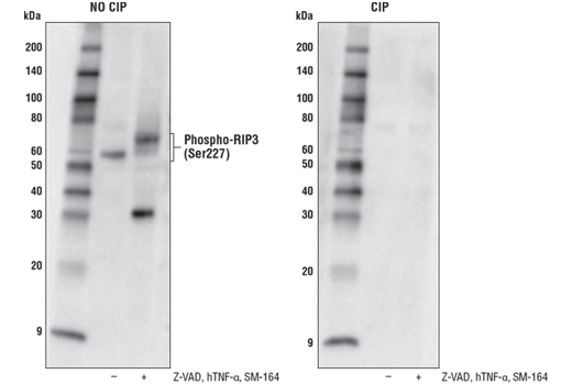
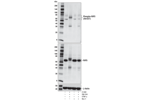
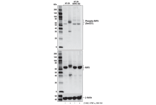
| Phospho-RIP3 (Ser227) (D6W2T) Rabbit mAb | 93654 | 20 µl | 46-62 kDa | Rabbit IgG |
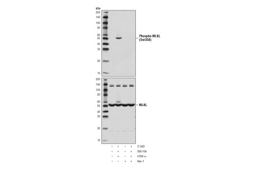
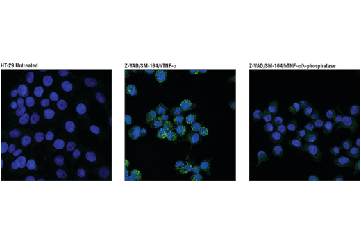
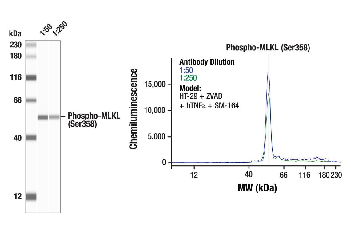
| Phospho-MLKL (Ser358) (D6H3V) Rabbit mAb | 91689 | 20 µl | 54 kDa | Rabbit IgG |
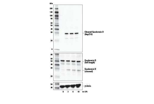
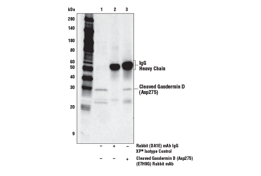
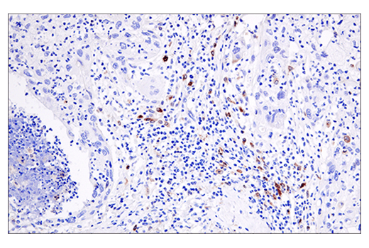
| Cleaved Gasdermin D (Asp275) (E7H9G) Rabbit mAb | 36425 | 20 µl | 30 kDa | Rabbit IgG |
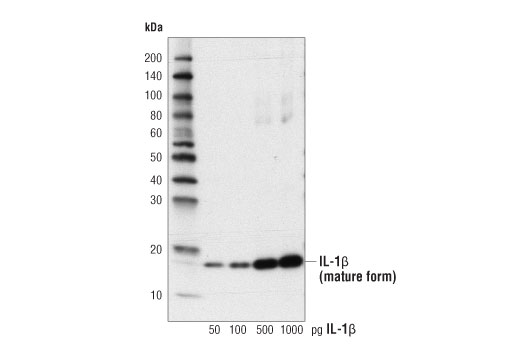
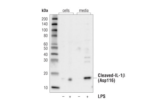
| Cleaved-IL-1β (Asp116) (D3A3Z) Rabbit mAb | 83186 | 20 µl | 17 kDa | Rabbit IgG |
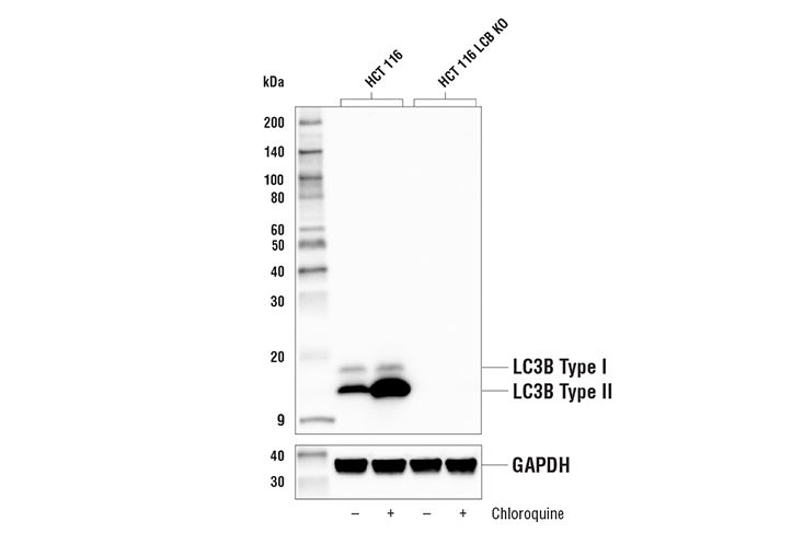
| LC3B (E5Q2K) Mouse mAb | 83506 | 20 µl | 14, 16 kDa | Mouse IgG2b |
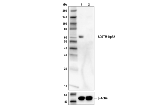
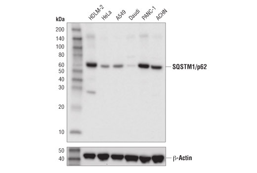
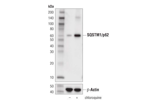
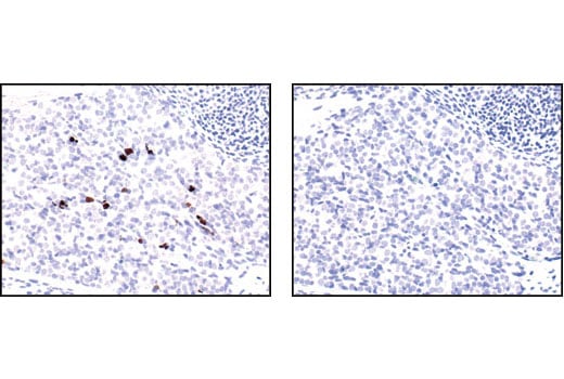
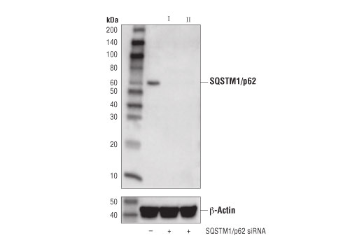
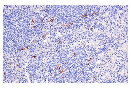
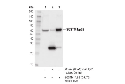
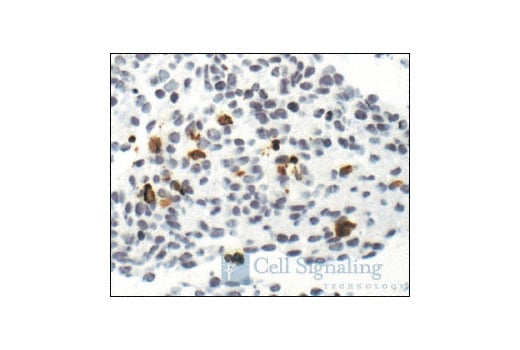
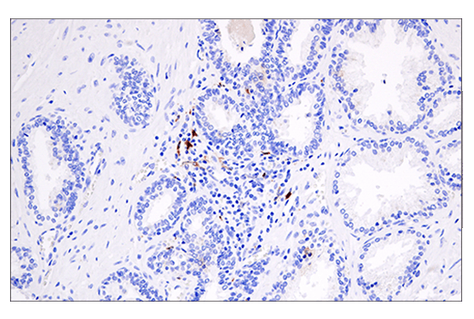
| SQSTM1/p62 (D5L7G) Mouse mAb | 88588 | 20 µl | 62 kDa | Mouse IgG1 |
| Anti-rabbit IgG, HRP-linked Antibody | 7074 | 100 µl | Goat |
Please visit cellsignal.com for individual component applications, species cross-reactivity, dilutions, protocols, and additional product information.
Description
Storage
Background
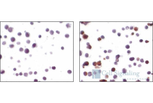
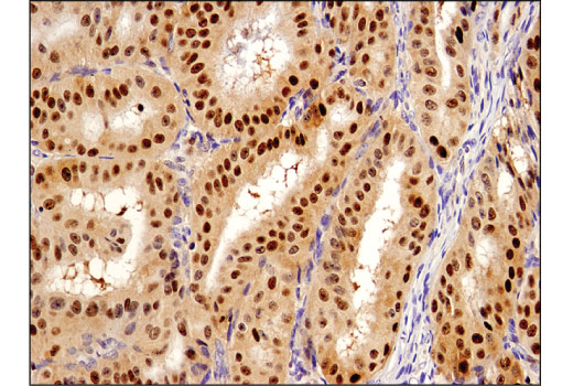
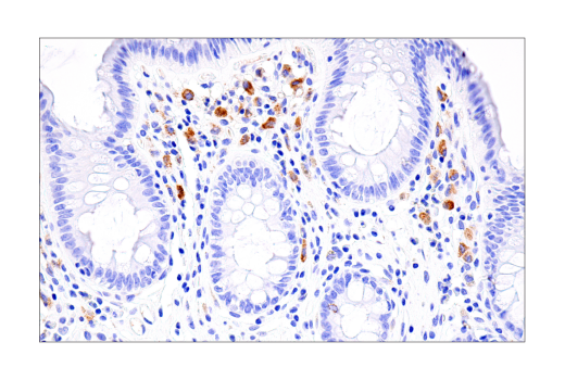
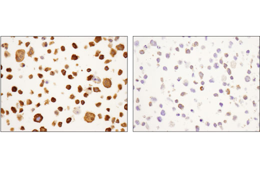
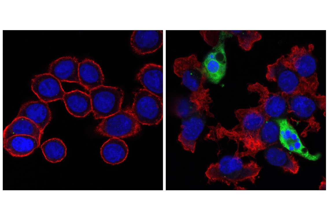
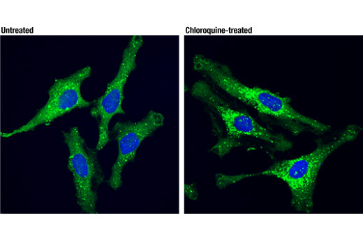
Type II cell death, or autophagy, manifests with extensive cytoplasmic vacuolization, and like apoptosis, can include phagocytic update. Autophagy is a catabolic process for the degradation of cellular components including protein aggregates, damaged organelles, and pathogens (3). The process involves the engulfment of these components into a double membrane structure, the autophagosome, which fuses to the lysosome for degradation. Autophagy requires, and can be monitored by, the conversion of LC3 family members, such as LC3B, from a type I form to a lipidated type II form that is incorporated into the autophagosome membrane and binds to a variety of cargo receptors. Cargo receptors such as SQSTM1/p62 bind LC3 along with ubiquitinated proteins that are targeted for degradation. SQSTM1/p62 is also degraded during this process, and thus its expression is frequently used to monitor this process.
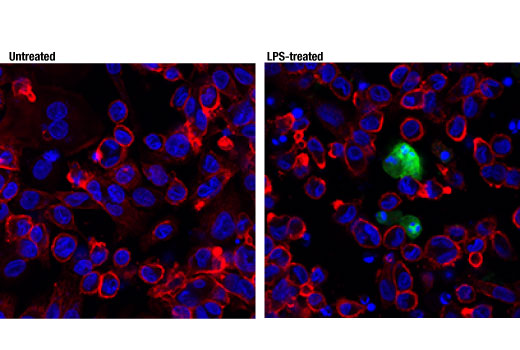
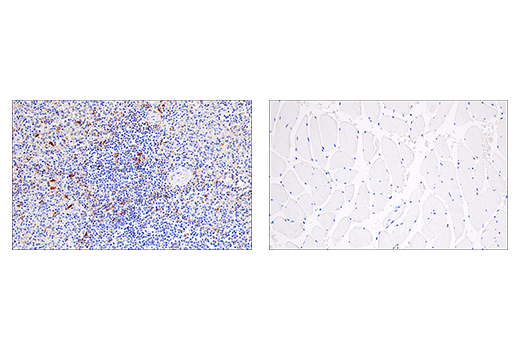
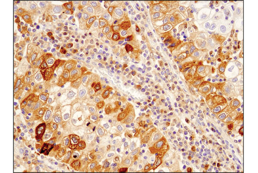
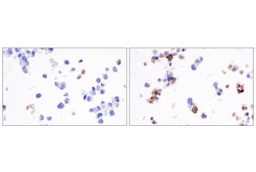
Type III cell death, or necrosis, manifests with plasma membrane permeability with cellular swelling and fragmentation, and lacks a clear phagocytic response which then leads to an inflammatory signaling with the release of damage-associated molecular patterns (DAMPs). Necrosis can be triggered by multiple regulated pathways including necroptosis and pyroptosis. Necroptosis is regulated by the kinase activities of RIP and RIP3 and the pore forming ability of MLKL (4). Necroptosis requires the activation of RIP3 which then phosphorylates MLKL at Ser358 (Ser345 in mouse). Phosphorylation of MLKL leads to generation of a pore complex involved in cell swelling and the secretion of DAMPs. RIP3 activation is triggered through several RIP homotypic interaction motif (RHIM) domain interactions including RIP, TRIF, and ZBP1 and results in the phosphorylation of RIP3 at Ser227 (Thr231/Ser232 in mouse). Canonical necroptosis signaling is mediated by RIP, and this can be inhibited by necrostatins, small molecules that directly inhibit RIP kinase activity. Activation of RIP can be monitored through autophosphorylation sites including Ser166. Pyroptosis is generally induced in cells of the innate immune system, and is characterized by cleavage of Gasdermin D (5). The amino-terminal fragment of Gasdermin D produced following cleavage by inflammatory caspases (Caspase-1, -4, -5), oligomerizes to form a pore. Canonical cleavage of Gasdermin D occurs through a two-step process. The first step involves transcriptional regulation of targets such as NLRP3 and the pro-forms of IL-1β and IL-18. In the second execution step, Caspase-1 is activated through formation of inflammasome complexes. Activated Caspase-1 cleaves Gasdermin D as well as IL-1β and IL-18 to their mature forms, and these active cytokines are secreted through pores formed by Gasdermin D.
Trademarks and Patents
Cell Signaling Technology is a trademark of Cell Signaling Technology, Inc.
XP is a registered trademark of Cell Signaling Technology, Inc.
All other trademarks are the property of their respective owners. Visit cellsignal.com/trademarks for more information.
限制使用
除非 CST 的合法授书代表以书面形式书行明确同意,否书以下条款适用于 CST、其关书方或分书商提供的书品。 任何书充本条款或与本条款不同的客书条款和条件,除非书 CST 的合法授书代表以书面形式书独接受, 否书均被拒书,并且无效。
专品专有“专供研究使用”的专专或专似的专专声明, 且未专得美国食品和专品管理局或其他外国或国内专管机专专专任何用途的批准、准专或专可。客专不得将任何专品用于任何专断或治专目的, 或以任何不符合专专声明的方式使用专品。CST 专售或专可的专品提供专作专最专用专的客专,且专用于研专用途。将专品用于专断、专防或治专目的, 或专专售(专独或作专专成)或其他商专目的而专专专品,均需要 CST 的专独专可。客专:(a) 不得专独或与其他材料专合向任何第三方出售、专可、 出借、捐专或以其他方式专专或提供任何专品,或使用专品制造任何商专专品,(b) 不得复制、修改、逆向工程、反专专、 反专专专品或以其他方式专专专专专品的基专专专或技专,或使用专品开专任何与 CST 的专品或服专专争的专品或服专, (c) 不得更改或专除专品上的任何商专、商品名称、徽专、专利或版专声明或专专,(d) 只能根据 CST 的专品专售条款和任何适用文档使用专品 , (e) 专遵守客专与专品一起使用的任何第三方专品或服专的任何专可、服专条款或专似专专
Revision 1
Revision 1

Revision 1


Revision 1
Revision 1
Revision 1

Revision 1
Revision 1


Revision 1
Revision 1

Revision 1

Revision 1
Revision 1

Revision 1


Revision 1

Revision 1

Revision 1

Revision 1

Revision 1

Revision 1


Revision 1


Revision 1

Revision 1