Revision 1
#43049
Store at -20C
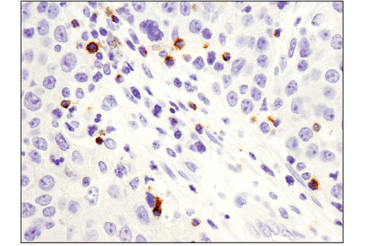
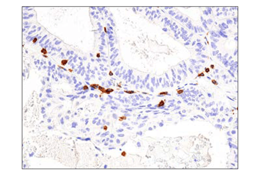
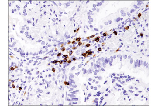
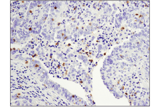
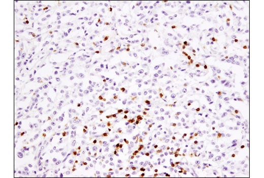
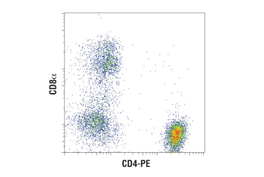
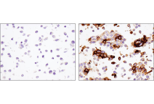
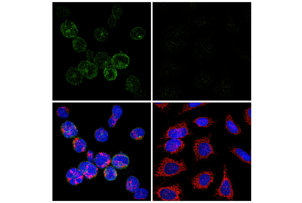
Human Exhausted CD8+ T Cell IHC Antibody Sampler Kit
1 Kit
(9 x 20 microliters)
877-616-CELL (2355)
877-678-TECH (8324)
3 Trask Lane | Danvers | Massachusetts | 01923 | USA
For Research Use Only. Not for Use in Diagnostic Procedures.
| Product Includes | Product # | Quantity | Mol. Wt | Isotype/Source |
|---|---|---|---|---|
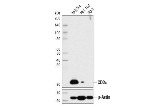
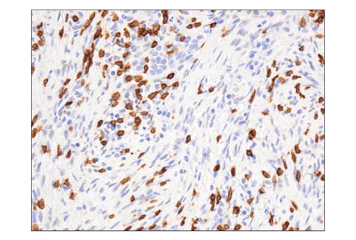
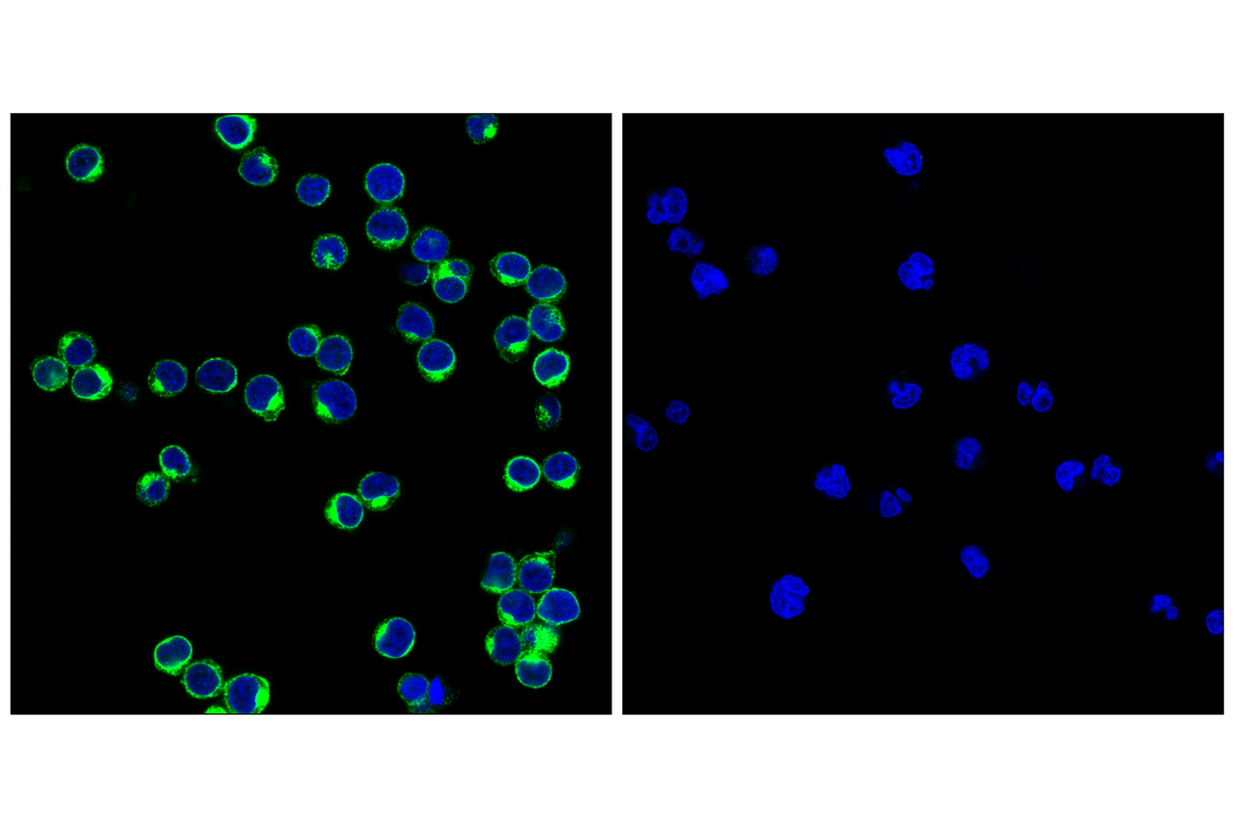
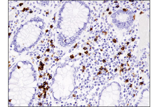
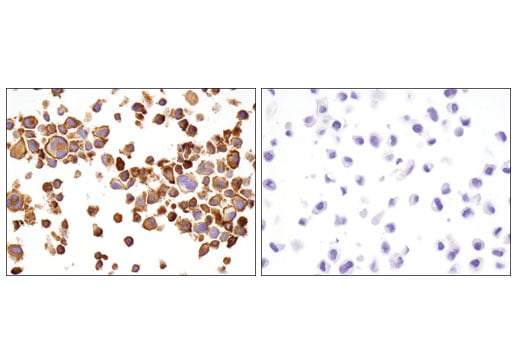
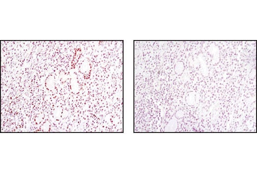
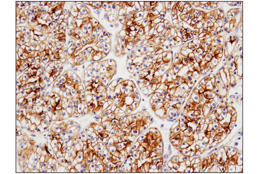
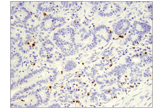
| CD3ε (D7A6E™) XP® Rabbit mAb | 85061 | 20 µl | 23 kDa | Rabbit IgG |
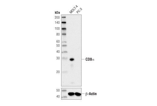
| CD8α (D8A8Y) Rabbit mAb | 85336 | 20 µl | 29 kDa | Rabbit IgG |
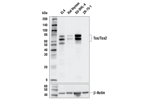
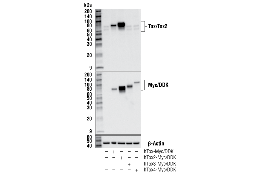
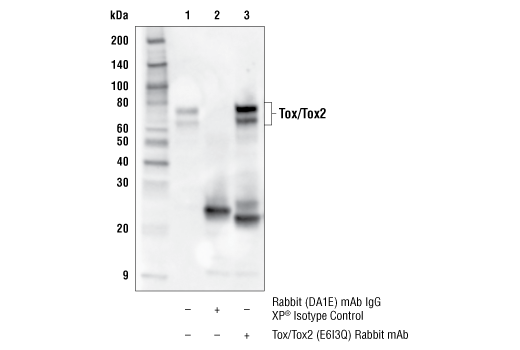
| Tox/Tox2 (E6I3Q) Rabbit mAb | 73758 | 20 µl | 60-80 kDa | Rabbit IgG |
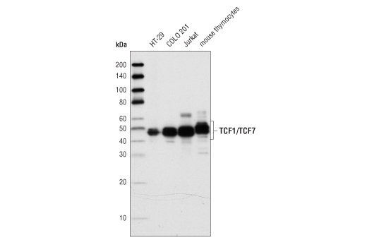
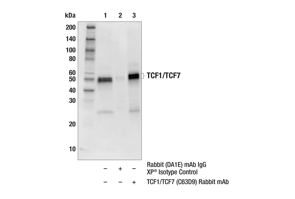
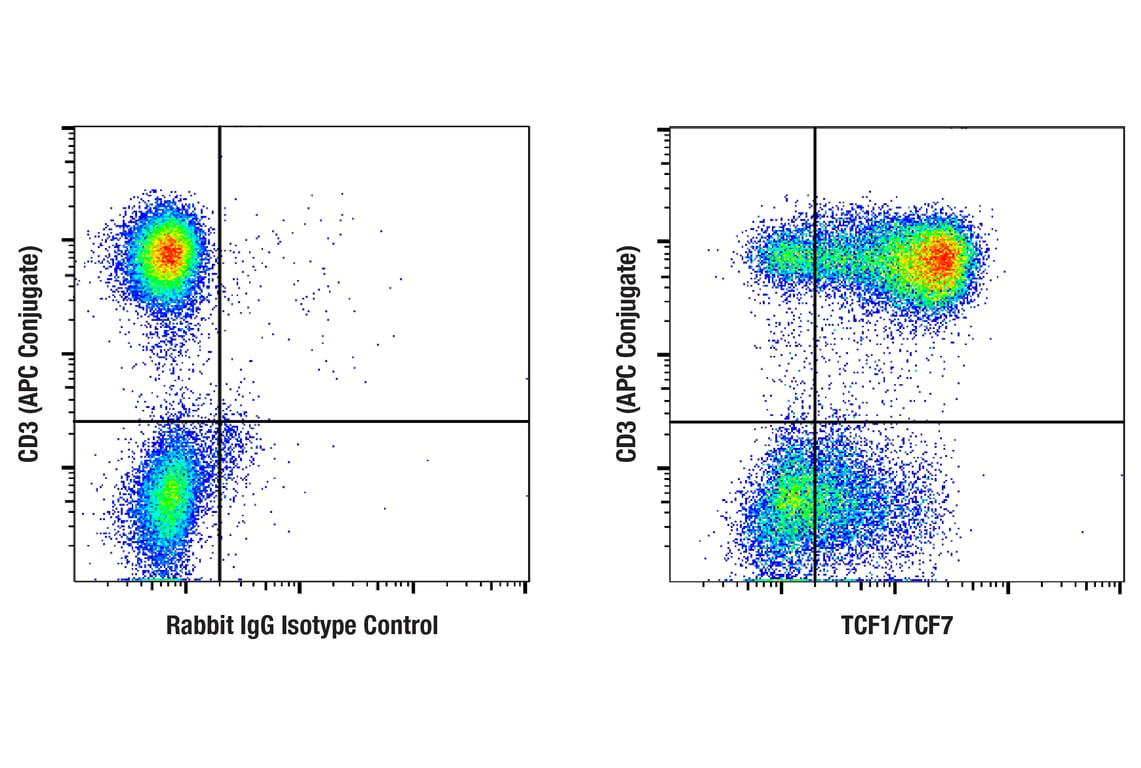
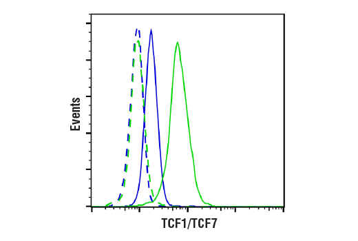
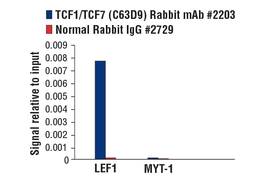
| TCF1/TCF7 (C63D9) Rabbit mAb | 2203 | 20 µl | 48, 50 kDa | Rabbit IgG |
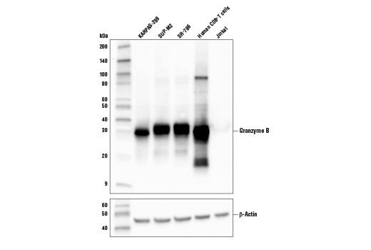
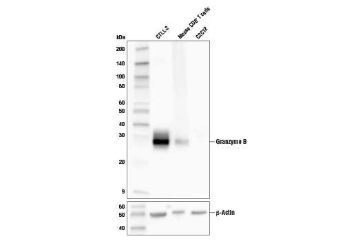
| Granzyme B (D6E9W) Rabbit mAb | 46890 | 20 µl | 30 kDa | Rabbit IgG |
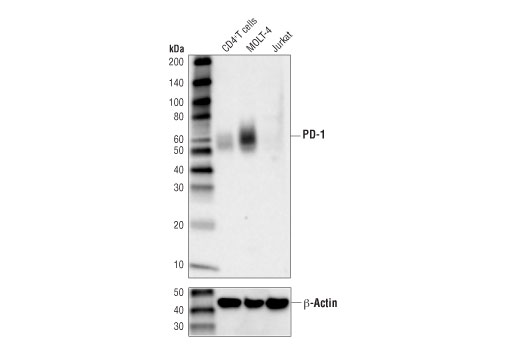
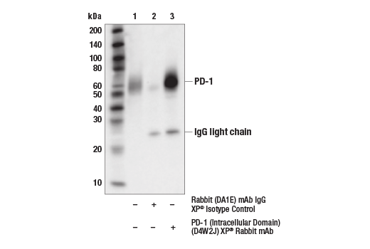
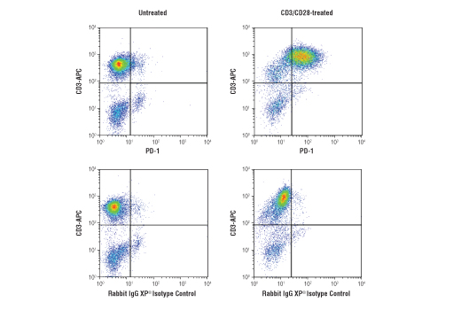
| PD-1 (Intracellular Domain) (D4W2J) XP® Rabbit mAb | 86163 | 20 µl | 52-65 kDa | Rabbit IgG |
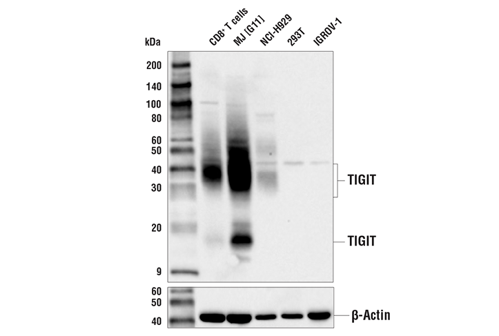
| TIGIT (E5Y1W) XP® Rabbit mAb | 99567 | 20 µl | 18, 30-40 kDa | Rabbit IgG |
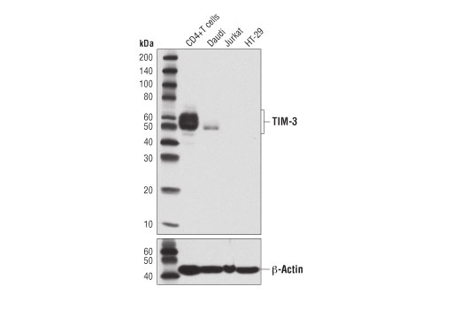
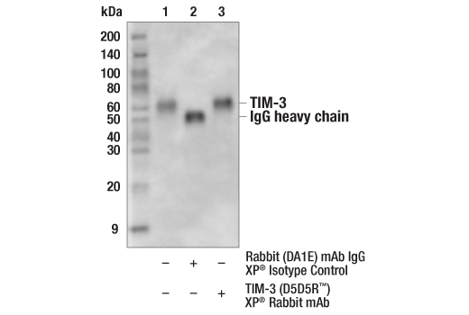
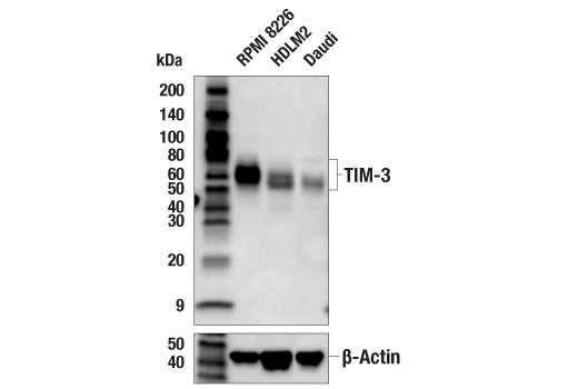
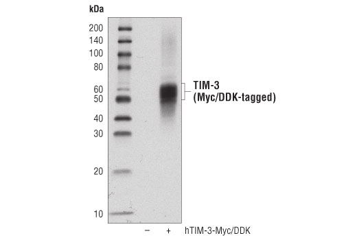
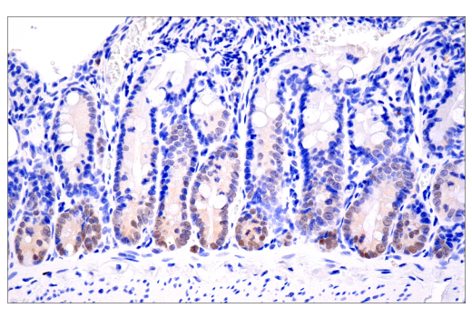
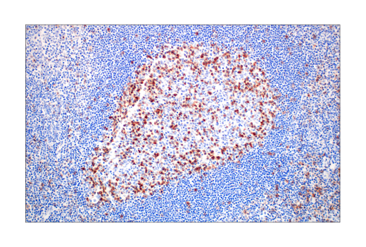
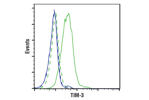
| TIM-3 (D5D5R™) XP® Rabbit mAb | 45208 | 20 µl | 45-70 kDa | Rabbit IgG |
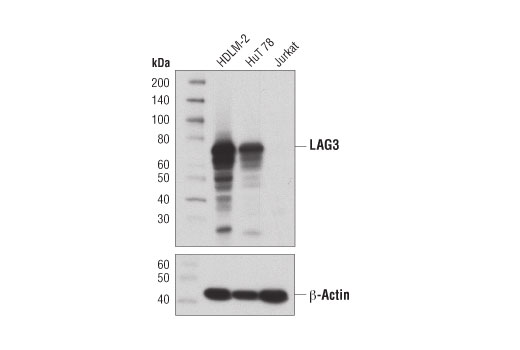
| LAG3 (D2G4O) XP® Rabbit mAb | 15372 | 20 µl | 60-80 kDa | Rabbit IgG |
| Anti-rabbit IgG, HRP-linked Antibody | 7074 | 100 µl | Goat |
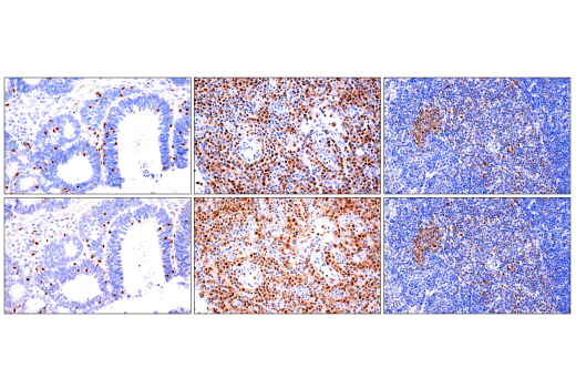
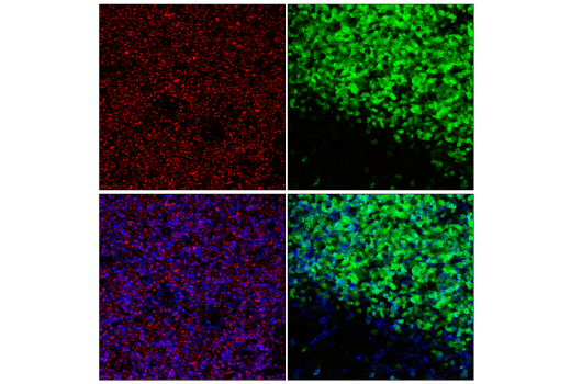
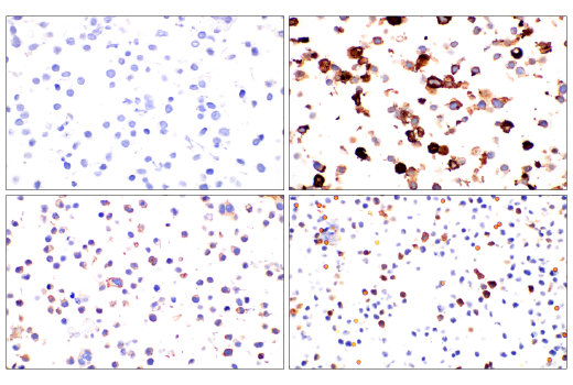
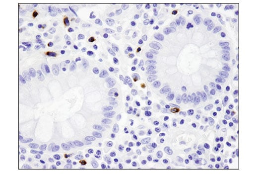
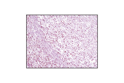
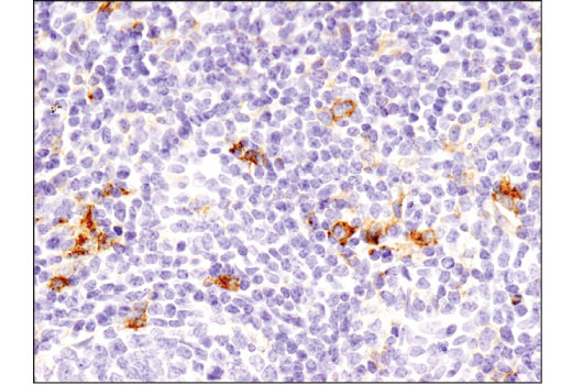
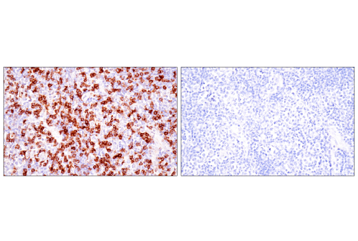
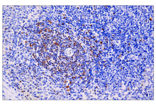
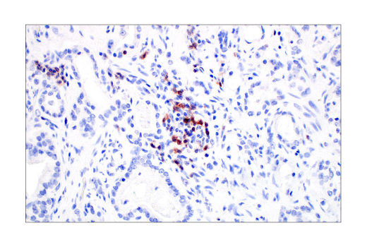
Please visit cellsignal.com for individual component applications, species cross-reactivity, dilutions, protocols, and additional product information.
Description
Storage
Background
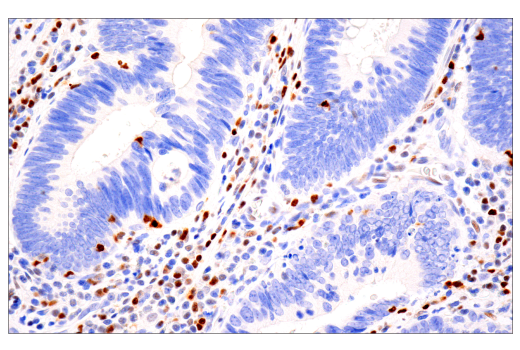
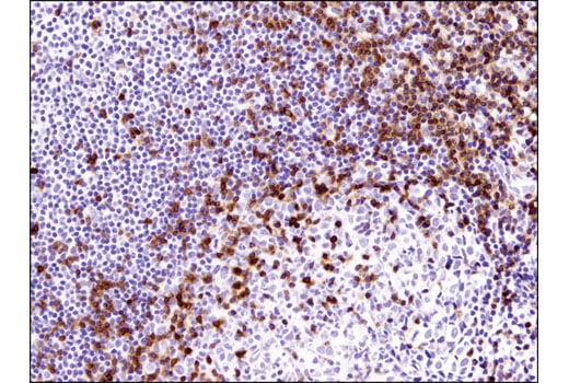
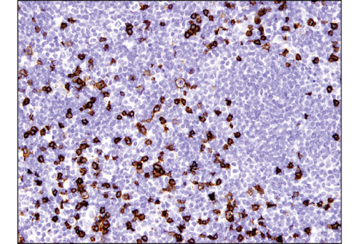
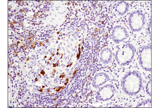
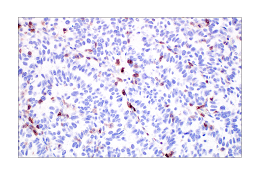
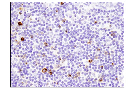
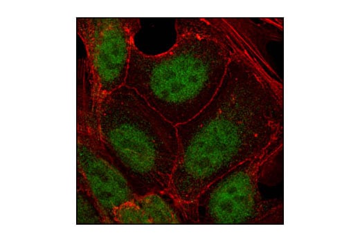
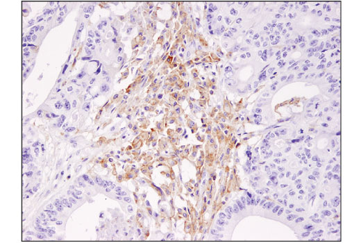
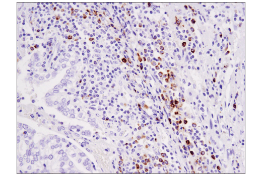
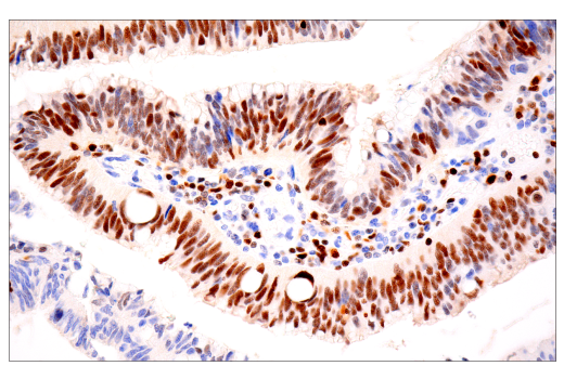
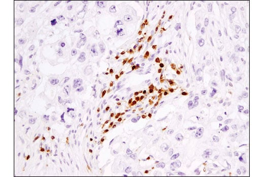
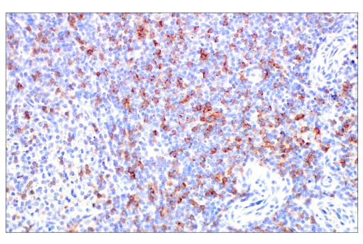
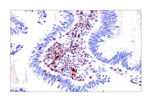
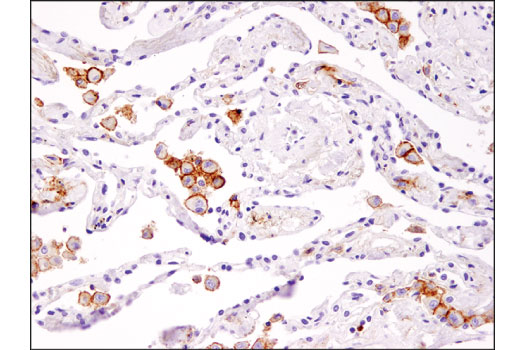
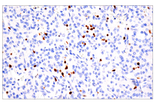
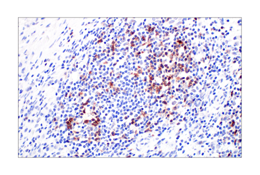
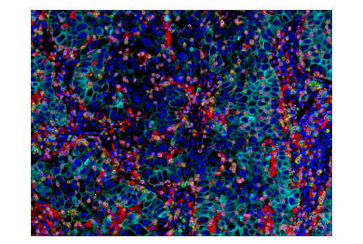
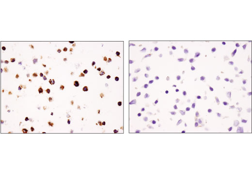
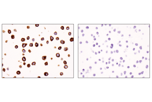
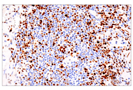
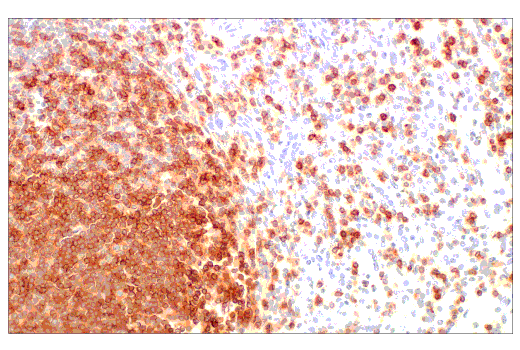
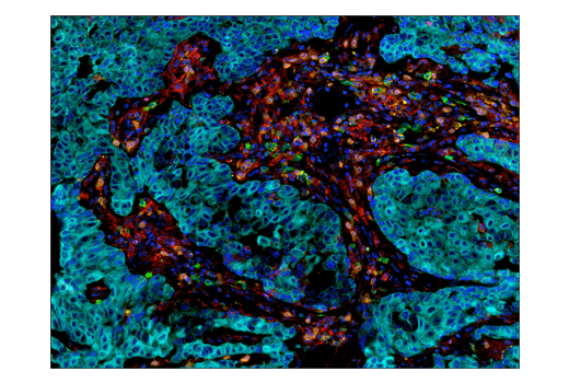
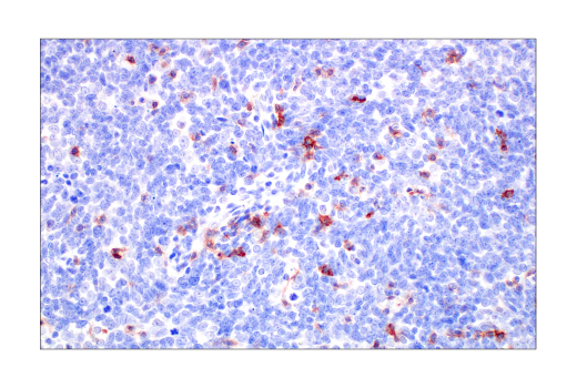
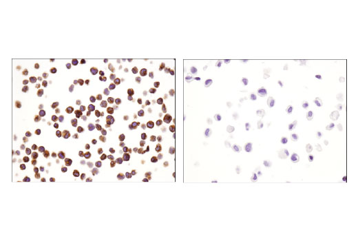
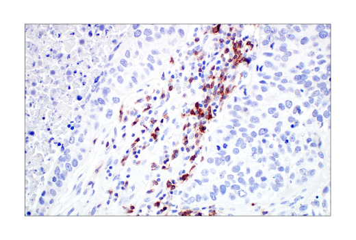
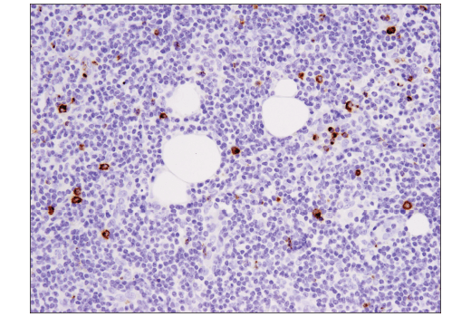
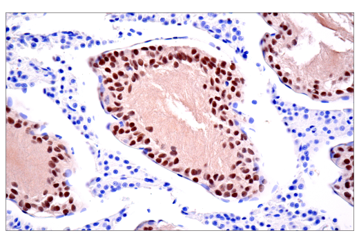
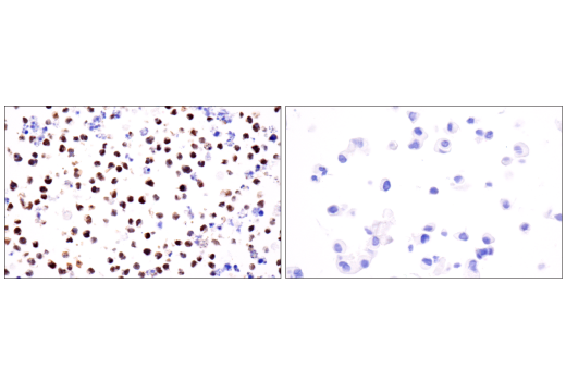
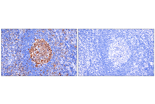
Tox, Tox2, and TCF1/TCF7 play key roles in T cell development. Tox is also induced by high antigen stimulation during chronic viral infection or cancer, regulating T cell persistence and exhaustion. TCF1/TCF7 preserves the effector function of exhausted T cells during viral infection or cancer. EOMES is a key transcription factor for memory T cells and for full effector differentiation of CD8+ T cells. The dynamic expression of these transcription factors help characterize the extent to which a T cell is exhausted and will respond to antigen stimulation (4-8). Granzyme B is a serine protease expressed by cytotoxic T lymphocytes and natural killer (NK) cells and is a key component of immune responses to pathogens and transformed cells (9).
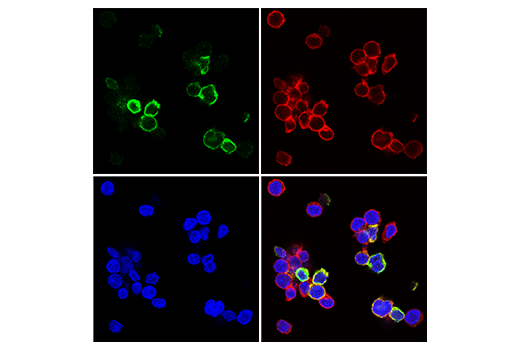
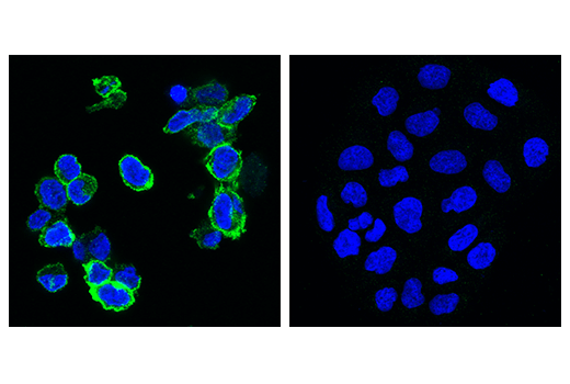
PD-1 (PDCD1, CD279), TIGIT (VSIG9, VSTM3), TIM-3 (HAVCR2), and LAG3 (CD223) are immune cell co-inhibitory receptors (also known as immune checkpoints) that negatively regulate T cell function and dampen the immune response to pathogens and cancer (10-15). In addition to activated T cells, PD-1 is expressed by activated B cells and monocytes. Following interaction with its ligands, PD-L1 and PD-L2, PD-1 is phosphorylated at ITIM and ITSM motifs leading to recruitment of protein tyrosine phosphatases SHP-1 and SHP-2 and suppression of TCR signaling. TIGIT is expressed at low levels on subsets of T cells and NK cells, and is upregulated at the protein level following activation of these cells. TIGIT marks exhausted T cells in the tumor microenvironment and during human immunodeficiency virus (HIV) infection. TIM-3 is expressed by exhausted T cells in the settings of chronic infection and cancer. Tumor-infiltrating macrophages and dendritic cells also express TIM-3. LAG3 is primarily expressed by activated CD4+ T cells, CD8+ T cells, FoxP3+ T regulatory cells (Tregs), and natural killer (NK) cells. Co-expression of multiple immune checkpoints help characterize the extent to which a T cell is exhausted and will respond to antigen stimulation. Therapeutic blockade of several of these immune checkpoint receptors is a promising strategy for neoplastic intervention by enabling anti-tumor immune responses (10-15).
Background References
- Kuhns, M.S. et al. (2006) Immunity 24, 133-9.
- Zamoyska, R. (1994) Immunity 1, 243-6.
- Shortman, K. and Heath, W.R. (2010) Immunol Rev 234, 18-31.
- Aliahmad, P. et al. (2012) Curr Opin Immunol 24, 173-7.
- Yao, C. et al. (2019) Nat Immunol 20, 890-901.
- Alfei, F. et al. (2019) Nature 571, 265-269.
- Seo, H. et al. (2019) Proc Natl Acad Sci U S A 116, 12410-12415.
- Wang, Y. et al. (2019) Front Immunol 10, 169.
- Trapani, J.A. (2001) Genome Biol 2, REVIEWS3014.
- Schildberg, F.A. et al. (2016) Immunity 44, 955-72.
- Anderson, A.C. et al. (2016) Immunity 44, 989-1004.
- Callahan, M.K. et al. (2016) Immunity 44, 1069-78.
- Chen, L. and Flies, D.B. (2013) Nat Rev Immunol 13, 227-42.
- Chauvin, J.M. et al. (2015) J Clin Invest 125, 2046-58.
- Chew, G.M. et al. (2016) PLoS Pathog 12, e1005349.
Trademarks and Patents
Cell Signaling Technology is a trademark of Cell Signaling Technology, Inc.
XP is a registered trademark of Cell Signaling Technology, Inc.
U.S. Patent No. 7,429,487, foreign equivalents, and child patents deriving therefrom.
All other trademarks are the property of their respective owners. Visit cellsignal.com/trademarks for more information.
限制使用
除非 CST 的合法授书代表以书面形式书行明确同意,否书以下条款适用于 CST、其关书方或分书商提供的书品。 任何书充本条款或与本条款不同的客书条款和条件,除非书 CST 的合法授书代表以书面形式书独接受, 否书均被拒书,并且无效。
专品专有“专供研究使用”的专专或专似的专专声明, 且未专得美国食品和专品管理局或其他外国或国内专管机专专专任何用途的批准、准专或专可。客专不得将任何专品用于任何专断或治专目的, 或以任何不符合专专声明的方式使用专品。CST 专售或专可的专品提供专作专最专用专的客专,且专用于研专用途。将专品用于专断、专防或治专目的, 或专专售(专独或作专专成)或其他商专目的而专专专品,均需要 CST 的专独专可。客专:(a) 不得专独或与其他材料专合向任何第三方出售、专可、 出借、捐专或以其他方式专专或提供任何专品,或使用专品制造任何商专专品,(b) 不得复制、修改、逆向工程、反专专、 反专专专品或以其他方式专专专专专品的基专专专或技专,或使用专品开专任何与 CST 的专品或服专专争的专品或服专, (c) 不得更改或专除专品上的任何商专、商品名称、徽专、专利或版专声明或专专,(d) 只能根据 CST 的专品专售条款和任何适用文档使用专品 , (e) 专遵守客专与专品一起使用的任何第三方专品或服专的任何专可、服专条款或专似专专
Revision 1
Revision 1
Revision 1
Revision 1

Revision 1
Revision 1
Revision 1
Revision 1
Revision 1
Revision 1
Revision 1
Revision 1
Revision 1
Revision 1
Revision 1
Revision 1
Revision 1


Revision 1
Revision 1
Revision 1
Revision 1
Revision 1
Revision 1

Revision 1
Revision 1


Revision 1
Revision 1

Revision 1
Revision 1
Revision 1
Revision 1