WB
H M
Endogenous
550
Rabbit
#Q14517
2195
Product Information
Product Usage Information
| Application | Dilution |
|---|---|
| Western Blotting | 1:1000 |
Storage
Specificity / Sensitivity
Species Reactivity:
Human, Mouse
Species predicted to react based on 100% sequence homology
The antigen sequence used to produce this antibody shares
100% sequence homology with the species listed here, but
reactivity has not been tested or confirmed to work by CST.
Use of this product with these species is not covered under
our
Product Performance Guarantee.
Rat
Source / Purification
Polyclonal antibodies are produced by immunizing animals with a synthetic peptide corresponding to residues near the carboxy terminus of human FAT1 protein. Antibodies are purified by peptide affinity chromatography.
Background
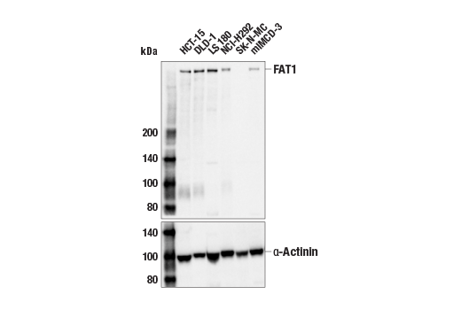
FAT1 is a member of the FAT atypical cadherin (FAT) subfamily of cadherin proteins (1). FAT1 is a single-pass transmembrane protein, first identified in a screen for tumor suppressor proteins in Drosophila (2). FAT1 is expressed primarily in epithelial cells, where it plays a prominent role in regulating cell growth and migration, in large part through the regulation of cell-cell adhesion dynamics (3). The intracellular cytoplasmic tail of FAT1 contains multiple functional motif/domains that regulate FAT1 functions, including a proline rich EVH1 binding motif that regulates actin cytoskeleton components (e.g., Ena/VASP proteins) at both cell-cell contact points and leading edges of migrating cells (4,5). FAT1 appears to play a role in linking cell adhesion events to intracellular signaling pathways. For example, FAT1 was capable of inhibiting the nuclear translocation of β-catenin through its cytoplasmic FC1 domain interaction with β-catenin (6), and activating the Hippo signaling pathway, suppressing YAP signaling by its N-terminal cytoplasmic region interaction with MST1 (7). Research studies have revealed that the tumor suppressor functions identified in Drosophila are conserved in vertebrate FAT1 homologs (8). For example, studies in human cancer cells showed that loss-of-function mutations in the gene encoding FAT1 promoted a hybrid epithelial-to-mesenchymal transition, which further enabled the development of cancer drug resistance (9). Notably, studies have also revealed an oncogenic function for FAT1 in some contexts (10).
- Tanoue, T. and Takeichi, M. (2005) J Cell Sci 118, 2347-53.
- Mahoney, P.A. et al. (1991) Cell 67, 853-68.
- Moeller, M.J. et al. (2004) EMBO J 23, 3769-79.
- Tanoue, T. and Takeichi, M. (2004) J Cell Biol 165, 517-28.
- Hou, R. et al. (2006) J Cell Biol 173, 417-29.
- Martin, D. et al. (2018) Nat Commun 9, 2372.
- Morris, L.G. et al. (2013) Nat Genet 45, 253-61.
- Pastushenko, I. et al. (2021) Nature 589, 448-455.
- Peng, Z. et al. (2021) Oncol Lett 21, 398.
Species Reactivity
Species reactivity is determined by testing in at least one approved application (e.g., western blot).
Western Blot Buffer
IMPORTANT: For western blots, incubate membrane with diluted primary antibody in 5% w/v BSA, 1X TBS, 0.1% Tween® 20 at 4°C with gentle shaking, overnight.
Applications Key
WB: Western Blotting
Cross-Reactivity Key
H: human M: mouse R: rat Hm: hamster Mk: monkey Vir: virus Mi: mink C: chicken Dm: D. melanogaster X: Xenopus Z: zebrafish B: bovine Dg: dog Pg: pig Sc: S. cerevisiae Ce: C. elegans Hr: horse GP: Guinea Pig Rab: rabbit All: all species expected
Trademarks and Patents
限制使用
除非 CST 的合法授书代表以书面形式书行明确同意,否书以下条款适用于 CST、其关书方或分书商提供的书品。 任何书充本条款或与本条款不同的客书条款和条件,除非书 CST 的合法授书代表以书面形式书独接受, 否书均被拒书,并且无效。
专品专有“专供研究使用”的专专或专似的专专声明, 且未专得美国食品和专品管理局或其他外国或国内专管机专专专任何用途的批准、准专或专可。客专不得将任何专品用于任何专断或治专目的, 或以任何不符合专专声明的方式使用专品。CST 专售或专可的专品提供专作专最专用专的客专,且专用于研专用途。将专品用于专断、专防或治专目的, 或专专售(专独或作专专成)或其他商专目的而专专专品,均需要 CST 的专独专可。客专:(a) 不得专独或与其他材料专合向任何第三方出售、专可、 出借、捐专或以其他方式专专或提供任何专品,或使用专品制造任何商专专品,(b) 不得复制、修改、逆向工程、反专专、 反专专专品或以其他方式专专专专专品的基专专专或技专,或使用专品开专任何与 CST 的专品或服专专争的专品或服专, (c) 不得更改或专除专品上的任何商专、商品名称、徽专、专利或版专声明或专专,(d) 只能根据 CST 的专品专售条款和任何适用文档使用专品, (e) 专遵守客专与专品一起使用的任何第三方专品或服专的任何专可、服专条款或专似专专