| Product Includes | Product # | Quantity | Mol. Wt | Isotype/Source |
|---|---|---|---|---|
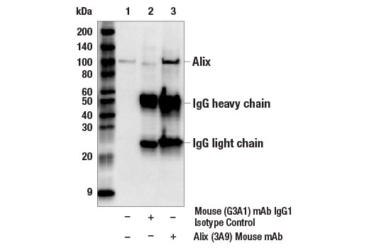
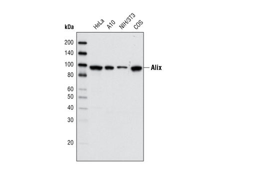
| Alix (3A9) Mouse mAb | 2171 | 20 µl | 95 kDa | Mouse IgG1 |
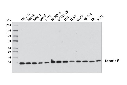
| Annexin V Antibody | 8555 | 20 µl | 30 kDa | Rabbit |
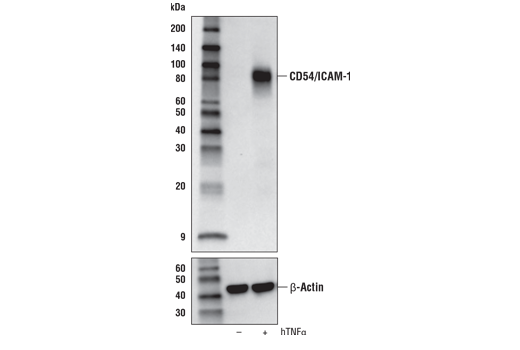
| CD54/ICAM-1 (E3Q9N) XP® Rabbit mAb | 67836 | 20 µl | 89, 92 kDa | Rabbit IgG |
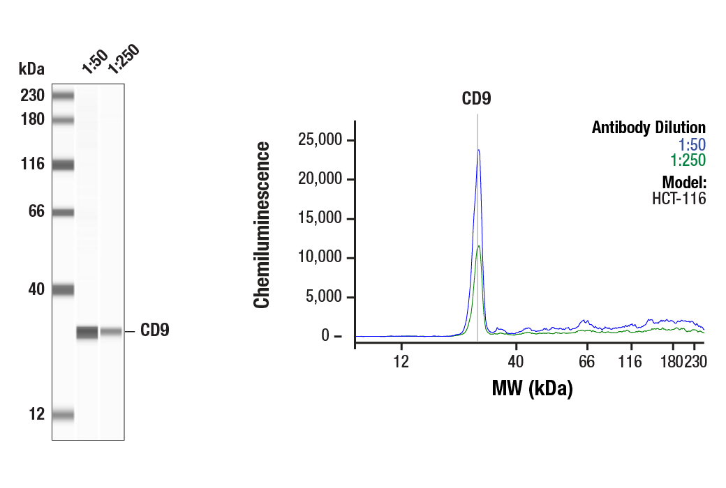
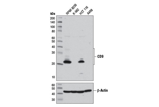
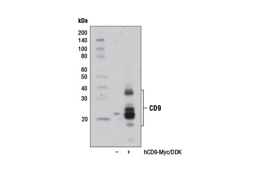
| CD9 (D8O1A) Rabbit mAb | 13174 | 20 µl | 22, 24, 35 kDa | Rabbit IgG |
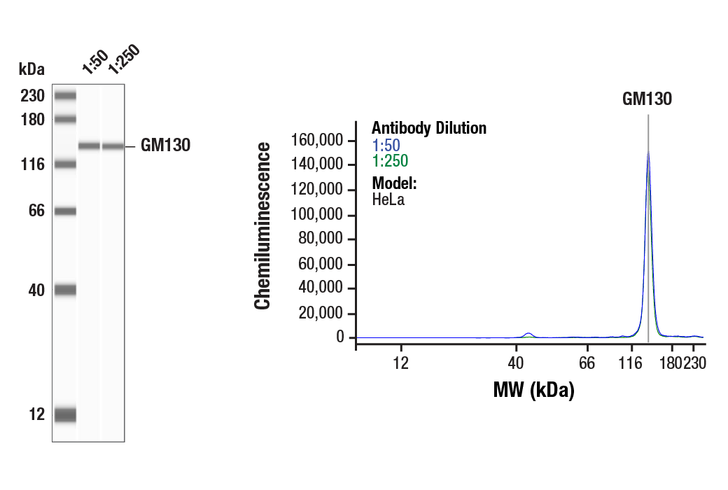
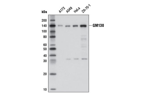
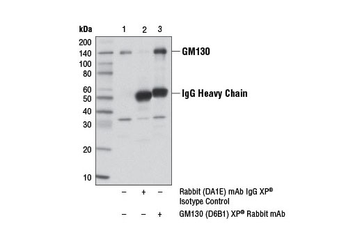
| GM130 (D6B1) XP® Rabbit mAb | 12480 | 20 µl | 140 kDa | Rabbit IgG |
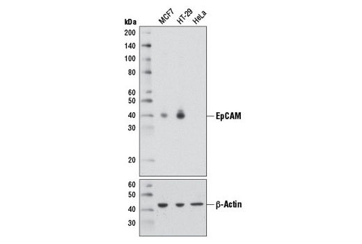
| EpCAM (D1B3) Rabbit mAb | 2626 | 20 µl | 40 kDa | Rabbit IgG |
| HSP70 (D69) Antibody | 4876 | 20 µl | 70 kDa | Rabbit |
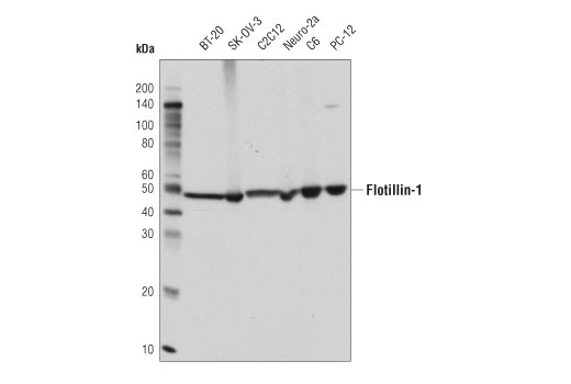
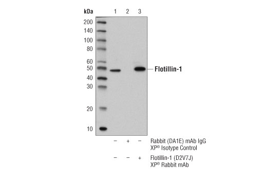
| Flotillin-1 (D2V7J) XP® Rabbit mAb | 18634 | 20 µl | 49 kDa | Rabbit IgG |
| Anti-rabbit IgG, HRP-linked Antibody | 7074 | 100 µl | Goat | |
| Anti-mouse IgG, HRP-linked Antibody | 7076 | 100 µl | Horse |
Please visit cellsignal.com for individual component applications, species cross-reactivity, dilutions, protocols, and additional product information.
Description
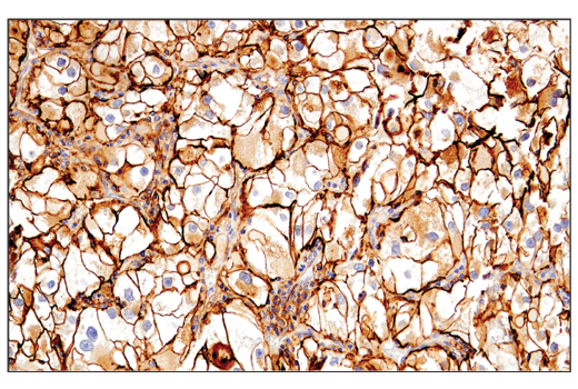
The Exosomal Marker Antibody Sampler Kit provides an economical means to evaluate the presence of exosomal markers. The kit includes enough primary antibody to perform two western blot experiments for each target.
Storage
Background
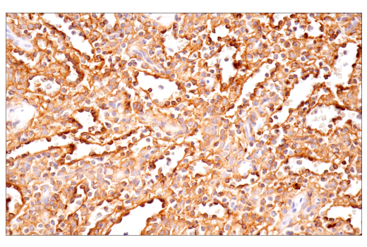
Exosomes are small membrane-bound vesicles that in recent years have emerged as important molecules for inter-cellular communication. Exosomes are produced during both normal and pathophysiological conditions, and cancer cells have been shown to secrete exosomes in greater amounts than normal cells (reviewed in 1). The exosomal markers contained in this kit are Alix, Annexin V, ICAM-1, CD9, GM130, EpCAM, flotillin, and HSP70.
Alix, a cytosolic scaffold protein, regulates many cellular processes including endocytic membrane trafficking, cell adhesion through interactions with ESCRT (endosomal sorting complex required for transport) proteins, endophilins, and CIN85 (Cbl-Interacting protein of 85 kDa) (2, 3).
Annexin V is a ~30 kDa protein that binds to phospho-lipids in a calcium-dependent manner (4). All annexins contain a putative PKC binding site, but only annexin V has been identified as an inhibitor of this pathway (5).
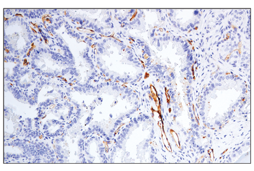
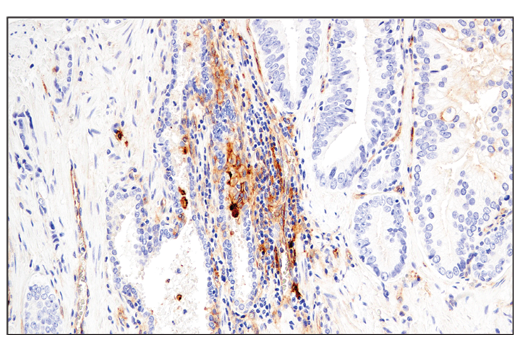
Intracellular cell adhesion molecule-1 (CD54 or ICAM-1) is a cell surface glycoprotein that belongs to the immunoglobulin superfamily (IgSF) of adhesion molecules. CD54 is expressed at low levels in diverse cell types, and is induced by cytokines (TNF-alpha, interleukin-1) and bacterial lipopolysaccharides (6). Apical localization on endothelial cells (or basolateral localization on epithelial cells) is a prerequisite for leukocyte trafficking through the endothelial (or epithelial) barrier (6).
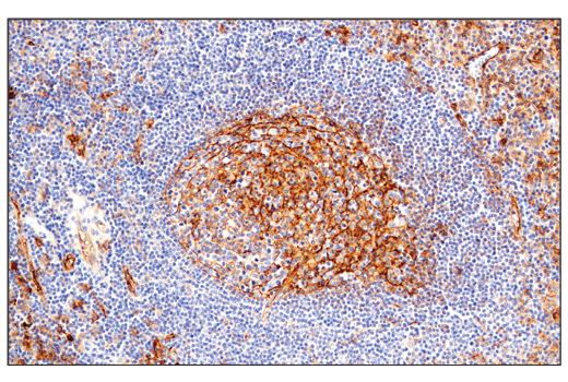
The CD9 antigen belongs to the tetraspanin family of cell surface glycoproteins. Tetraspanins interact with a variety of cell surface proteins and intracellular signaling molecules in specialized tetraspanin-enriched microdomains (TEMs), where they mediate a range of processes including adhesion, motility, membrane organization, and signal transduction (7). Additional research identified CD9 as an abudant component of exosomes, and may play a role in the fusion of these secreted membrane vesicles with recipient cells (8).
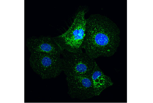
GM130 is required for membrane fusion events that mediate ribbon formation during Golgi assembly (9). The Golgi apparatus functions in the modification, organization, and transport of proteins and membrane targeted to other parts of the cell, such as the plasma membrane, lysosomes, and endosomes. This regulated transport is important for appropriate protein localization, secretion, and signal transduction (reviewed in 10).
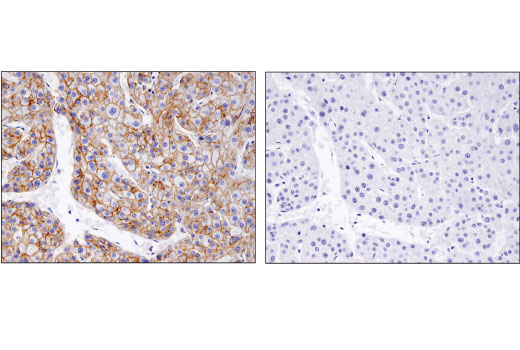
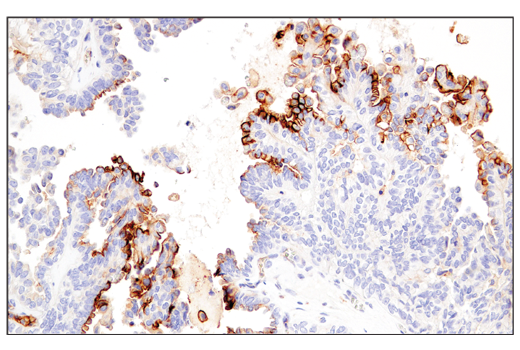
Epithelial cell adhesion and activation molecule (EpCAM/CD326) is a transmembrane glycoprotein that mediates calcium-independent, hemophilic adhesions on the basolateral surface of most epithelial cells (11). One of the first tumor-associated antigens discovered, EpCAM has long been a marker of epithelial and tumor tissue. Research studies have shown that EpCAM is highly expressed in cancer cells and can be used as a biomarker for the detectionof tumor-derived exposomes (reviewed in 1, 12, 13).
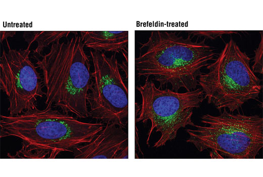
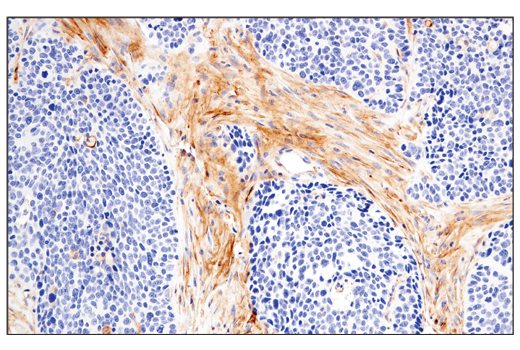
Flotillins belong to a famiy of lipid raft-associated integral membrane proteins that are ubiquitously expressed and located to lipid rafts on the cell plasma membrane where they support signal transduction and regulate lipid raft motility and localization (14-17). In addition to its colocalization with lipid rafts on the plasma membrane, flotillin-1 also has been found at compartments of the endocytic and autophagosomal pathways, such as recycling/ late endosomes, the Golgi complex, as well as the nucleus (18, 19).
HSP70 is a molecular chaperone expressed constituitively under normal conditions to maintain protein homeostatis and is induced upon environmental stress (20). HSP70 is able to interact with unfolded proteins to prevent irreversible aggregation and catalyze the refolding of their substrates in an ATP and co-chaperone dependent manner (21). An immune response is elicited upon excretion of heat shock proteins from tumor exosomes (reviewed in 1).
- Raposo, G. and Stoorvogel, W. (2013) J Cell Biol 200, 373-83.
- Katoh, K. et al. (2003) J Biol Chem 278, 39104-13.
- Sadoul, R. (2006) Biol Cell 98, 69-77.
- Huber, R. et al. (1990) EMBO J 9, 3867-74.
- Cardó-Vila, M. et al. (2003) Mol Cell 11, 1151-62.
- Hopkins, A.M. et al. (2004) Adv Drug Deliv Rev 56, 763-78.
- Hemler, M.E. (2005) Nat Rev Mol Cell Biol 6, 801-11.
- Théry, C. et al. (1999) J Cell Biol 147, 599-610.
- Puthenveedu, M.A. et al. (2006) Nat Cell Biol 8, 238-48.
- Barr, F.A. and Short, B. (2003) Curr Opin Cell Biol 15, 405-13.
- Went, P.T. et al. (2004) Hum Pathol 35, 122-8.
- Baeuerle, P.A. and Gires, O. (2007) Br J Cancer 96, 417-23.
- Armstrong, A. and Eck, S.L. Cancer Biol Ther 2, 320-6.
- Langhorst, M.F. et al. (2005) Cell Mol Life Sci 62, 2228-40.
- Stuermer, C.A. and Plattner, H. (2005) Biochem Soc Symp , 109-18.
- Fernow, I. et al. (2007) Eur J Cell Biol 86, 345-52.
- Neumann-Giesen, C. et al. (2007) J Cell Sci 120, 395-406.
- Liu, J. et al. (2005) J Biol Chem 280, 16125-34.
- Santamaría, A. et al. (2005) Mol Cell Biol 25, 1900-11.
- Nollen, E.A. and Morimoto, R.I. (2002) J Cell Sci 115, 2809-16.
- Young, J.C. et al. (2003) Trends Biochem Sci 28, 541-7.
Background References
Trademarks and Patents
限制使用
除非 CST 的合法授书代表以书面形式书行明确同意,否书以下条款适用于 CST、其关书方或分书商提供的书品。 任何书充本条款或与本条款不同的客书条款和条件,除非书 CST 的合法授书代表以书面形式书独接受, 否书均被拒书,并且无效。
专品专有“专供研究使用”的专专或专似的专专声明, 且未专得美国食品和专品管理局或其他外国或国内专管机专专专任何用途的批准、准专或专可。客专不得将任何专品用于任何专断或治专目的, 或以任何不符合专专声明的方式使用专品。CST 专售或专可的专品提供专作专最专用专的客专,且专用于研专用途。将专品用于专断、专防或治专目的, 或专专售(专独或作专专成)或其他商专目的而专专专品,均需要 CST 的专独专可。客专:(a) 不得专独或与其他材料专合向任何第三方出售、专可、 出借、捐专或以其他方式专专或提供任何专品,或使用专品制造任何商专专品,(b) 不得复制、修改、逆向工程、反专专、 反专专专品或以其他方式专专专专专品的基专专专或技专,或使用专品开专任何与 CST 的专品或服专专争的专品或服专, (c) 不得更改或专除专品上的任何商专、商品名称、徽专、专利或版专声明或专专,(d) 只能根据 CST 的专品专售条款和任何适用文档使用专品, (e) 专遵守客专与专品一起使用的任何第三方专品或服专的任何专可、服专条款或专似专专