| Product Includes | Product # | Quantity | Mol. Wt | Isotype/Source |
|---|---|---|---|---|
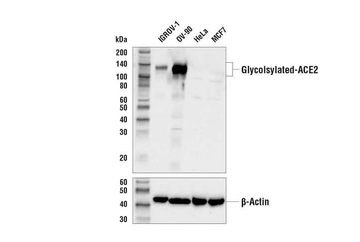
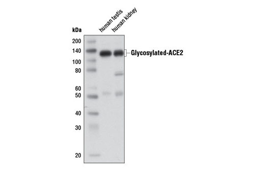
| ACE2 Antibody | 4355 | 20 µl | 120-135 kDa | Rabbit |
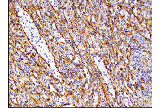
| DPP4/CD26 (D6D8K) Rabbit mAb | 67138 | 20 µl | 90, 120 kDa | Rabbit IgG |
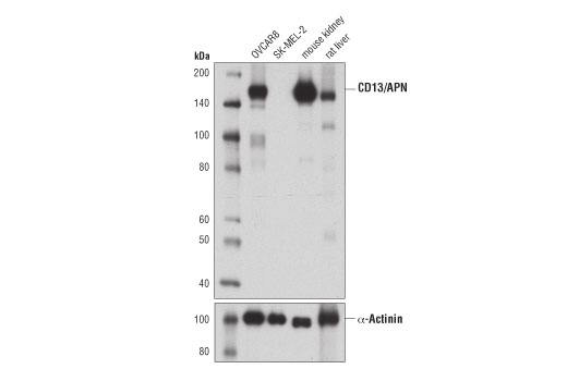
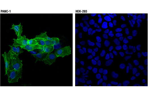
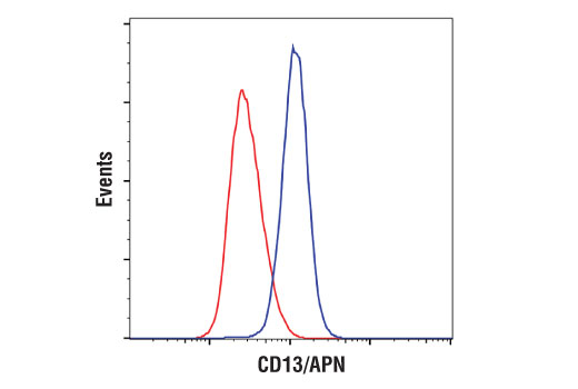
| CD13/APN (D6V1W) Rabbit mAb | 32720 | 20 µl | 160 kDa | Rabbit IgG |
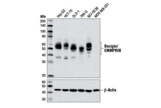
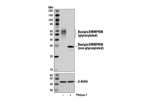
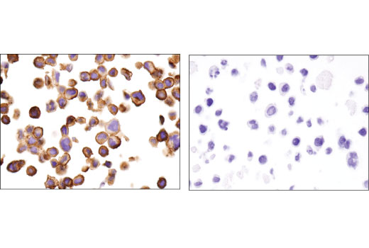
| Basigin/EMMPRIN (E1S1V) Rabbit mAb | 13287 | 20 µl | 38-58 kDa | Rabbit IgG |
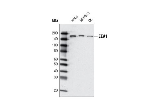
| EEA1 (C45B10) Rabbit mAb | 3288 | 20 µl | 170 kDa | Rabbit IgG |
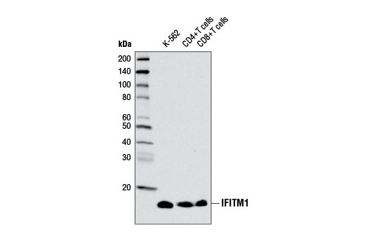
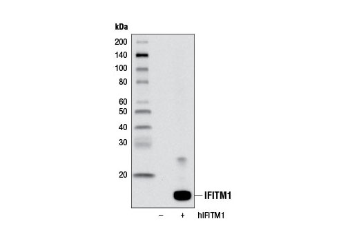
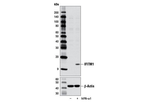
| IFITM1 Antibody | 13126 | 20 µl | 14 kDa | Rabbit |
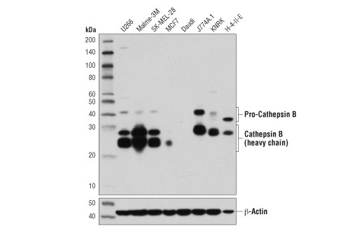
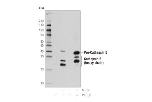
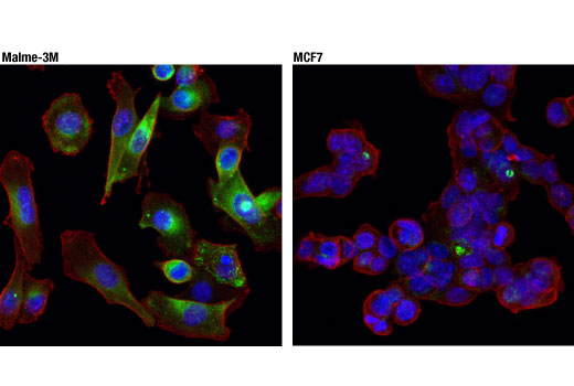
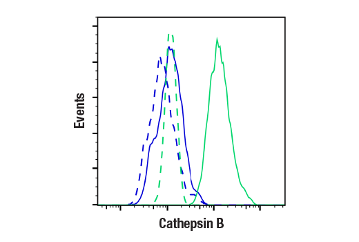
| Cathepsin B (D1C7Y) XP® Rabbit mAb | 31718 | 20 µl | 44, 27, 24 kDa | Rabbit IgG |
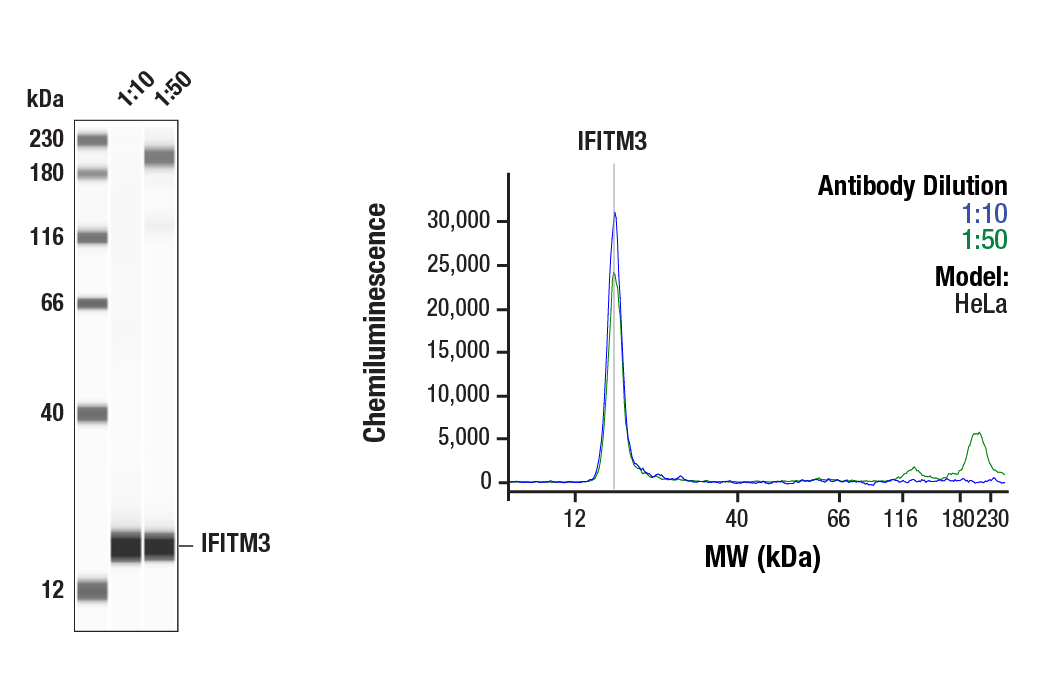
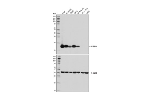
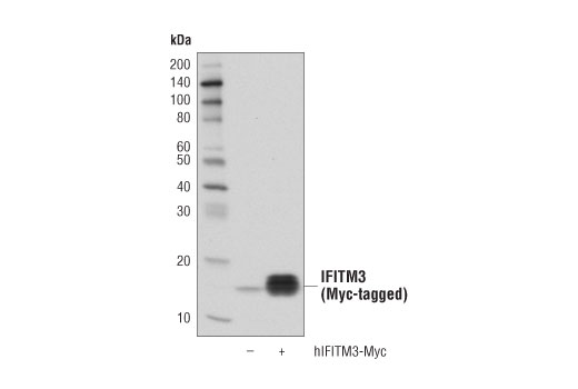
| IFITM3 (D8E8G) XP® Rabbit mAb | 59212 | 20 µl | 15 kDa | Rabbit IgG |
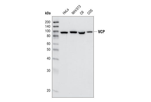
| VCP (7F3) Rabbit mAb | 2649 | 20 µl | 89 kDa | Rabbit IgG |
| Anti-rabbit IgG, HRP-linked Antibody | 7074 | 100 µl | Goat |
Please visit cellsignal.com for individual component applications, species cross-reactivity, dilutions, protocols, and additional product information.
Description
The Coronavirus Host Cell Attachment and Entry Antibody Sampler Kit provides an economical means of detecting key host cell proteins involved in the attachment and cellular entry of coronaviruses. The kit includes enough antibodies to perform two western blot experiments with each primary antibody.
Storage
Background
Coronaviruses are a group of viruses that contain single-stranded, positive-sense RNA genomes. Several members of this group, which include severe acute respiratory syndrome coronaviruses (SARS-CoV and SARS-CoV-2) and Middle East respiratory syndrome coronavirus (MERS-CoV), are highly pathogenic and have caused significant disease outbreaks in human hosts. In order for human coronaviruses to transcribe and replicate their genomes within host cells, they must first attach and gain entry into host cells using a variety of cell surface receptors and components of the endocytic machinery.
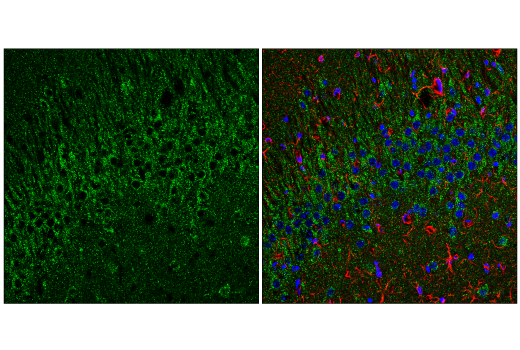
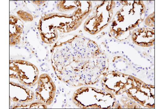
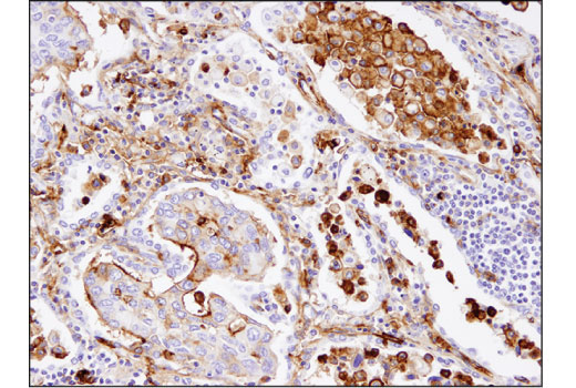
ACE2 is a carboxypeptidase that catalyses the conversion of angiotensin I to angiotensin 1-9, or of angiotensin II to the vasodilator angiotensin 1-7 (1). Research studies have identified ACE2 as the receptor for SARS and SARS-CoV-2 coronaviruses (2-4).
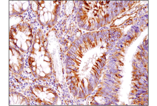
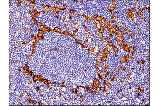
DPP4 (CD26) is a type II transmembrane glycoprotein expressed ubiquitously in most tissues and different cell types (5,6). In addition to its peptidase activity, DPP4 interacts with multiple important cell surface ligands, such as adenosine deaminase, fibronectin, and IGF2 receptor to influence processes like T cell activation, cell migration, and proliferation (7). Research studies have shown that DPP4 serves as a cellular receptor for the MERS-CoV spike protein (8).
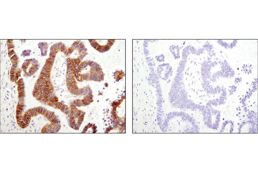
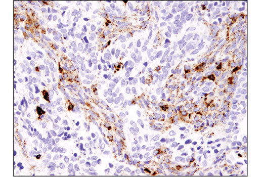
Aminopeptidase N (APN, CD13) is a widely expressed, membrane-bound proteolytic enzyme that breaks down peptides during digestion, cleaves cell surface antigens during antigen presentation, and acts as a receptor for human viruses, including several coronaviruses. This multifunctional protein is implicated in the regulation of many biological processes, including angiogenesis, cell proliferation, cell migration, inflammation, and immune response (9,10).
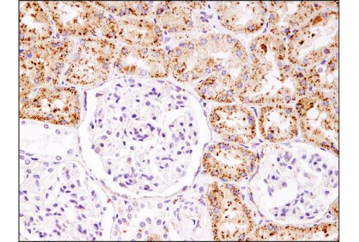
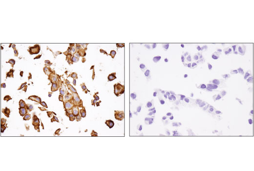
Basigin (EMMPRIN, CD147) is a type I integral membrane receptor protein belonging to the immunoglobulin superfamily (11). Multiple functions have been ascribed to Basigin; foremost among these is stimulating the secretion of extracellular matrix metalloproteinases by adjacent fibroblasts, a function which has been implicated in promoting tumor progression (12-14). Research studies have suggested that Basigin serves as a novel host cell surface receptor for SARS-CoV-2 (15).
EEA1 is an early endosomal marker and a Rab5 effector protein essential for early endosomal membrane fusion and trafficking (16,17). Research studies have shown that efficient coronavirus host cell entry and replication relies upon early endosomes containing EEA1 (18).
Interferon-induced transmembrane protein (IFITM) family members are composed of short amino- and carboxy-termini, two transmembrane domains, and a cytoplasmic domain (19). The primary function of IFITM family proteins appears to be viral restriction, as IFITM proteins inhibit cytosolic entry of coronaviruses by preventing fusion of viral and host membranes (20,21).
Valosin-containing protein (VCP) is a highly conserved and abundant 97 kDa protein that belongs to the AAA family of proteins. These protein complexes participate in many cellular functions, including vesicle transport and fusion, fragmentation and reassembly of the golgi stacks during mitosis, nuclear envelope formation and spindle disassembly following mitosis, cell cycle regulation, DNA damage repair, apoptosis, B and T cell activation, NF-κB-mediated transcriptional regulation, endoplasmic reticulum (ER)-associated degradation, and protein degradation (22). Research studies have shown that VCP facilitates the release of some coronaviruses from the early endosomal compartment (23).
Cathepsin B, part of the papain family of proteases, is a widely expressed lysosomal cysteine endopeptidase (24,25). Research studies have suggested that cathepsin B facilitates host cell entry of SARS-CoV by promoting fusion of viral and endosomal membranes (26).
- Schmidt, B.L. et al. (2000) J Clin Microbiol 38, 1279-82.
- Li, W. et al. (2005) EMBO J 24, 1634-43.
- Hoffmann, M. et al. (2020) Cell 181, 271-280.e8.
- Lan, J. et al. (2020) Nature 581, 215-220.
- Mentzel, S. et al. (1996) J Histochem Cytochem 44, 445-61.
- Röhrborn, D. et al. (2015) Front Immunol 6, 386.
- Zhong, J. et al. (2015) J Diabetes Res 2015, 606031.
- Wang, N. et al. (2013) Cell Res 23, 986-93.
- Luan, Y. and Xu, W. (2007) Curr Med Chem 14, 639-47.
- Mina-Osorio, P. (2008) Trends Mol Med 14, 361-71.
- Biswas, C. et al. (1995) Cancer Res 55, 434-9.
- Liao, C.G. et al. (2011) Mol Cell Biol 31, 2591-604.
- Sweeny, L. et al. (2012) Exp Cell Res 318, 1788-98.
- Lescaille, G. et al. (2012) BMC Cancer 12, 115.
- Wang, K. et al. (2020) Signal Transduct Target Ther 5, 283.
- Mu, F.T. et al. (1995) J Biol Chem 270, 13503-11.
- Christoforidis, S. et al. (1999) Nature 397, 621-5.
- Burkard, C. et al. (2014) PLoS Pathog 10, e1004502.
- Diamond, M.S. and Farzan, M. (2013) Nat Rev Immunol 13, 46-57.
- Brass, A.L. et al. (2009) Cell 139, 1243-54.
- Feeley, E.M. et al. (2011) PLoS Pathog 7, e1002337.
- Wang, Q. et al. J Struct Biol 146, 44-57.
- Wong, H.H. et al. (2015) J Virol 89, 11116-28.
- Chan, S.J. et al. (1986) Proc Natl Acad Sci U S A 83, 7721-5.
- Fong, D. et al. (1986) Proc Natl Acad Sci U S A 83, 2909-13.
- Simmons, G. et al. (2005) Proc Natl Acad Sci U S A 102, 11876-81.
Background References
Trademarks and Patents
限制使用
除非 CST 的合法授书代表以书面形式书行明确同意,否书以下条款适用于 CST、其关书方或分书商提供的书品。 任何书充本条款或与本条款不同的客书条款和条件,除非书 CST 的合法授书代表以书面形式书独接受, 否书均被拒书,并且无效。
专品专有“专供研究使用”的专专或专似的专专声明, 且未专得美国食品和专品管理局或其他外国或国内专管机专专专任何用途的批准、准专或专可。客专不得将任何专品用于任何专断或治专目的, 或以任何不符合专专声明的方式使用专品。CST 专售或专可的专品提供专作专最专用专的客专,且专用于研专用途。将专品用于专断、专防或治专目的, 或专专售(专独或作专专成)或其他商专目的而专专专品,均需要 CST 的专独专可。客专:(a) 不得专独或与其他材料专合向任何第三方出售、专可、 出借、捐专或以其他方式专专或提供任何专品,或使用专品制造任何商专专品,(b) 不得复制、修改、逆向工程、反专专、 反专专专品或以其他方式专专专专专品的基专专专或技专,或使用专品开专任何与 CST 的专品或服专专争的专品或服专, (c) 不得更改或专除专品上的任何商专、商品名称、徽专、专利或版专声明或专专,(d) 只能根据 CST 的专品专售条款和任何适用文档使用专品, (e) 专遵守客专与专品一起使用的任何第三方专品或服专的任何专可、服专条款或专似专专