Revision 1
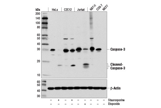
Western blot analysis of various cell lines, untreated (-) or treated with Staurosporine #9953 (1 μM; 3 hr) or with Etoposide #2200 (25 μM, overnight), using Caspase-3 (D3R6Y) Rabbit mAb (upper) or β-Actin (D6A8) Rabbit mAb #8457 (lower). MCF7 cells are negative for caspase-3 expression.
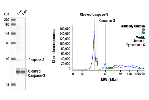
Simple Western™ analysis of lysates (1 mg/mL) from Jurkat cells treated with Cytochrome C using Caspase-3 (D3R6Y) Rabbit mAb #14220. The virtual lane view (left) shows the target bands (as indicated) at 1:10 and 1:50 dilutions of primary antibody. The corresponding electropherogram view (right) plots chemiluminescence by molecular weight along the capillary at 1:10 (blue line) and 1:50 (green line) dilutions of primary antibody. This experiment was performed under reducing conditions on the Jess™ Simple Western instrument from ProteinSimple, a BioTechne brand, using the 12-230 kDa separation module.
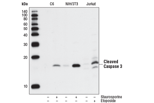
Western blot analysis of extracts from C6 (rat), NIH/3T3 (mouse), and Jurkat (human) cells, untreated or treated with staurosporine #9953 (1uM, 3hrs) or etoposide #2200 (25uM, 5hrs) as indicated, using Cleaved Caspase-3 (Asp175) (5A1E) Rabbit mAb.
Orders: 877-616-CELL (2355) • [email protected] • Support: 877-678-TECH (8324) • [email protected] •
Web:
cellsignal.com For Research Use Only. Not for Use in Diagnostic Procedures.
Revision 1
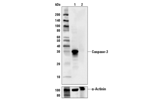
Western blot analysis of extracts from HCT116 cells (lane 1) or CASP3 knock-out cells (lane 2) using Caspase-3 (D3R6Y) Rabbit mAb #14220 (upper), and α-Actinin (D6F6) XP® Rabbit mAb #6487 (lower). The absence of signal in the CASP3 knock-out HCT116 cells confirms specificity of the antibody for CASP3.
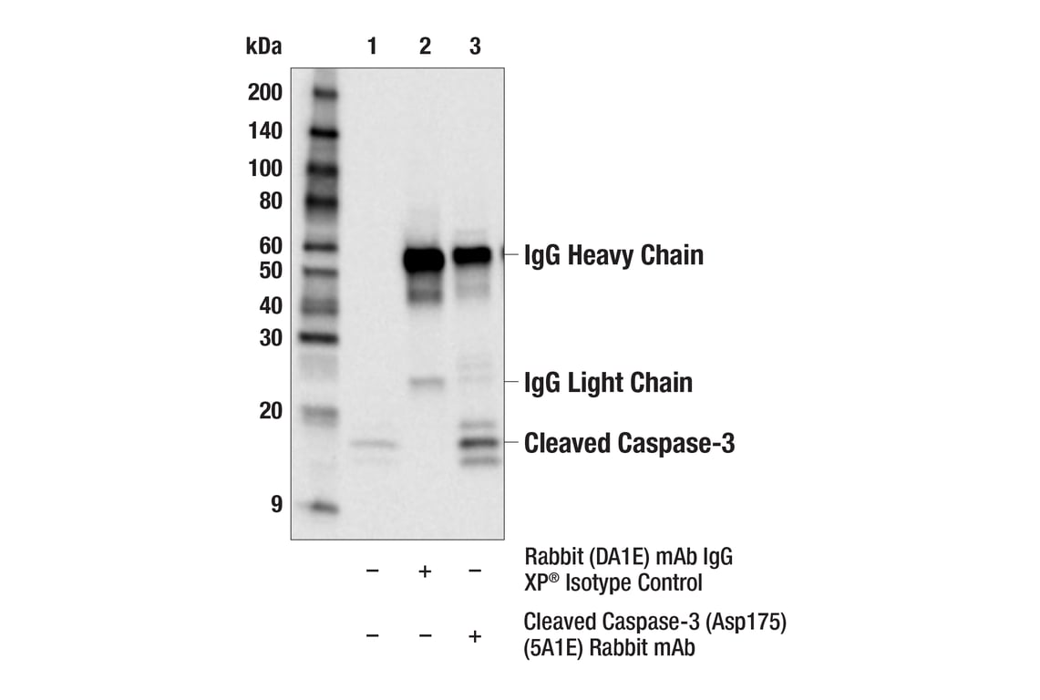
Immunoprecipitation of Cleaved Caspase-3 from Jurkat cells treated with etoposide (25uM, 5hrs). Lane 1 is 10% input, lane 2 is Rabbit (DA1E) mAb IgG XP® Isotype Control #3900, and lane 3 is Cleaved Caspase-3 (Asp175) (5A1E) Rabbit mAb. Western blot was performed using Cleaved Caspase-3 (Asp175) (5A1E) Rabbit mAb. Anti-rabbit IgG, HRP-linked Antibody #7074 was used as a secondary antibody.
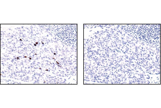
Immunohistochemical analysis of paraffin-embedded mouse embryo, using Cleaved Caspase-3 (Asp175) (5A1E) Rabbit mAb in the presence of control peptide (left) or Cleaved Caspase-3 (Asp175) Blocking Peptide (#1050) (right).
Orders: 877-616-CELL (2355) • [email protected] • Support: 877-678-TECH (8324) • [email protected] •
Web:
cellsignal.com For Research Use Only. Not for Use in Diagnostic Procedures.
Revision 1
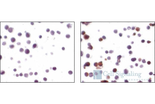
Immunohistochemical analysis using Cleaved Caspase-3 (Asp175) (5A1E) Rabbit mAb on SignalSlide® Cleaved Caspase-3 IHC Controls #8104 (paraffin-embedded Jurkat cells, untreated (left) or etoposide-treated (right)).
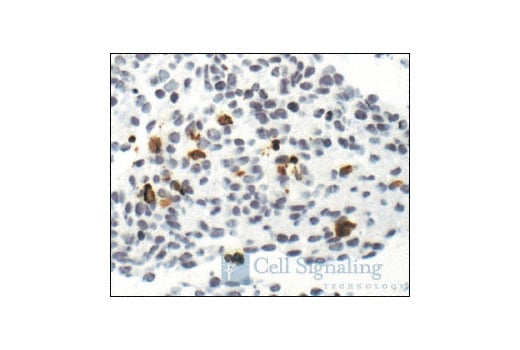
Immunohistochemical staining of paraffin-embedded mouse embryo, showing cytoplasmic localization in apoptotic cells, using Cleaved Caspase-3 (Asp175) (5A1E) Rabbit mAb.
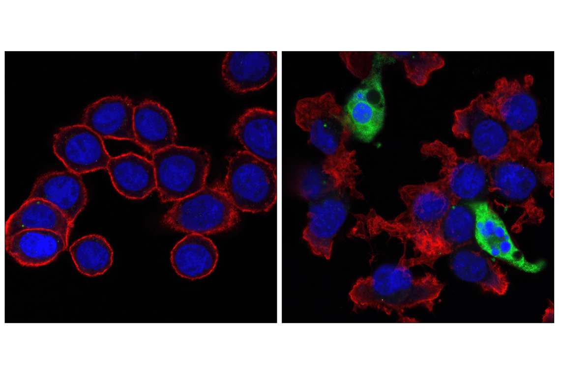
Confocal immunofluorescent images of HT-29 cells, untreated (left) or Staurosporine #9953 treated (right) labeled with Cleaved Caspase-3 (Asp175) (5A1E) Rabbit mAb (green). Actin filaments have been labeled with Alexa Fluor® 555 phalloidin #8953 (red). Blue pseudocolor = DRAQ5® #4084 (fluorescent DNA dye).
Orders: 877-616-CELL (2355) • [email protected] • Support: 877-678-TECH (8324) • [email protected] •
Web:
cellsignal.com For Research Use Only. Not for Use in Diagnostic Procedures.
Revision 1
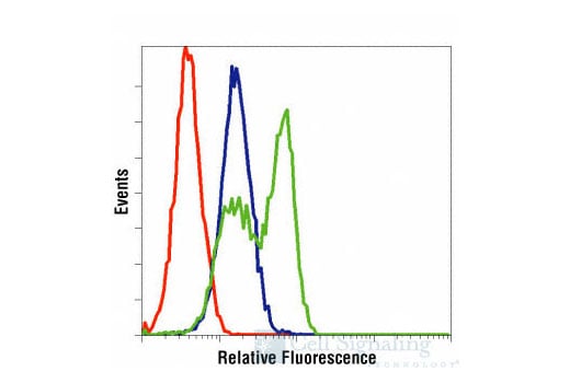
Flow cytometric analysis of Jurkat cells, untreated (blue) or treated with etoposide #2200 (green), using Cleaved Caspase-3(Asp175) (5A1E) Rabbit mAb compared to a nonspecific negative control antibody (red).
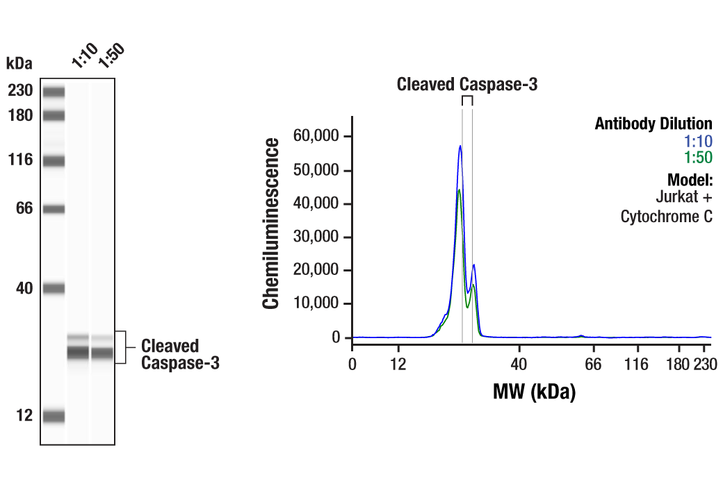
Simple Western™ analysis of lysates (0.1 mg/mL) from Jurkat cells treated with Cytochrome C using Cleaved Caspase-3 (Asp175) (5A1E) Rabbit mAb #9664. The virtual lane view (left) shows the target bands (as indicated) at 1:10 and 1:50 dilutions of primary antibody. The corresponding electropherogram view (right) plots chemiluminescence by molecular weight along the capillary at 1:10 (blue line) and 1:50 (green line) dilutions of primary antibody. This experiment was performed under reducing conditions on the Jess™ Simple Western instrument from ProteinSimple, a BioTechne brand, using the 12-230 kDa separation module.
Orders: 877-616-CELL (2355) • [email protected] • Support: 877-678-TECH (8324) • [email protected] •
Web:
cellsignal.com For Research Use Only. Not for Use in Diagnostic Procedures.