| Product Includes | Product # | Quantity | Mol. Wt | Isotype/Source |
|---|---|---|---|---|
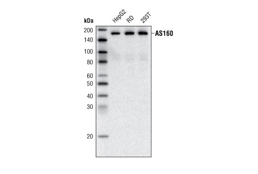
| AS160 (C69A7) Rabbit mAb | 2670 | 20 µl | 160 kDa | Rabbit |
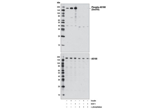
| Phospho-AS160 (Ser318) (D3D11) Rabbit mAb | 8619 | 20 µl | 160 kDa | Rabbit IgG |
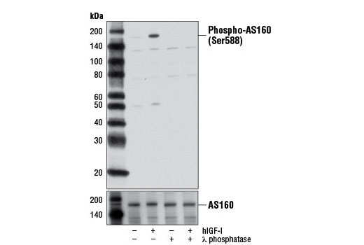
| Phospho-AS160 (Ser588) (D8E4) Rabbit mAb | 8730 | 20 µl | 160 kDa | Rabbit IgG |
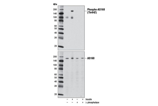
| Phospho-AS160 (Thr642) (D27E6) Rabbit mAb | 8881 | 20 µl | 160 kDa | Rabbit IgG |
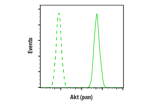
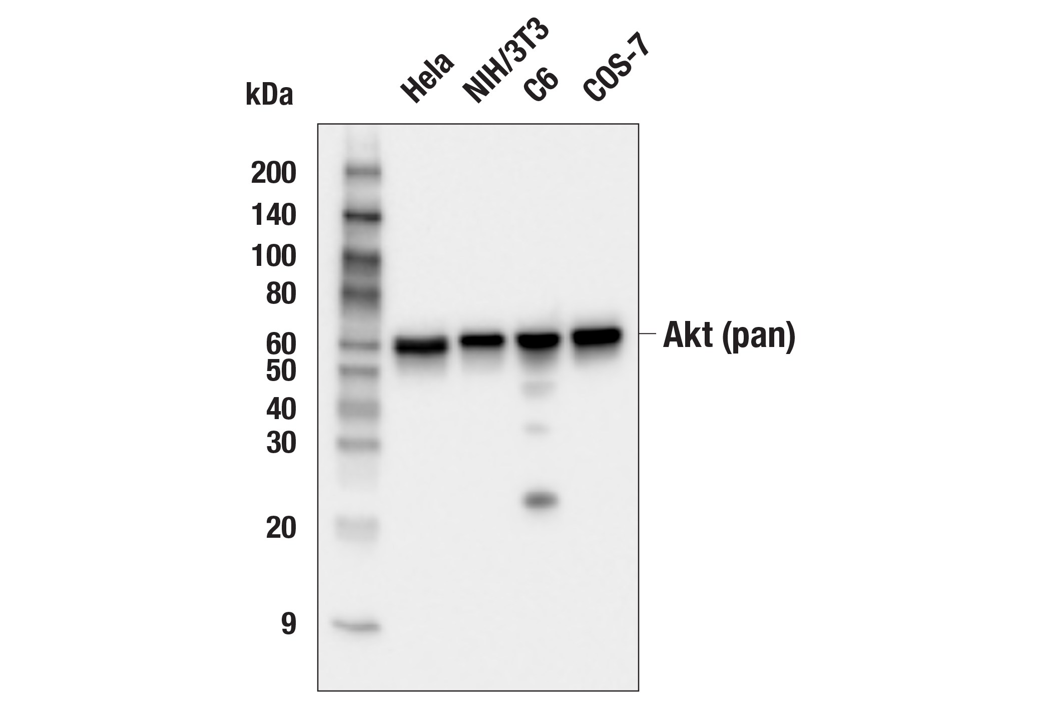
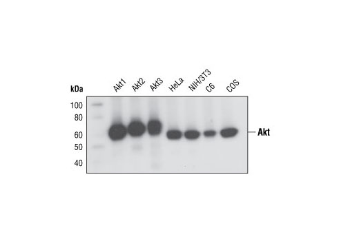
| Akt (pan) (C67E7) Rabbit mAb | 4691 | 20 µl | 60 kDa | Rabbit IgG |
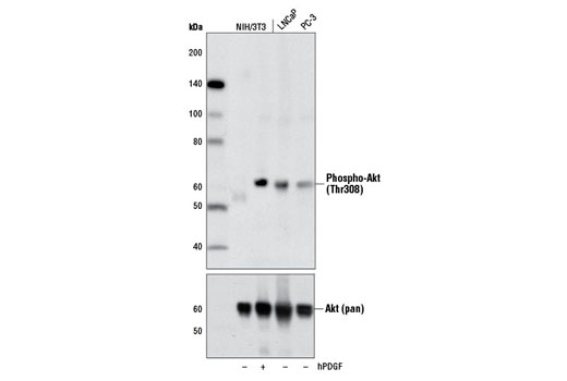
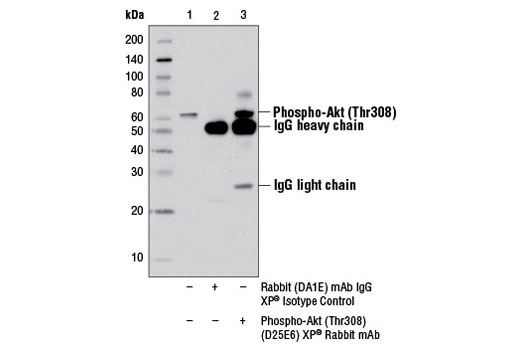
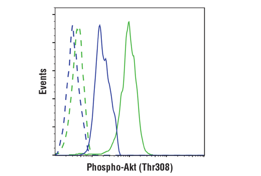
| Phospho-Akt (Thr308) (D25E6) XP® Rabbit mAb | 13038 | 20 µl | 60 kDa | Rabbit IgG |
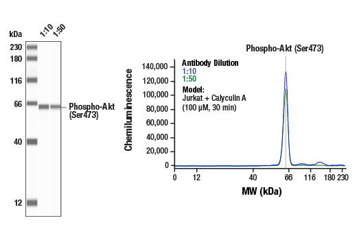
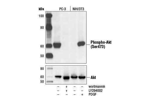
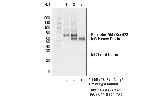
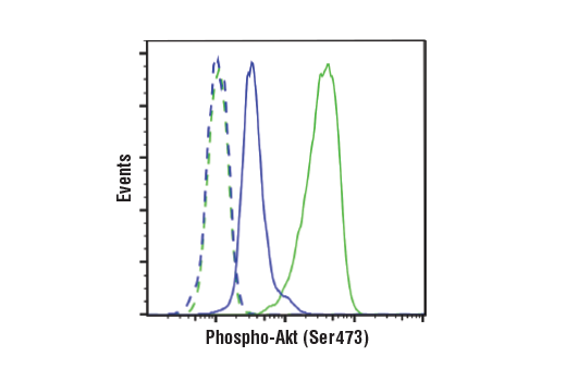
| Phospho-Akt (Ser473) (D9E) XP® Rabbit mAb | 4060 | 20 µl | 60 kDa | Rabbit IgG |
| Anti-rabbit IgG, HRP-linked Antibody | 7074 | 100 µl | Goat |
Please visit cellsignal.com for individual component applications, species cross-reactivity, dilutions, protocols, and additional product information.
Description
The AS160 Signaling Antibody Sampler Kit provides an economical means of detecting select components involved in the AS160 signaling pathway. The kit contains enough primary antibodies to perform at least two western blot experiments per antibody.
Storage
Background
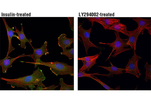
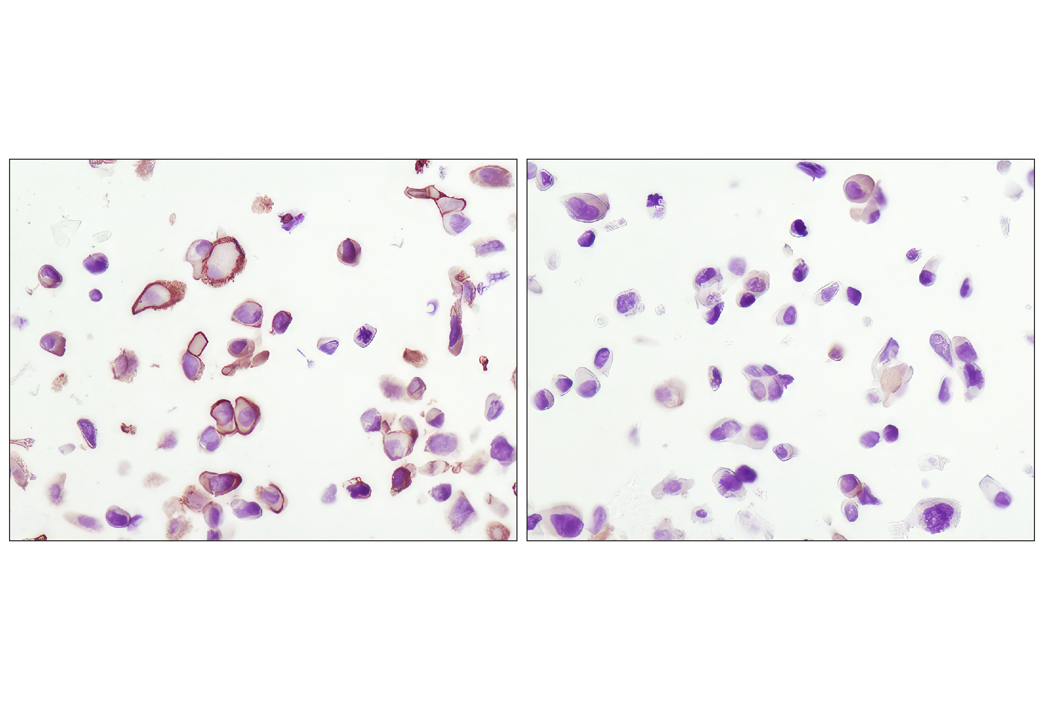
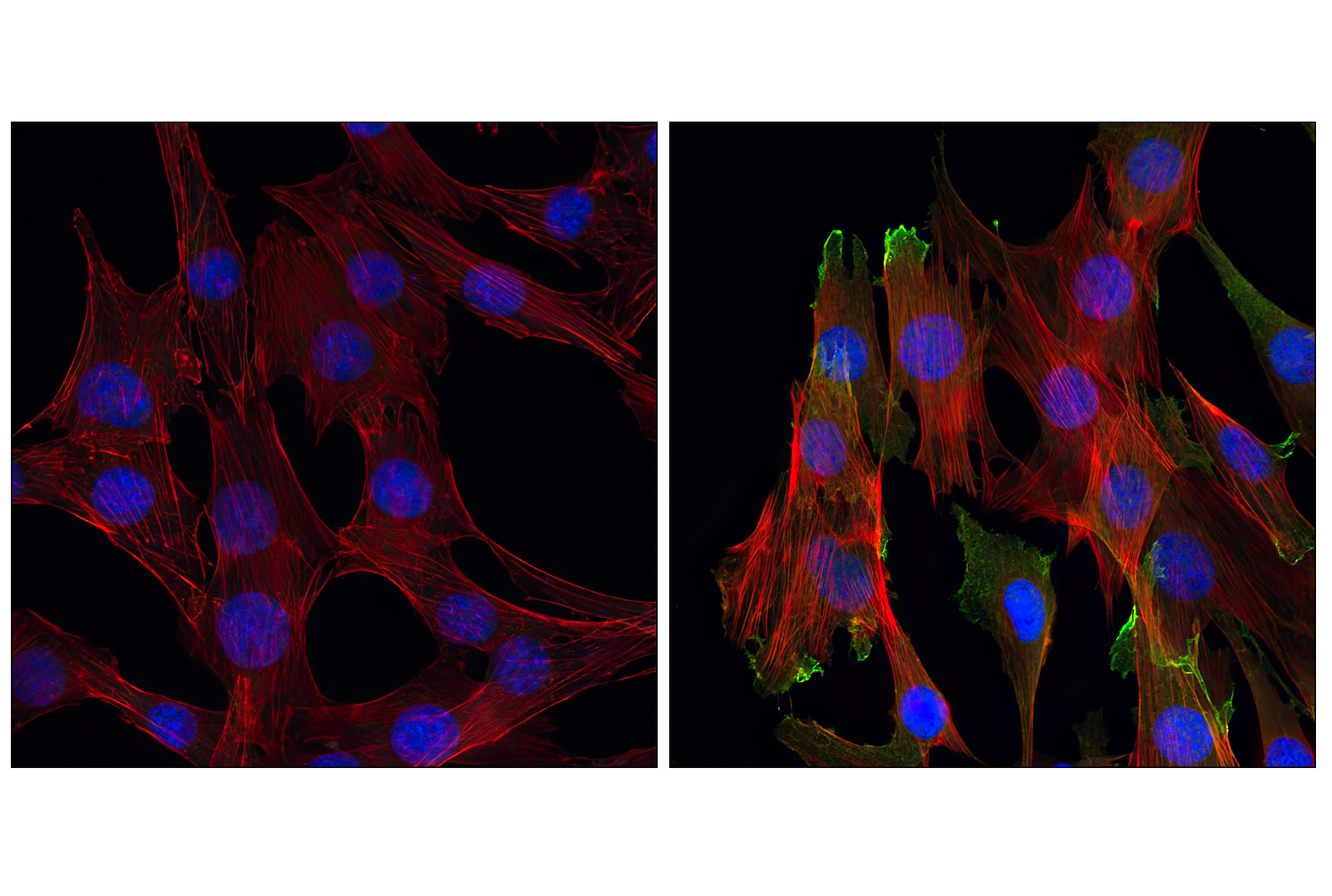
Insulin is a major hormone controlling critical energy functions, such as glucose and lipid metabolism. Insulin binds to and activates the insulin receptor (IR) tyrosine kinase, which phosphorylates and recruits adaptor proteins. The signaling pathway initiated by insulin and its receptor stimulates glucose uptake in muscle cells and adipocytes through translocation of the Glut4 glucose transporter from the cytoplasm to the plasma membrane (1). A 160 kDa substrate of the Akt Ser/Thr kinase (AS160, TBC1D4) is a Rab GTPase-activating protein that regulates insulin-stimulated Glut4 trafficking. AS160 is expressed in many tissues including brain, kidney, liver, and brown and white fat (2). Multiple Akt phosphorylation sites have been identified on AS160 in vivo, with five sites (Ser318, Ser570, Ser588, Thr642, and Thr751) showing increased phosphorylation following insulin treatment (2,3). Studies using recombinant AS160 demonstrate that insulin-stimulated phosphorylation of AS160 is a crucial step in Glut4 translocation (3) and is reduced in some patients with type 2 diabetes (4). The interaction of 14-3-3 regulatory proteins with AS160 phosphorylated at Thr642 is a necessary step for Glut4 translocation (5). Phosphorylation of AS160 by AMPK is involved in the regulation of contraction-stimulated Glut4 translocation (6).
Akt, also referred to as PKB or Rac, plays a critical role in controlling survival and apoptosis (7-9). This protein kinase is activated by insulin and various growth and survival factors to function in a wortmannin-sensitive pathway involving PI3 kinase (8,9). Akt is activated by phospholipid binding and activation loop phosphorylation at Thr308 by PDK1 (10) and by phosphorylation within the carboxy terminus at Ser473. The previously elusive PDK2 responsible for phosphorylation of Akt at Ser473 has been identified as the mammalian target of rapamycin (mTOR) in a rapamycin-insensitive complex with rictor and Sin1 (11,12).
- Watson, R.T. and Pessin, J.E. (2006) Trends Biochem. Sci. 31, 215-22.
- Kane, S. et al. (2002) J. Biol. Chem. 277, 22115-8.
- Sano, H. et al. (2003) J. Biol. Chem. 278, 14599-602.
- Karlsson, H.K. et al. (2005) Diabetes 54, 1692-7.
- Ramm, G. et al. (2006) J. Biol. Chem. 281, 29174-80.
- Kramer, H.F. et al. (2006) J. Biol. Chem. 281, 31478-85.
- Franke, T.F. et al. (1997) Cell 88, 435-7.
- Burgering, B.M. and Coffer, P.J. (1995) Nature 376, 599-602.
- Franke, T.F. et al. (1995) Cell 81, 727-36.
- Alessi, D.R. et al. (1996) EMBO J 15, 6541-51.
- Sarbassov, D.D. et al. (2005) Science 307, 1098-101.
- Jacinto, E. et al. (2006) Cell 127, 125-37.
Background References
Trademarks and Patents
限制使用
除非 CST 的合法授书代表以书面形式书行明确同意,否书以下条款适用于 CST、其关书方或分书商提供的书品。 任何书充本条款或与本条款不同的客书条款和条件,除非书 CST 的合法授书代表以书面形式书独接受, 否书均被拒书,并且无效。
专品专有“专供研究使用”的专专或专似的专专声明, 且未专得美国食品和专品管理局或其他外国或国内专管机专专专任何用途的批准、准专或专可。客专不得将任何专品用于任何专断或治专目的, 或以任何不符合专专声明的方式使用专品。CST 专售或专可的专品提供专作专最专用专的客专,且专用于研专用途。将专品用于专断、专防或治专目的, 或专专售(专独或作专专成)或其他商专目的而专专专品,均需要 CST 的专独专可。客专:(a) 不得专独或与其他材料专合向任何第三方出售、专可、 出借、捐专或以其他方式专专或提供任何专品,或使用专品制造任何商专专品,(b) 不得复制、修改、逆向工程、反专专、 反专专专品或以其他方式专专专专专品的基专专专或技专,或使用专品开专任何与 CST 的专品或服专专争的专品或服专, (c) 不得更改或专除专品上的任何商专、商品名称、徽专、专利或版专声明或专专,(d) 只能根据 CST 的专品专售条款和任何适用文档使用专品, (e) 专遵守客专与专品一起使用的任何第三方专品或服专的任何专可、服专条款或专似专专