| Product Includes | Product # | Quantity | Mol. Wt | Isotype/Source |
|---|---|---|---|---|
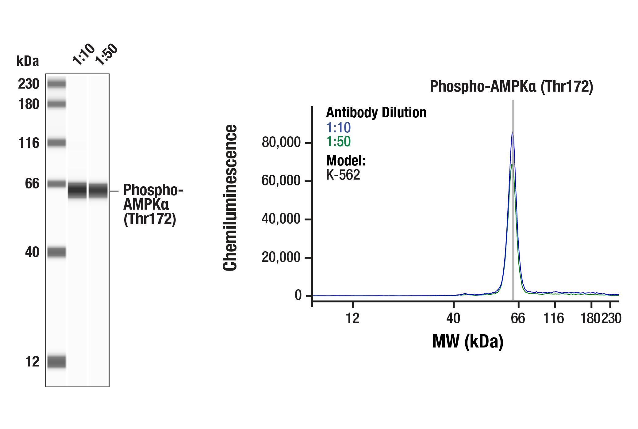
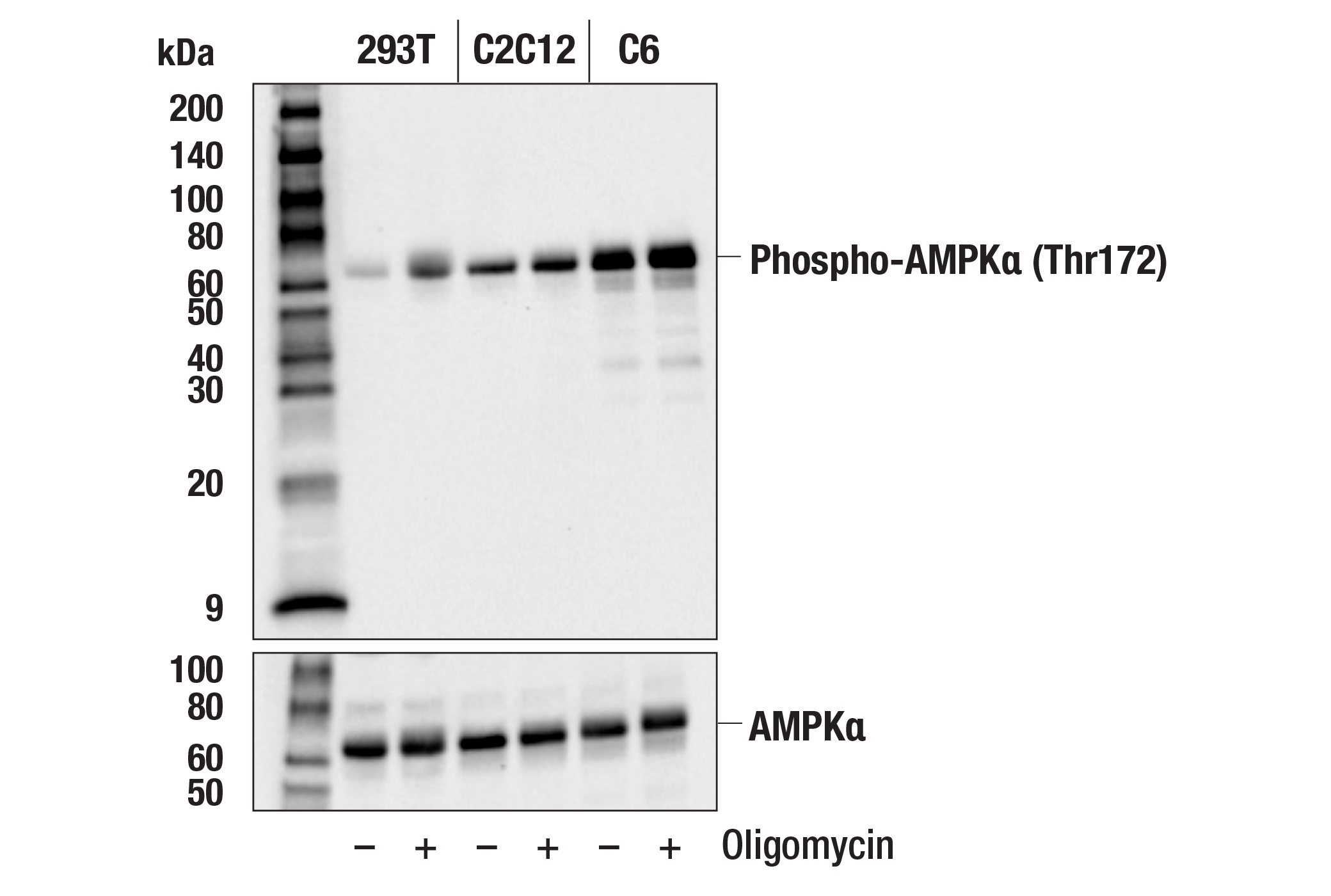
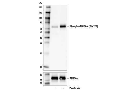
| Phospho-AMPKα (Thr172) (D4D6D) Rabbit mAb | 50081 | 20 µl | 62 kDa | Rabbit IgG |
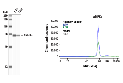
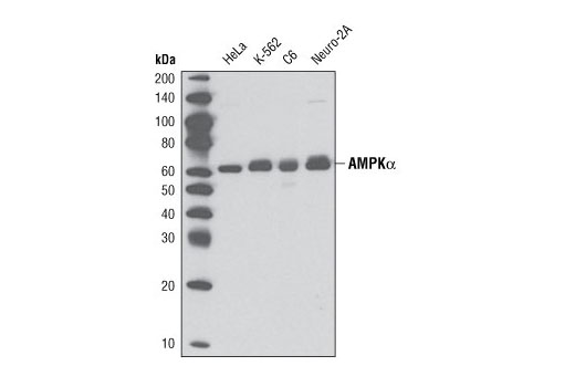
| AMPKα (D5A2) Rabbit mAb | 5831 | 20 µl | 62 kDa | Rabbit IgG |
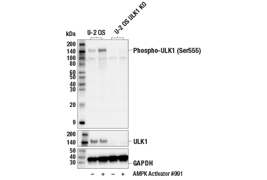
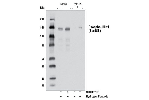
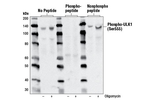
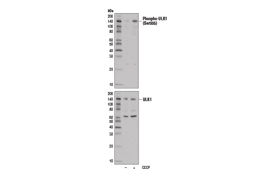
| Phospho-ULK1 (Ser555) (D1H4) Rabbit mAb | 5869 | 20 µl | 140-150 kDa | Rabbit IgG |
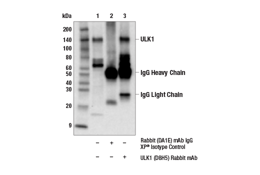
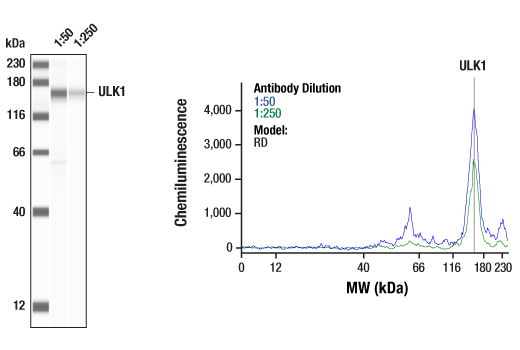
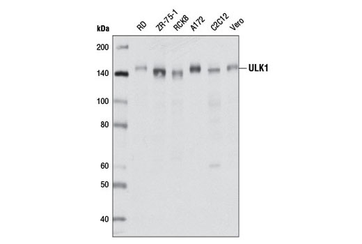
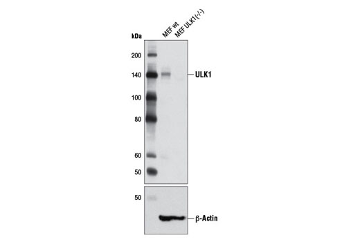
| ULK1 (D8H5) Rabbit mAb | 8054 | 20 µl | 150 kDa | Rabbit IgG |
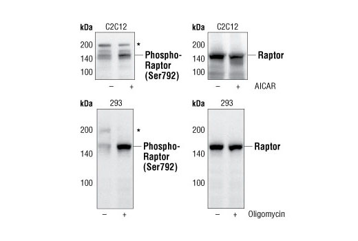
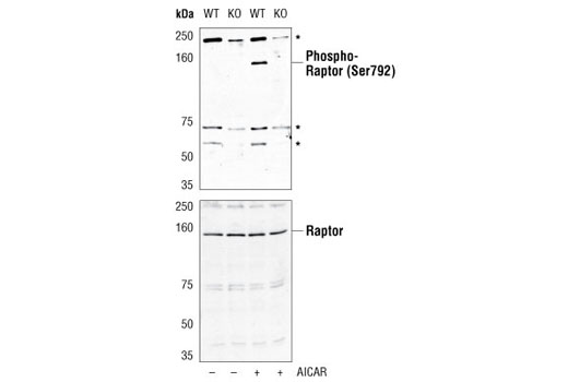
| Phospho-Raptor (Ser792) Antibody | 2083 | 20 µl | 150 kDa | Rabbit |
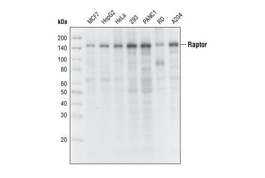
| Raptor (24C12) Rabbit mAb | 2280 | 20 µl | 150 kDa | Rabbit |
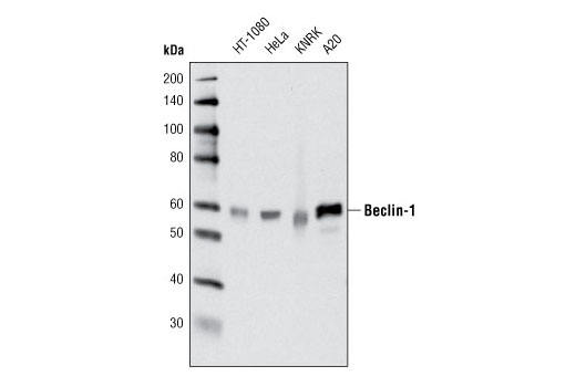
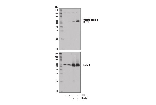
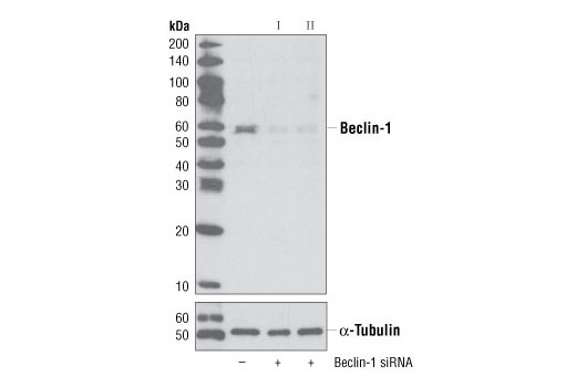
| Beclin-1 (D40C5) Rabbit mAb | 3495 | 20 µl | 60 kDa | Rabbit IgG |
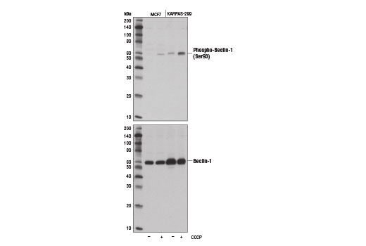
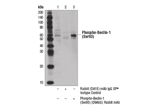
| Phospho-Beclin-1 (Ser93) (D9A5G) Rabbit mAb | 14717 | 20 µl | 60 kDa | Rabbit IgG |
| Anti-rabbit IgG, HRP-linked Antibody | 7074 | 100 µl | Goat |
Please visit cellsignal.com for individual component applications, species cross-reactivity, dilutions, protocols, and additional product information.
Description
The AMPK Substrate Antibody Sampler Kit provides an economical means of detecting total and phosphorylated substrates of AMPK. The kit provides enough antibody to perform two western blots with each primary antibody.
Storage
Background
AMP-activated protein kinase (AMPK) is highly conserved from yeast to plants and animals and plays a key role in the regulation of energy homeostasis (1). AMPK is a heterotrimeric complex composed of a catalytic α subunit and regulatory β and γ subunits, each of which is encoded by two or three distinct genes (α1, 2; β1, 2; γ1, 2, 3) (2). The kinase is activated by an elevated AMP/ATP ratio due to cellular and environmental stress, such as heat shock, hypoxia, and ischemia (1). The tumor suppressor LKB1, in association with accessory proteins STRAD and MO25, phosphorylates AMPKα at Thr172 in the activation loop, and this phosphorylation is required for AMPK activation (3-5).
AMPK phosphorylates a number of targets controlling cellular processes such as metabolism, cell growth, and autophagy (6). It suppresses the activity of the mammalian target of rapamycin (mTOR), that plays a key role in promoting cell growth. The regulatory associated protein of mTOR (Raptor) was identified as an mTOR binding partner that mediates mTOR signaling to downstream targets (7,8). Raptor binds to mTOR substrates, including 4E-BP1 and p70 S6 kinase, through their TOR signaling (TOS) motifs and is required for mTOR-mediated phosphorylation of these substrates (9,10). AMPK directly phosphorylates Raptor at Ser722/Ser792, and this phosphorylation is essential for inhibition of the raptor-containing mTOR complex 1 (mTORC1) and induces cell cycle arrest when cells are stressed for energy (11). AMPK also promotes autophagy by directly phosphorylating ULK1 (11,12). ULK1 is a Ser/Thr kinase required for the Initiation and formation of the autophagosome. AMPK, activated during low nutrient conditions, directly phosphorylates ULK1 at multiple sites including Ser317, Ser555, and Ser777 (11,12). Conversely, mTOR, which is a regulator of cell growth and an inhibitor of autophagy, phosphorylates ULK1 at Ser757 and disrupts the interaction between ULK1 and AMPK (11). AMPK can also directly phosphorylate Beclin-1, a component of the complex downstream of ULK1 in autophagosome formation that activates the class III phosphatidylinositol 3-kinase VPS34. AMPK phosphorylates Beclin-1 at Ser93 and Ser96 residues in human, which correspond to murine Ser91 and Ser94 (14).
- Hardie, D.G. (2004) J Cell Sci 117, 5479-87.
- Carling, D. (2004) Trends Biochem Sci 29, 18-24.
- Hawley, S.A. et al. (1996) J Biol Chem 271, 27879-87.
- Lizcano, J.M. et al. (2004) EMBO J 23, 833-43.
- Shaw, R.J. et al. (2004) Proc Natl Acad Sci U S A 101, 3329-35.
- Mihaylova, M.M. and Shaw, R.J. (2011) Nat Cell Biol 13, 1016-23.
- Hara, K. et al. (2002) Cell 110, 177-89.
- Kim, D.H. et al. (2002) Cell 110, 163-75.
- Beugnet, A. et al. (2003) J Biol Chem 278, 40717-22.
- Nojima, H. et al. (2003) J Biol Chem 278, 15461-4.
- Gwinn, D.M. et al. (2008) Mol Cell 30, 214-26.
- Kim, J. et al. (2011) Nat Cell Biol 13, 132-41.
- Egan, D.F. et al. (2011) Science 331, 456-61.
- Kim, J. et al. (2013) Cell 152, 290-303.
Background References
Trademarks and Patents
限制使用
除非 CST 的合法授书代表以书面形式书行明确同意,否书以下条款适用于 CST、其关书方或分书商提供的书品。 任何书充本条款或与本条款不同的客书条款和条件,除非书 CST 的合法授书代表以书面形式书独接受, 否书均被拒书,并且无效。
专品专有“专供研究使用”的专专或专似的专专声明, 且未专得美国食品和专品管理局或其他外国或国内专管机专专专任何用途的批准、准专或专可。客专不得将任何专品用于任何专断或治专目的, 或以任何不符合专专声明的方式使用专品。CST 专售或专可的专品提供专作专最专用专的客专,且专用于研专用途。将专品用于专断、专防或治专目的, 或专专售(专独或作专专成)或其他商专目的而专专专品,均需要 CST 的专独专可。客专:(a) 不得专独或与其他材料专合向任何第三方出售、专可、 出借、捐专或以其他方式专专或提供任何专品,或使用专品制造任何商专专品,(b) 不得复制、修改、逆向工程、反专专、 反专专专品或以其他方式专专专专专品的基专专专或技专,或使用专品开专任何与 CST 的专品或服专专争的专品或服专, (c) 不得更改或专除专品上的任何商专、商品名称、徽专、专利或版专声明或专专,(d) 只能根据 CST 的专品专售条款和任何适用文档使用专品, (e) 专遵守客专与专品一起使用的任何第三方专品或服专的任何专可、服专条款或专似专专