Revision 5
Cell Signaling Technology

Orders: 877-616-CELL (2355) [email protected]

Support: 877-678-TECH (8324)

Web: [email protected] cellsignal.com

3 Trask LaneDanversMassachusetts01923USA
For Research Use Only. Not for Use in Diagnostic Procedures.
Applications:

FC-FP

:

:

:

:

UniProt ID:

#P07766, #P01579, #P01732, #P01730, #P01375

Entrez-Gene Id:

916, 3458, 925, 920, 7124

Product Information

Product Usage Information

Cytokine production by T cells can be induced by activating cells through a variety of methods. These include stimulation through the T cell receptor by incubation with an agonist CD3 antibody such as Human CD3ε Activating (OKT3) Mouse mAb (Low Endotoxin, Azide-free), or treatment with a combination of PMA (phorbol-12-myristate-13-acetate) and Ionomycin. The addition of Brefeldin A and Monensin during the cell stimulation will limit cytokine secretion and facilitate detection of intracellular cytokines. All antibodies in this kit are compatible with the Flow Cytometry TritonTM X-100 Permeabilization Protocol for Directly Conjugated Antibodies and can be used in a single staining mix on fixed and permeabilized cells. Prior to fixation and antibody incubation, we recommend adding a fixable viability dye such as the Ghost Dye Violet 510 Viability Dye to enable identification and exclusion of dead cells from analysis.

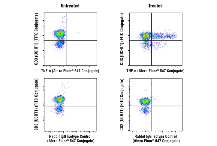
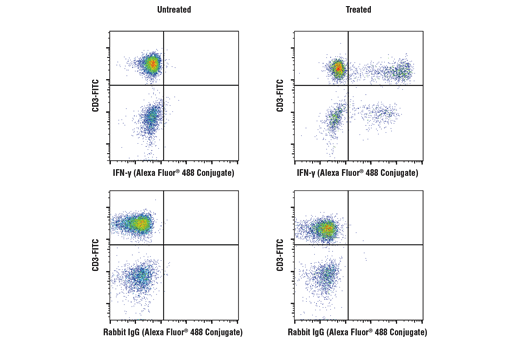
Gating strategy for observing production of Th1 cytokines by T cells:
If a fixable viability dye was used, first gate on viable cells. Next, gate on lymphocytes based on forward scatter and side scatter. To observe cytokine production by helper T cells, gate on the CD3+CD4+ cells within the lymphocyte gate and then identify cells within this gate that are positive for staining of IFN-γ and/or TNF-α. To observe cytokine production by cytotoxic T cells, gate on the CD3+CD8+ cells within the lymphocyte gate and then identify cells within this gate that are positive for staining of IFN-γ and/or TNF-α.

Storage

CD3 (UCHT1) Mouse mAb (PE Conjugate), CD4 (RPA-T4) Mouse mAb (violetFluor 450 Conjugate), and CD8α (RPA-T8) Mouse mAb (PE-Cy7® Conjugate) are supplied in 10 mM NaH2PO4, 150 mM NaCl, 0.09% NaN3, 0.1% gelatin, pH7.2. TNF-α (D1G2) Rabbit mAb (Alexa Fluor® 647 Conjugate) and IFN-γ (D3H2) XP® Rabbit mAb (Alexa Fluor® 488 Conjugate) are supplied in PBS (pH 7.2), less than 0.1% sodium azide and 2 mg/ml BSA. Store at 4oC. Do not aliquot the antibodies. Protect from light. Do not freeze.

All components in this kit are stable in accordance with the date printed on the outer packaging label when stored at the recommended temperature. Please refer to product labels, datasheets, or web pages for specific "Best By" dates for each individual component.

Specificity / Sensitivity

Each antibody in the Human T Cell Th1 Cytokine Response Flow Cytometry Panel detects endogenous levels of its target protein. CD3 (UCHT1) Mouse mAb (PE Conjugate) recognizes CD3ε. CD3 (UCHT1) Mouse mAb (PE Conjugate), CD4 (RPA-T4) Mouse mAb (violetFluor 450 Conjugate), and CD8α (RPA-T8) Mouse mAb (PE-Cy7® Conjugate) detect epitopes within the extracellular domains. TNF-α (D1G2) Rabbit mAb (Alexa Fluor® 647 Conjugate) and IFN-γ (D3H2) XP® Rabbit mAb (Alexa Fluor® 488 Conjugate) detect epitopes within the intracellular domains.

Species Reactivity:

Human

Source / Purification

Monoclonal antibodies were purified from tissue culture supernatant via affinity chromatography. The purified antibodies were conjugated under optimal conditions, with unreacted dye removed from the preparation.
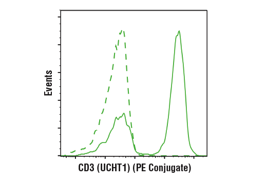
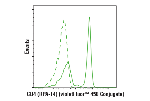
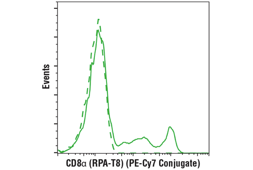
Product Description

The Human T Cell Th1 Cytokine Response Flow Cytometry Panel can be used to observe production of signature Th1 cytokines including IFN-γ and TNF-α.

T cells are identified by expression of CD3. There are two major subsets of conventional T cells: helper T cells which express CD4, and cytotoxic T cells which express CD8. Th1 cells are one of several subsets of helper T cell subsets. Th1 cells promote cell-mediated immunity to combat viral infection, intracellular bacteria, and tumor cells. Both Th1 and cytotoxic T cells can produce IFN-γ and TNF-α.

Species Reactivity

Species reactivity is determined by testing in at least one approved application (e.g., western blot).

Applications Key

FC-FP: Flow Cytometry (Fixed/Permeabilized)

Cross-Reactivity Key

H: human M: mouse R: rat Hm: hamster Mk: monkey Vir: virus Mi: mink C: chicken Dm: D. melanogaster X: Xenopus Z: zebrafish B: bovine Dg: dog Pg: pig Sc: S. cerevisiae Ce: C. elegans Hr: horse GP: Guinea Pig Rab: rabbit All: all species expected

Trademarks and Patents

Cell Signaling Technology is a trademark of Cell Signaling Technology, Inc.
Cy and CyDye are registered trademarks of GE Healthcare.
This product is provided under an intellectual property license from Life Technologies Corporation. The transfer of this product is conditioned on the buyer using the purchased product solely in research conducted by the buyer, excluding contract research or any fee for service research, and the buyer must not (1) use this product or its components for (a) diagnostic, therapeutic or prophylactic purposes; (b) testing, analysis or screening services, or information in return for compensation on a per-test basis; or (c) manufacturing or quality assurance or quality control, and/or (2) sell or transfer this product or its components for resale, whether or not resold for use in research. For information on purchasing a license to this product for purposes other than as described above, contact Life Technologies Corporation, 5791 Van Allen Way, Carlsbad, CA 92008 USA or [email protected].
All other trademarks are the property of their respective owners. Visit cellsignal.com/trademarks for more information.

限制使用

除非 CST 的合法授书代表以书面形式书行明确同意,否书以下条款适用于 CST、其关书方或分书商提供的书品。 任何书充本条款或与本条款不同的客书条款和条件,除非书 CST 的合法授书代表以书面形式书独接受, 否书均被拒书,并且无效。

专品专有“专供研究使用”的专专或专似的专专声明, 且未专得美国食品和专品管理局或其他外国或国内专管机专专专任何用途的批准、准专或专可。客专不得将任何专品用于任何专断或治专目的, 或以任何不符合专专声明的方式使用专品。CST 专售或专可的专品提供专作专最专用专的客专,且专用于研专用途。将专品用于专断、专防或治专目的, 或专专售(专独或作专专成)或其他商专目的而专专专品,均需要 CST 的专独专可。客专:(a) 不得专独或与其他材料专合向任何第三方出售、专可、 出借、捐专或以其他方式专专或提供任何专品,或使用专品制造任何商专专品,(b) 不得复制、修改、逆向工程、反专专、 反专专专品或以其他方式专专专专专品的基专专专或技专,或使用专品开专任何与 CST 的专品或服专专争的专品或服专, (c) 不得更改或专除专品上的任何商专、商品名称、徽专、专利或版专声明或专专,(d) 只能根据 CST 的专品专售条款和任何适用文档使用专品, (e) 专遵守客专与专品一起使用的任何第三方专品或服专的任何专可、服专条款或专似专专

Revision 5
#28572

Human T Cell Th1 Cytokine Response Flow Cytometry Panel

Flow Cytometry Image 1: Human T Cell Th1 Cytokine Response Flow Cytometry Panel Expand Image
与浓度匹配的 Mouse (MOPC-21) mAb IgG1 Isotype Control (PE Conjugate) #63630(虚线)比较,使用 CD3 (UCHT1) Mouse mAb (PE Conjugate)(实线)对活的人外周血单核细胞进行流式细胞分析。
Flow Cytometry Image 2: Human T Cell Th1 Cytokine Response Flow Cytometry Panel Expand Image
与浓度匹配的 Mouse (MOPC-21) mAb IgG1 Isotype Control (violetFluor 450 Conjugate) #40282(虚线)比较,使用 CD4 (RPA-T4) Mouse mAb (violetFluor 450 Conjugate)(实线)对活的人外周血单核细胞进行流式细胞分析。
Flow Cytometry Image 3: Human T Cell Th1 Cytokine Response Flow Cytometry Panel Expand Image
与浓度匹配的 Mouse (MOPC-21) mAb IgG1 Isotype Control (PE-Cy7® Conjugate) #79339(虚线)比较,使用 CD8α (RPA-T8) Mouse mAb (PE-Cy7® Conjugate)(实线)对活的人外周血单核细胞进行流式细胞分析。
Flow Cytometry Image 4: Human T Cell Th1 Cytokine Response Flow Cytometry Panel Expand Image
使用 TNF-α (D1G2) Rabbit mAb (Alexa Fluor® 647 Conjugate)(顶行)或浓度匹配的 Rabbit (DA1E) mAb IgG XP® Isotype Control (Alexa Fluor® 647 Conjugate) #2985(底行)并用 CD3 (UCHT1) Mouse mAb (FITC Conjugate) #86774共染色,对未经处理的(左栏)或经过 TPA (12-O-Tetradecanoylphorbol-13-Acetate) #4174(40 nM,4小时)、Ionomycin, Calcium Salt #9995(2 μM,4 小时)和 Brefeldin A #9972(1 μg/ml,最后 3 小时的处理)(右栏)处理的人外周血淋巴细胞进行流式细胞分析。
Flow Cytometry Image 5: Human T Cell Th1 Cytokine Response Flow Cytometry Panel Expand Image
使用 IFN-γ (D3H2) XP® 兔单克隆抗体 (Alexa Fluor® 488 Conjugate)(顶行)或浓度匹配的兔 (DA1E) 单克隆抗体 IgG XP® Isotype Control (Alexa Fluor® 488Conjugate) #2975(底行),对未经处理的(左栏)或经 TPA #4174(40 nM,4 小时)、Ionomycin #9995(2 μM,4 小时)和 Brefeldin A #9972(1 μg/mL,最后 3 小时刺激)(右栏)处理的人外周血单核细胞进行流式细胞分析。将细胞用 #19881 CD3 (UCHT1) Mouse mAb (APC Conjugate) 共染色。