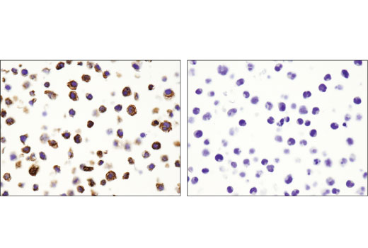
Immunohistochemistry (Paraffin)
A. Solutions and Reagents
NOTE: Prepare solutions with reverse osmosis deionized (RODI) or equivalent grade water.
- Xylene.
- Ethanol, anhydrous denatured, histological grade (100% and 95%).
- Deionized water (dH2O).
- Hematoxylin (optional).
- Wash Buffer:
- 1X Tris Buffered Saline with Tween® 20 (TBST): To prepare 1L 1X TBST add 100
ml 10X Tris Buffered Saline with Tween® 20 (#9997) to 900 ml dH20, mix.
- SignalStain® Antibody Diluent (#8112).
- 1X Citrate Unmasking Solution: To prepare 250 mL of 1X citrate unmasking solution, dilute 25 ml
of SignalStain® Citrate Unmasking Solution (10X) (#14746) with 225 mL of dH2O.
- 3% Hydrogen Peroxide: To prepare 100 ml, add 10 ml 30% H2O2 to 90 ml
dH2O.
- Blocking Solution: TBST/5% Normal Goat Serum or 1X Animal-Free Blocking Solution.
- TBST/5% Normal Goat Serum: to 5 ml 1X TBST, add 250 µl Normal Goat Serum (#5425).
- 1X Animal-Free Blocking Solution: to 4 mL of dH2O add 1 ml of Animal-Free Blocking
Solution (5X) (#15019).
- Detection System: SignalStain® Boost IHC Detection Reagents (HRP, Rabbit #8114).
- Substrate: SignalStain® DAB Substrate Kit (#8059).
- Hematoxylin: Hematoxylin (#14166).
- Mounting Medium: SignalStain® Mounting Medium (#14177).
B. Deparaffinization/Rehydration
NOTE: Do not allow slides to dry at any time during this procedure.
- Deparaffinize/hydrate sections:
- Incubate sections in three washes of xylene for 5 min each.
- Incubate sections in two washes of 100% ethanol for 10 min each.
- Incubate sections in two washes of 95% ethanol for 10 min each.
- Wash sections two times in dH2O for 5 min each.
C. Antigen Unmasking
For Citrate: Heat slides in a microwave submersed in 1X citrate unmasking solution until boiling is
initiated; follow with 10 min at a sub-boiling temperature (95°-98°C). Cool slides on bench top for 30 min.
D. Staining
- Wash sections in dH2O three times for 5 min each.
- Incubate sections in 3% hydrogen peroxide for 10 min.
- Wash sections in dH2O two times for 5 min each.
- Wash sections in wash buffer for 5 min.
- Block each section with 100–400 µl of preferred blocking solution for 1 hr at room temperature.
- Remove blocking solution and add 100–400 µl primary antibody diluted in SignalStain® Antibody
Diluent (#8112) to each section. Incubate
overnight at 4°C.
- Equilibrate SignalStain® Boost Detection Reagent (HRP, Rabbit #8114) to room temperature.
- Remove antibody solution and wash sections with wash buffer three times for 5 min each.
- Cover section with 1–3 drops SignalStain® Boost Detection Reagent (HRP, Rabbit #8114) as needed. Incubate in a humidified
chamber for 30 min at room temperature.
- Wash sections three times with wash buffer for 5 min each.
- Add 1 drop (30 µl) SignalStain® DAB Chromogen Concentrate to 1 ml SignalStain® DAB
Diluent and mix well before use.
- Apply 100–400 µl SignalStain® DAB to each section and monitor closely. 1–10 min generally
provides an acceptable staining intensity.
- Immerse slides in dH2O.
- If desired, counterstain sections with hematoxylin (#14166).
- Wash sections in dH2O two times for 5 min each.
- Dehydrate sections:
- Incubate sections in 95% ethanol two times for 10 sec each.
- Repeat in 100% ethanol, incubating sections two times for 10 sec each.
- Repeat in xylene, incubating sections two times for 10 sec each.
- Mount sections with coverslips and mounting medium (#14177).
| DETECTION REAGENT/SUBSTRATE COMPATIBILITY |
RECOMMENDED
DETECTION REAGENTS |
SignalStain® Boost IHC Detection Reagent (HRP, Rabbit) #8114 |
SignalStain® Boost IHC Detection Reagent (AP, Rabbit) #18653 |
COMPATIBLE
CHROMOGEN |
SignalStain® DAB Substrate Kit #8059 |
SignalStain® Vibrant Red Alkaline Phosphatase Substrate Kit #76713 |
|
SignalStain® Vivid Purple Peroxidase Substrate Kit #96632 |
SignalStain® Ultra Blue Alkaline Phosphatase Substrate Kit #12824 |
|
SignalStain® Deep Black Peroxidase Substrate Kit #72986 |
|
|
SignalStain® Radiant Yellow Peroxidase Substrate Kit #69644 |
|
NOTE: Use of detection reagents other than those specified in this protocol may require further optimization
of the primary antibody to account for the different sensitivities of the detection reagents.
posted February 2010
revised June 2020
Protocol Id: 283
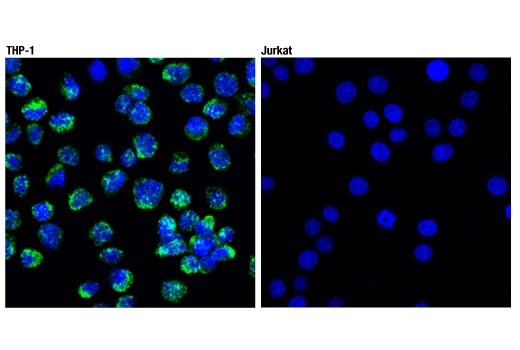
Immunofluorescence (Immunocytochemistry)
A. Solutions and Reagents
NOTE: Prepare solutions with reverse osmosis deionized (RODI) or equivalently purified water.
- 20X Phosphate Buffered Saline (PBS): (9808) To prepare 1L 1X PBS: add 50 ml 20X PBS to 950 ml dH2O, mix. Adjust pH to 8.0.
- Formaldehyde: 16%, methanol free, Polysciences, Inc. (cat# 18814), use fresh and store opened vials at 4°C in dark, dilute in 1X PBS for use.
- Methanol, 100%
- Blocking Buffer (1X PBS / 5% normal serum / 0.3% Triton™ X-100): To prepare 10 ml, add 0.5 ml normal serum from the same species as the secondary antibody (e.g., Normal Goat Serum (#5425)) and 0.5 mL 20X PBS to 9.0 mL dH2O, mix well. While stirring, add 30 µl Triton™ X-100.
- Antibody Dilution Buffer (1X PBS / 1% BSA / 0.3% Triton X-100): To prepare 10 ml, add 30 µl Triton™ X-100 to 10 ml 1X PBS. Mix well then add 0.1 g BSA (9998), mix.
Recommended Fluorochrome-conjugated Anti-Rabbit secondary antibodies:
- Prolong® Gold AntiFade Reagent (#9071), Prolong® Gold AntiFade Reagent with DAPI (#8961).
B. Specimen Preparation - Cultured Cell Lines (IF-IC)
NOTE: Cells should be grown, treated, fixed and stained directly in multiwell plates, chamber slides or on coverslips.
- Aspirate liquid, then cover cells to a depth of 2–3 mm with 4% formaldehyde in 1X PBS.
NOTE: Formaldehyde is toxic, use only in fume hood.
- Allow cells to fix for 15 minutes at room temperature.
- Aspirate fixative, rinse three times in 1X PBS for 5 minutes each.
- Proceed with Immunostaining (Section C).
C. Immunostaining
NOTE: All subsequent incubations should be carried out at room temperature unless otherwise noted in a humid light-tight box or covered dish/plate to prevent drying and fluorochrome fading.
- Methanol Permeabilization Step: Cover cells with ice-cold 100% methanol (use enough to cover completely to a depth of 3–5 mm, DO NOT LET DRY), incubate in methanol for 10 minutes at –20°C, rinse in 1X PBS for 5 minutes.
- Block specimen in Blocking Buffer for 60 minutes.
- While blocking, prepare primary antibody by diluting as indicated on product webpage in Antibody Dilution Buffer.
- Aspirate blocking solution, apply diluted primary antibody.
- Incubate overnight at 4°C.
- Rinse three times in 1X PBS for 5 minutes each.
- Incubate specimen in fluorochrome-conjugated secondary antibody diluted in Antibody Dilution Buffer for 1–2 hours at room temperature in dark.
- Rinse in 1X PBS as in step 6.
- Coverslip slides with Prolong® Gold Antifade Reagent (#9071), Prolong® Gold AntiFade Reagent with DAPI (#8961).
- For best results examine specimens immediately using appropriate excitation wavelength. For long term storage, store slides flat at 4°C protected from light.
posted November 2006
revised December 2010
Protocol Id: 32
Flow Cytometry, Methanol Permeabilization Protocol for Rabbit
Antibodies
A. Solutions and Reagents
All reagents required for this protocol may be efficiently purchased together in our Intracellular Flow
Cytometry Kit (Methanol) #13593, or
individually using the catalog numbers listed below.
NOTE: Prepare solutions with reverse osmosis deionized (RODI) or equivalent grade water.
- 1X Phosphate Buffered Saline (PBS): To prepare 1 L 1X PBS: add 100 ml 10X PBS (#12528) to 900 ml water mix.
- 4% Formaldehyde, Methanol-Free (#47746)
- 100% Methanol (#13604): Chill before use
- Antibody Dilution Buffer: Purchase ready-to-use Flow Cytometry Antibody Dilution Buffer (#13616), or prepare a 0.5% BSA PBS buffer by
dissolving 0.5 g Bovine Serum Albumin (BSA) (#9998) in 100 ml 1X PBS. Store at 4°C.
- Recommended Anti-Rabbit secondary antibodies::
- Anti-Rabbit IgG (H+L), F(ab')2 Fragment (Alexa Fluor® 488 Conjugate) #4412
- Anti-Rabbit IgG (H+L), F(ab')2 Fragment (Alexa Fluor® 594 Conjugate) #8889
- Anti-Rabbit IgG (H+L), F(ab')2 Fragment (Alexa Fluor® 647 Conjugate) #4414
- Anti-Rabbit IgG (H+L), F(ab')2 Fragment (PE Conjugate) #79408
NOTE: When including fluorescent cellular dyes in your experiment (including viability dyes, DNA dyes, etc.), please refer to the dye product page for the recommended protocol. Visit www.cellsignal.com for a full listing of cellular dyes validated for use in flow cytometry.
B. Fixation
NOTE: Adherent cells or tissue should be dissociated and in single-cell suspension prior to fixation.
NOTE: Optimal centrifugation conditions will vary depending upon cell type and reagent volume. Generally, 150-300g for 1-5 minutes will be sufficient to pellet the cells.
NOTE: If using whole blood, lyse red blood cells and wash by centrifugation prior to fixation.
NOTE: Antibodies targeting CD markers or other extracellular proteins may be added prior to fixation if the epitope is disrupted by formaldehyde and/or methanol. The antibodies will remain bound to the target of interest during the fixation and permeabilization process. However, note that some fluorophores (including PE and APC) are damaged by methanol and thus should not be added prior to permeabilization. Conduct a small-scale experiment if you are unsure.
- Pellet cells by centrifugation and remove supernatant.
- Resuspend cells in approximately 100 µl 4% formaldehyde per 1 million cells. Mix well to dissociate pellet and prevent cross-linking of individual cells.
- Fix for 15 min at room temperature (20-25°C).
- Wash by centrifugation with excess 1X PBS. Discard supernatant in appropriate waste container. Resuspend cells in 0.5-1 ml 1X PBS. Proceed to Permeabilization step.
- Alternatively, cells may be stored overnight at 4°C in 1X PBS.
C. Permeabilization
- Permeabilize cells by adding ice-cold 100% methanol slowly to pre-chilled cells, while gently vortexing, to a final concentration of 90% methanol.
- Permeabilize for a minimum of 10 min on ice.
- Proceed with immunostaining (Section D) or store cells at -20°C in 90% methanol.
D. Immunostaining
NOTE: Count cells using a hemocytometer or alternative method.
- Aliquot desired number of cells into tubes or wells. (Generally, 5x10⁵ to 1x10⁶ cells per assay.)
- Wash cells by centrifugation in excess 1X PBS to remove methanol. Discard supernatant in appropriate
waste container. Repeat if necessary.
- Resuspend cells in 100 µl of diluted primary antibody, prepared in Antibody Dilution Buffer at a recommended dilution or as determined via titration.
- Incubate for 1 hr at room temperature.
- Wash by centrifugation in Antibody Dilution Buffer or 1X PBS. Discard supernatant. Repeat.
- Resuspend cells in 100 µl of diluted fluorochrome-conjugated secondary antibody (prepared in Antibody Dilution Buffer at the recommended dilution).
- Incubate for 30 min at room temperature. Protect from light.
- Wash by centrifugation in Antibody Dilution Buffer or 1X PBS. Discard supernatant. Repeat.
- Resuspend cells in 200-500 µl of 1X PBS and analyze on flow cytometer.
posted July 2009
revised June 2020