Recombinant antibodies offer several key advantages compared to traditional antibodies. These include superior lot-to-lot consistency, continuous supply, and animal-free manufacturing. As such, recombinant antibodies are seeing increased use for scientific research, especially as a means of addressing the ongoing reproducibility crisis.
Traditional polyclonal and monoclonal antibodies are the product of normal B cell development and genetic recombination. They are generated by immunizing an animal with an antigen to elicit an immune response. While polyclonal antibodies are secreted by many different B cell clones and recognize multiple antigenic epitopes, monoclonals originate from a single B cell clone and are specific for just one epitope.
Recombinant antibodies are monoclonal, but their production involves in vitro genetic manipulation. After cloning the antibody genes into an expression vector, this is then transfected into an appropriate host cell line for antibody expression. Mammalian cell lines are most commonly used for recombinant antibody production, although cell lines of bacterial, yeast, or insect origin are also suitable.
Because recombinant antibody production involves sequencing the antibody light and heavy chains, it is a highly controlled and reliable process. In contrast, hybridoma-based systems for producing monoclonal antibodies are subject to genetic drift and instability, increasing the potential for lot-to-lot variability or loss of antibody expression. Recombinant antibodies are highly consistent from lot to lot, thereby ensuring reproducible experimental results.
In vitro methods for producing antibodies are amenable to large-scale production, meaning antibody availability is unlikely to become a limiting factor. Moreover, since the recombinant antibody sequence is known, continuity of supply is assured; in situations where an antibody will be used to support large, long-term studies, this can be an especially critical factor.
Unlike traditional methods for antibody production, recombinant approaches avoid the need to use animals. Where polyclonal antibodies are purified directly from the serum of the immunized host, and monoclonals are purified from either hybridoma-derived tissue culture supernatant or ascites, recombinant antibodies are instead purified from the tissue culture supernatants of transfected host cell lines. Regardless of whether an antibody is polyclonal, monoclonal or recombinant, it must always be properly validated in the intended application prior to experimental use. At CST, we adhere to the Hallmarks of Antibody Validation™, six complementary strategies for determining the specificity, sensitivity, and functionality of an antibody in any given assay. By carefully tailoring these strategies to each antibody product, we guarantee that CST antibodies will work as expected, to help you achieve results you can trust.

| Cat. # | Size | Price | Inventory |
|---|---|---|---|
| 93668T | 20 µl | ||
| 93668S | 100 µl |
| REACTIVITY | H M R |
| SENSITIVITY | Endogenous |
| MW (kDa) | 40 |
| Source/Isotype | Rabbit IgG |
Product Information
| Application | Dilution |
|---|---|
| Western Blotting | 1:1000 |
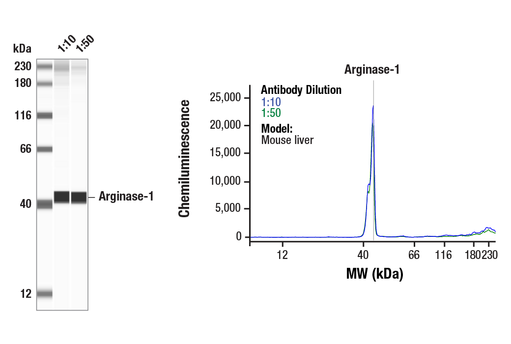
| Simple Western™ | 1:10 - 1:50 |
| IHC Leica Bond | 1:200 - 1:800 |
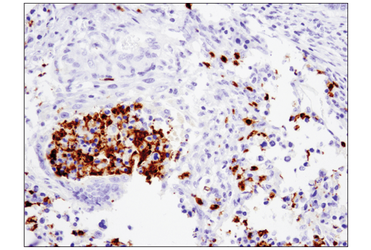
| Immunohistochemistry (Paraffin) | 1:50 - 1:200 |
| Immunofluorescence (Frozen) | 1:50 - 1:200 |
| Immunofluorescence (Immunocytochemistry) | 1:50 - 1:200 |
| Flow Cytometry (Fixed/Permeabilized) | 1:50 |
For western blots, incubate membrane with diluted primary antibody in 5% w/v nonfat dry milk, 1X TBS, 0.1% Tween® 20 at 4°C with gentle shaking, overnight.
NOTE: Please refer to primary antibody product webpage for recommended antibody dilution.
NOTE: Prepare solutions with reverse osmosis deionized (RODI) or equivalent grade water.
Load 20 µl onto SDS-PAGE gel (10 cm x 10 cm).
NOTE: Loading of prestained molecular weight markers (#59329, 10 µl/lane) to verify electrotransfer and biotinylated protein ladder (#7727, 10 µl/lane) to determine molecular weights are recommended.
NOTE: Volumes are for 10 cm x 10 cm (100 cm2) of membrane; for different sized membranes, adjust volumes accordingly.
* Avoid repeated exposure to skin.
posted June 2005
revised June 2020
Protocol Id: 263
NOTE: Please see product datasheet or product webpage for appropriate antibody dilution^.
| Step | Reagents | Time/Temperature | |
|---|---|---|---|
| 1 | Dewax | BOND™ Dewax Solution, 100% Alcohol, BOND™ Wash Solution | Pre-programmed Leica® BOND™ |
| 2 | Antigen Retrieval | BOND™ Epitope Retrieval ER2 Solution | 20 min., 100˚C | Protocol: HIER 20 min with ER2 |
| 3 | Peroxide Block | Refine Detection Kit Peroxide Block* | 5 min. |
| WASH | BOND™ Wash Solution | 3x 0:00 min. | |
| 4 | Protein Block (optional) | #5425 NGS or #15019 Animal-Free Blocking Solution | 20 min. |
| 5 | Primary Antibody^ | Dilute in #8112 SignalStain® Antibody Diluent | 30 min. |
| WASH | BOND™ Wash Solution | 3x 2:00 min. | |
| NA | Post Primary Mouse Linker | Refine Detection Kit Post Primary* | Not Applied |
| 6 | Secondary Detection | Refine Detection Kit Polymer* | 10 min. |
| WASH | BOND™ Wash Solution/Deionized Water | Custom (see below) | |
| 7a | Visualization | Refine Detection Kit Mixed DAB Refine* | 0:00 min. |
| 7b | Visualization | Refine Detection Kit Mixed DAB Refine* | 10 min. |
| WASH | Deionized Water | 3x 0:00 min. | |
| 8 | Counterstain | Refine Detection Kit Hematoxylin* | 5 min. |
| WASH | Deionized Water | 0:00 min. | |
| WASH | BOND™ Wash Solution | 0:00 min. | |
| WASH | Deionized Water | 0:00 min. | |
| 9 | Dehydration (Offline): | ||
| Incubate sections in 95% ethanol two times for 10 seconds each. | |||
| Repeat in 100% ethanol, incubating sections two times for 10 seconds each. | |||
| Repeat in xylene, incubating sections two times for 10 seconds each. | |||
| 10 | Mount sections with coverslips and #84583 SignalStain® Mounting Medium | ||
| Optional Custom wash: | BOND™ Wash Solution | 2:00 | |
| BOND™ Wash Solution | Dispenser Type: OPEN 0:00 | ||
| BOND™ Wash Solution | 2:00 | ||
| BOND™ Wash Solution | Dispenser Type: OPEN 0:00 | ||
| BOND™ Wash Solution | 0:00 | ||
| Deionized Water | 0:00 | ||
*Reagent included in BOND™ Polymer Refine Detection Kit (Catalog No: DS9800)
LEICA® is a registered trademark of Leica Microsystems IR GmbH.
BOND™ is a trademark of Leica Biosystems Melbourne Pty. Ltd. No affiliation or sponsorship between CST and Leica Microsystems IR GmbH or Leica Biosystems Melbourne Pty. Ltd is implied.
posted August 2018
revised September 2018
Protocol Id: 1444
NOTE: Prepare solutions with reverse osmosis deionized (RODI) or equivalent grade water.
NOTE: Do not allow slides to dry at any time during this procedure.
For Citrate: Heat slides in a microwave submersed in 1X citrate unmasking solution until boiling is initiated; follow with 10 min at a sub-boiling temperature (95°-98°C). Cool slides on bench top for 30 min.
|
RECOMMENDED DETECTION REAGENTS |
SignalStain® Boost IHC Detection Reagent (HRP, Rabbit) #8114 | SignalStain® Boost IHC Detection Reagent (AP, Rabbit) #18653 |
|---|---|---|
|
COMPATIBLE CHROMOGEN |
SignalStain® DAB Substrate Kit #8059 | SignalStain® Vibrant Red Alkaline Phosphatase Substrate Kit #76713 |
| SignalStain® Vivid Purple Peroxidase Substrate Kit #96632 | SignalStain® Ultra Blue Alkaline Phosphatase Substrate Kit #12824 | |
| SignalStain® Deep Black Peroxidase Substrate Kit #72986 | ||
| SignalStain® Radiant Yellow Peroxidase Substrate Kit #69644 |
NOTE: Use of detection reagents other than those specified in this protocol may require further optimization of the primary antibody to account for the different sensitivities of the detection reagents.
posted February 2010
revised June 2020
Protocol Id: 283
NOTE: Prepare solutions with reverse osmosis deionized (RODI) or equivalently purified water.
Recommended Fluorochrome-conjugated Anti-Rabbit secondary antibodies:
Cover sections with 4% formaldehyde dilute in 1X PBS.
NOTE: Formaldehyde is toxic, use only in fume hood.
NOTE: All subsequent incubations should be carried out at room temperature unless otherwise noted in a humid light-tight box or covered dish/plate to prevent drying and fluorochrome fading.
posted November 2006
revised December 2010
Protocol Id: 147
NOTE: Prepare solutions with reverse osmosis deionized (RODI) or equivalently purified water.
Recommended Fluorochrome-conjugated Anti-Rabbit secondary antibodies:
NOTE: Cells should be grown, treated, fixed and stained directly in multiwell plates, chamber slides or on coverslips.
NOTE: All subsequent incubations should be carried out at room temperature unless otherwise noted in a humid light-tight box or covered dish/plate to prevent drying and fluorochrome fading.
posted November 2006
revised December 2010
Protocol Id: 32
All reagents required for this protocol may be efficiently purchased together in our Intracellular Flow Cytometry Kit (Methanol) #13593, or individually using the catalog numbers listed below.
NOTE: Prepare solutions with reverse osmosis deionized (RODI) or equivalent grade water.
NOTE: When including fluorescent cellular dyes in your experiment (including viability dyes, DNA dyes, etc.), please refer to the dye product page for the recommended protocol. Visit www.cellsignal.com for a full listing of cellular dyes validated for use in flow cytometry.
NOTE: Adherent cells or tissue should be dissociated and in single-cell suspension prior to fixation.
NOTE: Optimal centrifugation conditions will vary depending upon cell type and reagent volume. Generally, 150-300g for 1-5 minutes will be sufficient to pellet the cells.
NOTE: If using whole blood, lyse red blood cells and wash by centrifugation prior to fixation.
NOTE: Antibodies targeting CD markers or other extracellular proteins may be added prior to fixation if the epitope is disrupted by formaldehyde and/or methanol. The antibodies will remain bound to the target of interest during the fixation and permeabilization process. However, note that some fluorophores (including PE and APC) are damaged by methanol and thus should not be added prior to permeabilization. Conduct a small-scale experiment if you are unsure.
NOTE: Count cells using a hemocytometer or alternative method.
posted July 2009
revised June 2020
实验步骤编号:404
人, 小鼠, 大鼠
使用与人精氨酸酶-1 蛋白的 Val47 周围残基相对应的合成肽对动物进行免疫接种来产生单克隆抗体。
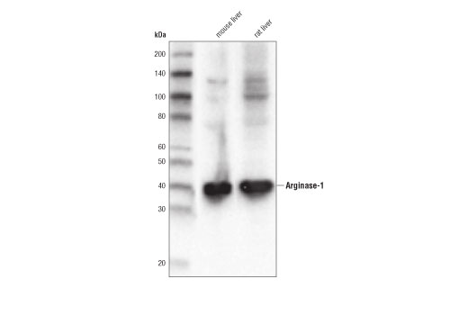
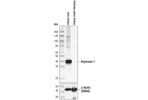
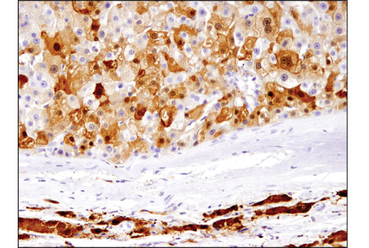
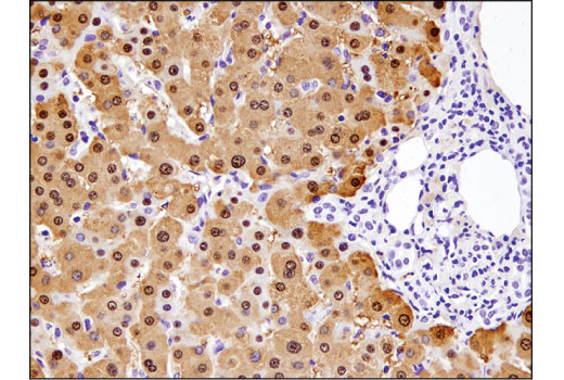
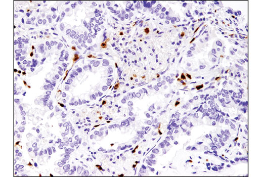
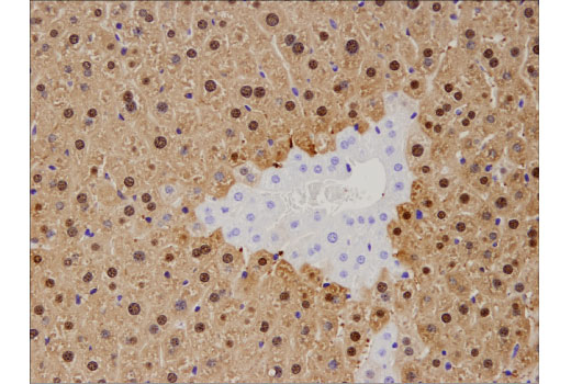
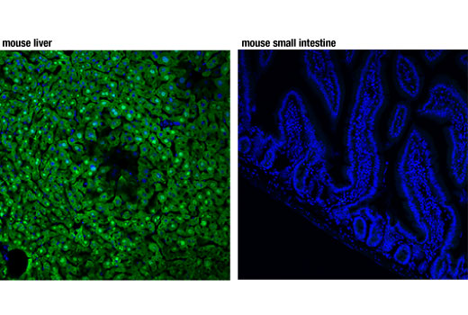
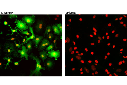
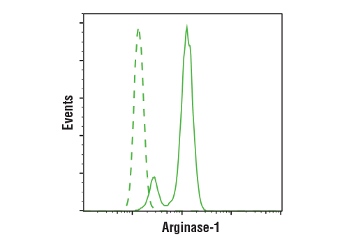
L-精氨酸在调节免疫系统方面发挥关键作用 (1-3)。在炎症、癌症和某些其他病理状况下,骨髓细胞分化被抑制,导致出现不成熟的骨髓细胞异质群体,称为骨髓源性抑制细胞 (MDSC)。MDSCs 被募集至癌症相关炎症的位点,并表达高水平的精氨酸酶-1 (4)。精氨酸酶-1 催化尿素循环的最后一步,即将 L- 精氨酸转化为 L-鸟氨酸和尿素 (5)。因此,MDSC 可增加 L-精氨酸的分解代谢,导致癌症炎症微环境中 L-精氨酸的耗减 (4,6)。左旋精氨酸的减少会抑制 T 细胞增殖和功能,因此导致肿瘤进展 (4,6)。精氨酸酶-1 引起了研究人员的极大兴趣,这些研究人员正在寻找治疗靶标以抑制 MDSCs 在癌症免疫治疗中的作用(7)。此外,研究表明,精氨酸酶-1 可将原发性肝细胞癌 (HCC) 与肝脏中的转移性肿瘤区分开来,表明其在 HCC 诊断中可作为潜在生物标记物的价值 (8,9)。
除非如以 CST 合法授权代表签署的书面形式另行明确同意,否则以下条款适用于 CST、其附属公司或其分销商提供的产品。除非 CST 合法授权代表以书面形式单独接受,否则任何附加于或异于此处所载条款和条件的客户条款和条件均被拒绝且无效。
产品用“仅供研究使用”或类似标示声明标示,并且尚未经 FDA 或其他国外或国内监管实体出于任何目的批准、准许或许可。客户不得出于任何诊断或治疗目的或以任何与产品标示声明相冲突的方式使用任何产品。CST 销售或许可的产品提供给作为最终用户的客户,且仅用于研究和开发用途。出于诊断、预防或治疗目的任何产品使用或出于转售(单独或作为成分)或其他商业目的的任何产品购买都要求来自 CST 的单独许可。客户 (a) 不得向任何第三方出售、许可、出借、捐赠或另行转让或提供任何本公司产品,无论单独或联合其他材料方式,或使用本公司产品制造任何商业产品,(b) 不得复制、修改、逆向工程、反编译、反汇编或另行尝试发现本公司产品的底层结构或技术,或出于开发与 CST 产品或服务竞争的任何产品或服务的目的使用本公司产品,(c) 不得从本公司产品改变或移除任何商标、商品名称、徽标、专利或版权声明或标记,(d) 仅应根据 CST 产品销售条款和任何适用文档使用本公司产品,以及 (e) 应就客户联系本公司产品所用的任何第三方产品或服务而言遵守任何许可、服务条款或类似协议。