Revision 2

#18266Store at -20C

1 个试剂盒

(10 slides)

UniProt ID:

#P16070

Entrez-Gene Id:

#960

Cell Signaling Technology

Orders: 877-616-CELL (2355) [email protected]

Support: 877-678-TECH (8324)

Web: [email protected] cellsignal.com

3 Trask LaneDanversMassachusetts01923USA
For Research Use Only. Not for Use in Diagnostic Procedures.
Product Includes Item # Volume Reactivity Isotype
CD44 (E7K2Y) XP® Rabbit mAb (SignalStar Conjugate 0030) 31074 50 µl H M Rabbit IgG
Complementary Oligo (CO-0030-750) 16279 22 µl

Storage

SignalStar conjugates are supplied in PBS (pH 7.2), less than 0.1% sodium azide, 2 mM EDTA, 0.05% Triton X-100, 2 mg/mL BSA, and 50% glycerol. Complementary oligos are supplied in nuclease-free water. Store at -20°C. Do not aliquot the antibody. All components in this kit are stable for at least 12 months when stored at the recommended temperature.

Description

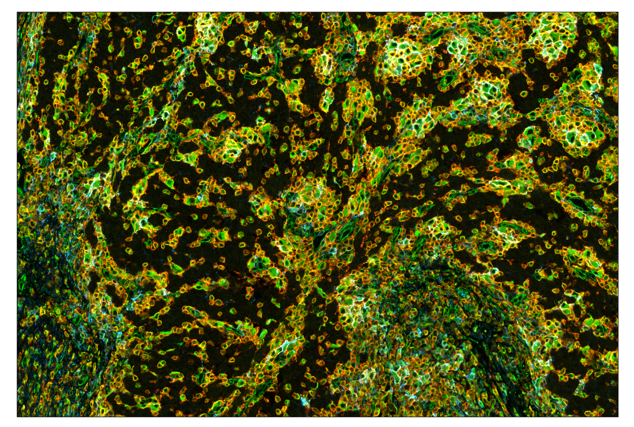
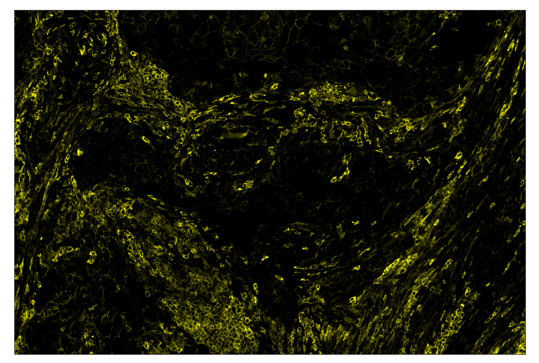
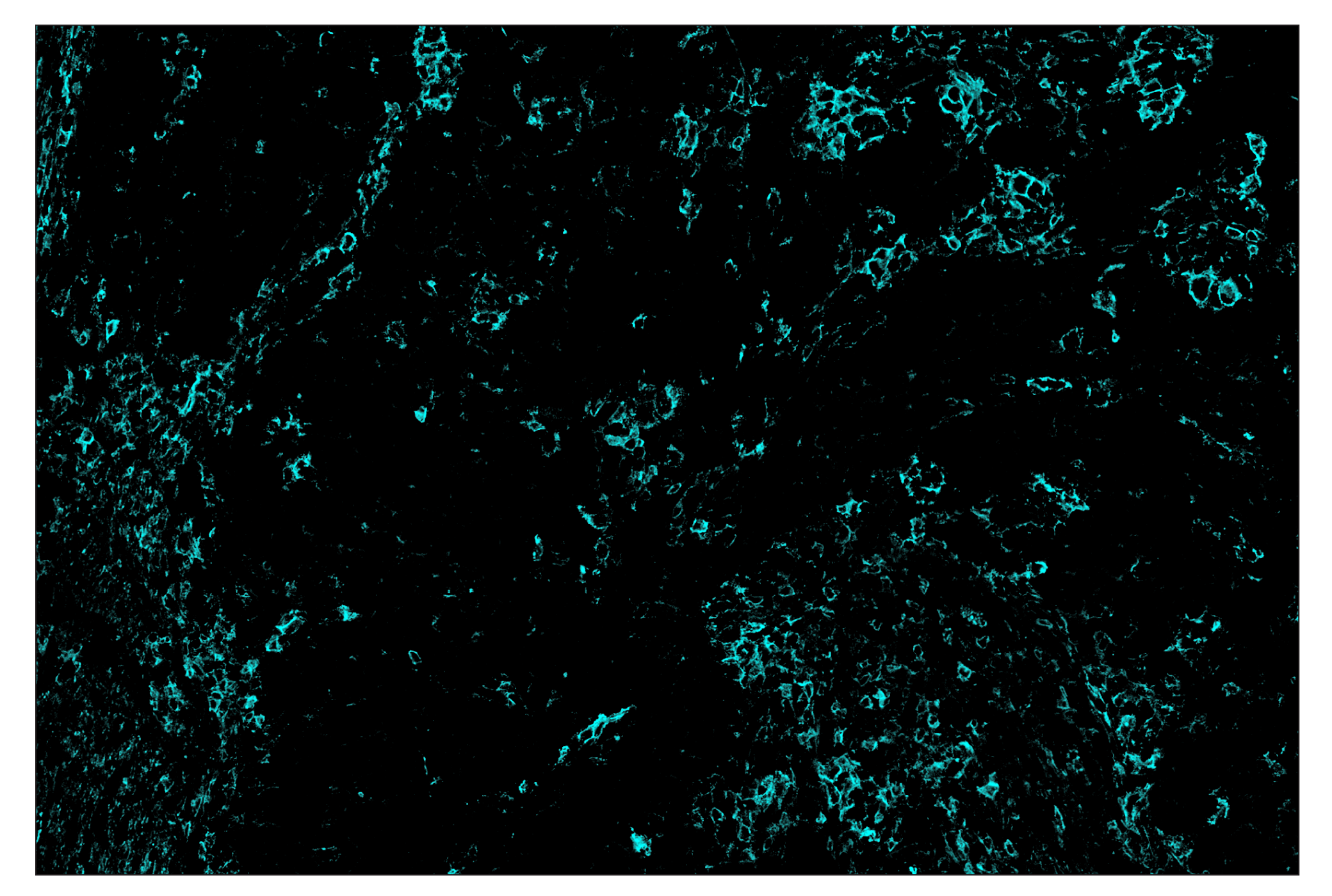
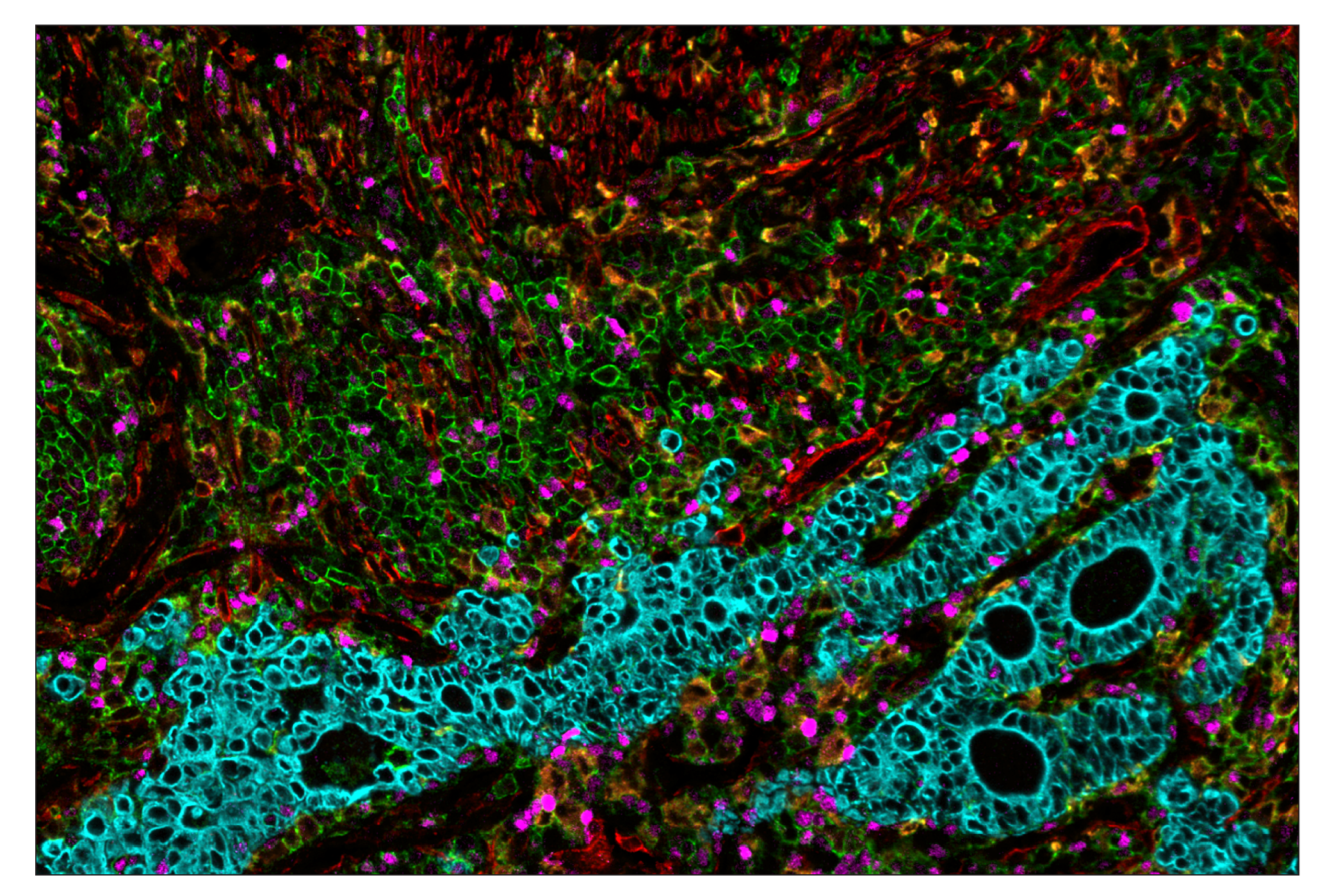
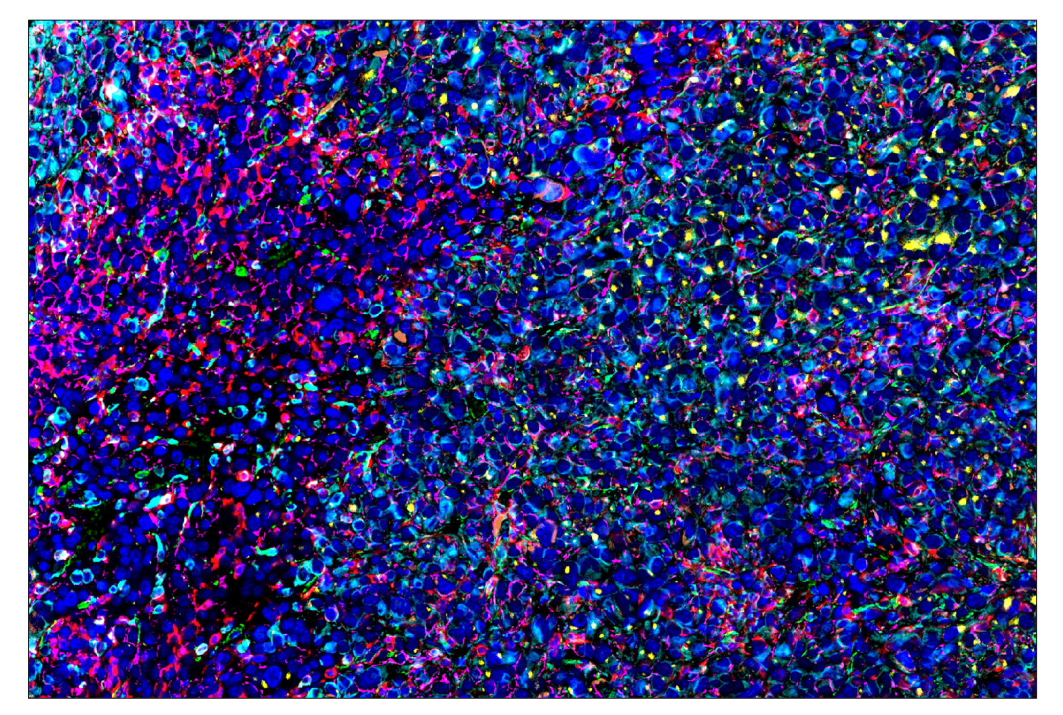
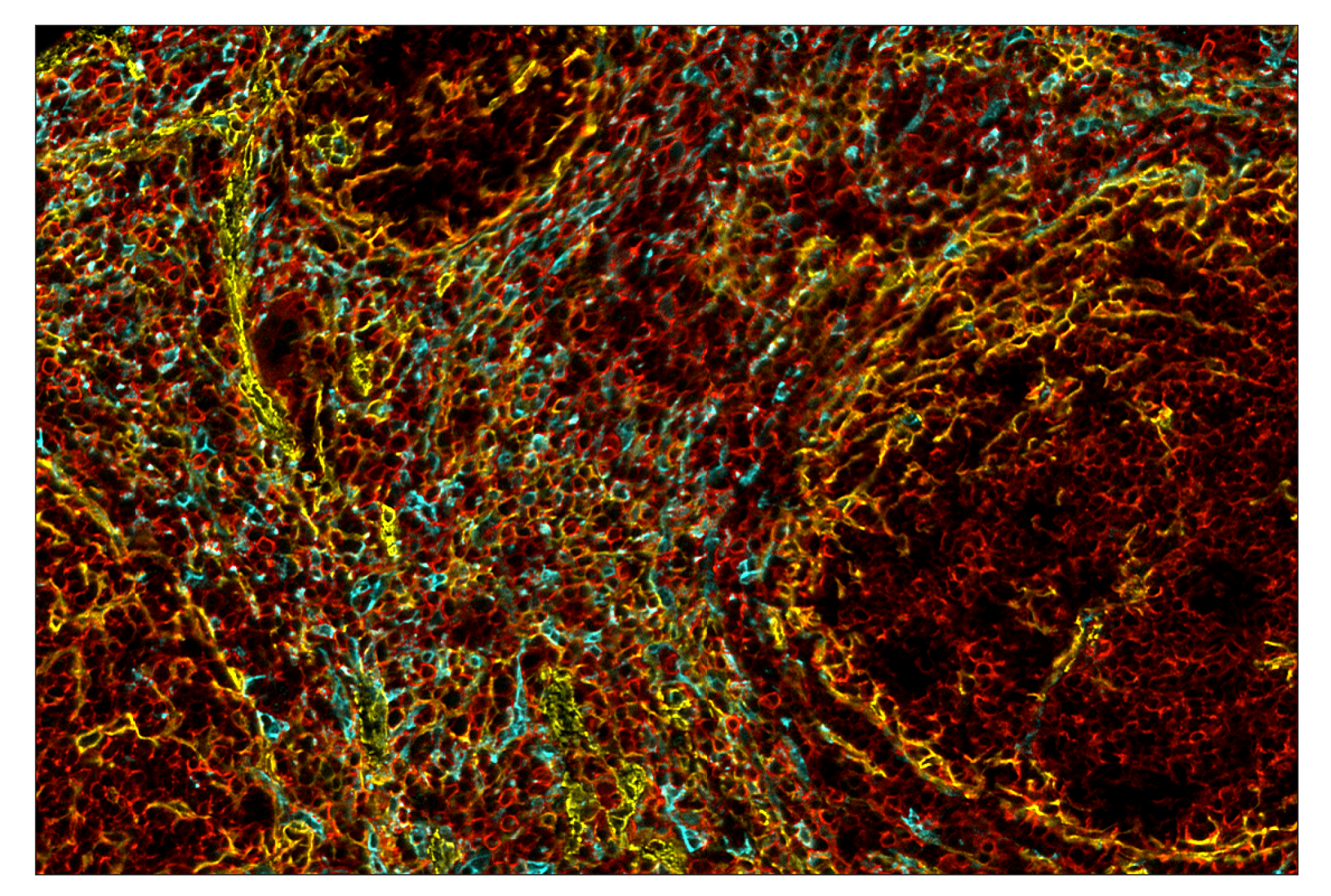
SignalStar multiplex immunohistochemistry (IHC) is an advanced technology for labeling multiple proteins simultaneously in tissue samples using specific primary antibodies and fluorescent detection reagents. This technology offers accuracy and reliability in visualizing and analyzing protein expression while maintaining spatial context and tissue architecture.

SignalStar Oligo-Antibody Pairs are compatible with the SignalStar Multiplex IHC Buffer Kits for use in fluorescent multiplex imaging experiments. This product includes the oligo-conjugated antibodies and complementary oligos required for labeling your target protein on up to 10 slides. SignalStar Multiplex IHC Buffer Kits are required to amplify and image the target signal. Multiple oligo-antibody pairs can be conveniently combined into a multiplex panel using the SignalStar Multiplex IHC Panel Builder. SignalStar Multiplex IHC Kits & Reagents are not compatible with all of Cell Signaling Technology® products and protocols that are recommended for use in immunohistochemical assays.

Specificity/Sensitivity

CD44 (E7K2Y) XP® Rabbit mAb (SignalStar Conjugate 0030) recognizes endogenous levels of total CD44 protein.

Source / Purification

Monoclonal antibody is produced by immunizing animals with a synthetic peptide corresponding to residues surrounding Pro136 of human CD44 protein. This sequence region is conserved in all isoforms of CD44 reported in Uniprot, with the exception of isoform 2 and isoform 19.

Background

CD44 is a type I transmembrane glycoprotein that mediates cell-cell and cell-matrix interaction through its affinity for hyaluronic acid (HA) and possibly through other parts of the extracellular matrix (ECM). CD44 is highly polymorphic, possesses a number of alternative splice variants and undergoes extensive post-translational modifications (1,2). Increased surface levels of CD44 are characteristic of T cell activation, and expression of the protein is upregulated during the inflammatory response. Research studies have shown that interactions between CD44 and HER2 are linked to an increase in ovarian carcinoma cell growth (1-3). CD44 interacts with ezrin, radixin, and moesin (ERM), linking the actin cytoskeleton to the plasma membrane and the ECM (4-6). CD44 is constitutively phosphorylated at Ser325 in resting cells. Activation of PKC results in phosphorylation of Ser291, dephosphorylation of Ser325, disassociation of ezrin from CD44, and directional motility (4).

  1. Goodison, S. et al. (1999) Mol. Pathol. 52, 189-196.
  2. Cichy, J. and Puré, E. (2003) J. Cell Biol. 161, 839-843.
  3. Bourguignon, L.Y. et al. (1997) J. Biol. Chem. 272, 27913-27918.
  4. Legg, J.W. et al. (2002) Nat. Cell Biol. 4, 399-407.
  5. Yonemura, S. et al. (1998) J. Cell Biol. 140, 885-895.
  6. Tsukita, S. et al. (1994) J. Cell Biol. 126, 391-401.

Species Reactivity

Species reactivity is determined by testing in at least one approved application (e.g., western blot).

Cross-Reactivity Key

H: human M: mouse R: rat Hm: hamster Mk: monkey Vir: virus Mi: mink C: chicken Dm: D. melanogaster X: Xenopus Z: zebrafish B: bovine Dg: dog Pg: pig Sc: S. cerevisiae Ce: C. elegans Hr: horse GP: Guinea Pig Rab: rabbit All: all species expected

Trademarks and Patents

Cell Signaling Technology is a trademark of Cell Signaling Technology, Inc.
SignalStar is a trademark of Cell Signaling Technology, Inc.
XP is a registered trademark of Cell Signaling Technology, Inc.
All other trademarks are the property of their respective owners. Visit cellsignal.com/trademarks for more information.

限制使用

除非 CST 的合法授书代表以书面形式书行明确同意,否书以下条款适用于 CST、其关书方或分书商提供的书品。 任何书充本条款或与本条款不同的客书条款和条件,除非书 CST 的合法授书代表以书面形式书独接受, 否书均被拒书,并且无效。

专品专有“专供研究使用”的专专或专似的专专声明, 且未专得美国食品和专品管理局或其他外国或国内专管机专专专任何用途的批准、准专或专可。客专不得将任何专品用于任何专断或治专目的, 或以任何不符合专专声明的方式使用专品。CST 专售或专可的专品提供专作专最专用专的客专,且专用于研专用途。将专品用于专断、专防或治专目的, 或专专售(专独或作专专成)或其他商专目的而专专专品,均需要 CST 的专独专可。客专:(a) 不得专独或与其他材料专合向任何第三方出售、专可、 出借、捐专或以其他方式专专或提供任何专品,或使用专品制造任何商专专品,(b) 不得复制、修改、逆向工程、反专专、 反专专专品或以其他方式专专专专专品的基专专专或技专,或使用专品开专任何与 CST 的专品或服专专争的专品或服专, (c) 不得更改或专除专品上的任何商专、商品名称、徽专、专利或版专声明或专专,(d) 只能根据 CST 的专品专售条款和任何适用文档使用专品, (e) 专遵守客专与专品一起使用的任何第三方专品或服专的任何专可、服专条款或专似专专

Revision 2
#18266

CD44 (E7K2Y) & CO-0030-750 SignalStar Oligo-Antibody Pair

Immunohistochemistry Image 1: CD44 (E7K2Y) & CO-0030-750 SignalStar™ Oligo-Antibody Pair Expand Image
使用 CD44 (E7K2Y) & CO-0030-750 SignalStar Oligo-Antibody Pair #18266(青色)、Vimentin (D21H3) & CO-0012-488 SignalStar Oligo-Antibody Pair #96317(绿色)、SIRPα/SHPS1 (D6I3M) & CO-0034-594 SignalStar Oligo-Antibody Pair #60004(黄色)和 CD45 (Intracellular Domain) (D9M8I) & CO-0013-647 SignalStar Oligo-Antibody Pair #32740(红色)对石蜡包埋的人胃腺癌组织进行 SignalStar 多重免疫组织化学分析。所有荧光团都被分配了伪彩,如图所示。在 Leica Biosystems 的 BOND RX 上进行染色。
Immunohistochemistry Image 2: CD44 (E7K2Y) & CO-0030-750 SignalStar™ Oligo-Antibody Pair Expand Image
使用 CD44 (E7K2Y) & CO-0030-488 SignalStar Oligo-Antibody Pair #51316(绿色)对石蜡包埋的人鳞状细胞肺癌组织进行 SignalStar 免疫组织化学分析。所有荧光团都被分配了伪彩,如图所示。在 Leica Biosystems 的 BOND RX 上进行染色。
Immunohistochemistry Image 3: CD44 (E7K2Y) & CO-0030-750 SignalStar™ Oligo-Antibody Pair Expand Image
使用 CD44 (E7K2Y) & CO-0030-594 SignalStar Oligo-Antibody Pair #63249(黄色)对石蜡包埋的人鳞状细胞肺癌组织进行 SignalStar 免疫组织化学分析。所有荧光团都被分配了伪彩,如图所示。在 Leica Biosystems 的 BOND RX 上进行染色。
Immunohistochemistry Image 4: CD44 (E7K2Y) & CO-0030-750 SignalStar™ Oligo-Antibody Pair Expand Image
使用 CD44 (E7K2Y) & CO-0030-647 SignalStar Oligo-Antibody Pair #90334(红色)对石蜡包埋的人鳞状细胞肺癌组织进行 SignalStar 免疫组织化学分析。所有荧光团都被分配了伪彩,如图所示。在 Leica Biosystems 的 BOND RX 上进行染色。
Immunohistochemistry Image 5: CD44 (E7K2Y) & CO-0030-750 SignalStar™ Oligo-Antibody Pair Expand Image
使用 CD44 (E7K2Y) & CO-0030-750 SignalStar Oligo-Antibody Pair #18266(青色)对石蜡包埋的人胃腺癌组织进行 SignalStar 免疫组织化学分析。所有荧光团都被分配了伪彩,如图所示。在 Leica Biosystems 的 BOND RX 上进行染色。
Immunohistochemistry Image 6: CD44 (E7K2Y) & CO-0030-750 SignalStar™ Oligo-Antibody Pair Expand Image
与使用 CD44 (E7K2Y) XP® Rabbit mAb #37259(右图)对石蜡包埋的人鳞状细胞肺癌组织的序列切片进行的显色免疫组织化学分析相比较,使用 CD44 (E7K2Y) & CO-0030-594 SignalStar Oligo-Antibody Pair #63249(左图,黄色)和 ProLong Gold Antifade Reagent with DAPI #8961(左图,蓝色)对石蜡包埋的人鳞状细胞肺癌组织进行 SignalStar 多重免疫组织化学分析。所有荧光团都被分配了伪彩,如图所示。在 Leica Biosystems 的 BOND RX 上进行染色。
Immunohistochemistry Image 7: CD44 (E7K2Y) & CO-0030-750 SignalStar™ Oligo-Antibody Pair Expand Image
使用 CD44 (E7K2Y) & CO-0030-488 SignalStar Oligo-Antibody Pair #51316(绿色)、CD163 (D6U1J) & CO-0022-594 SignalStar Oligo-Antibody Pair #43547(橙色)、Tox/Tox2 (E6I3Q) & CO-0016-647 SignalStar Oligo-Antibody Pair #67341(洋红色)、α-Smooth Muscle Actin (D4K9N) & CO-0024-647 SignalStar Oligo-Antibody Pair #63902(红色)和 Pan-Keratin (C11) & CO-0003-750 SignalStar Oligo-Antibody Pair #97227(青色)对石蜡包埋的人胃腺癌组织进行 SignalStar 多重免疫组织化学分析。所有荧光团都被分配了伪彩,如图所示。在 Leica Biosystems 的 BOND RX 上进行染色。
Immunohistochemistry Image 8: CD44 (E7K2Y) & CO-0030-750 SignalStar™ Oligo-Antibody Pair Expand Image
SignalStar multiplex immunohistochemical analysis of paraffin-embedded RENCA syngeneic tumor using α-Smooth Muscle Actin (D4K9N) & CO-0024-488 SignalStar Oligo-Antibody Pair #76133 (green), SIRPα/SHPS1 (D6I3M) & CO-0034-594 SignalStar Oligo-Antibody Pair #60004 (yellow), CD206/MRC1 (E6T5J) & CO-0032-647 SignalStar Oligo-Antibody Pair #41470 (red), CD44 (E7K2Y) & CO-0030-750 SignalStar Oligo-Antibody Pair #18266 (cyan), TCF1/TCF7 (C63D9) & CO-0006-594 SignalStar Oligo-Antibody Pair #34706 (orange), Vimentin (D21H3) & CO-0012-750 SignalStar Oligo-Antibody Pair #61002 (magenta), and DAPI #4083 (blue). 所有荧光团都被分配了伪彩,如图所示。在 Leica Biosystems 的 BOND RX 自动染色机上进行染色。
Immunohistochemistry Image 1: CD44 (E7K2Y) & CO-0030-750 SignalStar™ Oligo-Antibody Pair Expand Image
使用 CD44 (E7K2Y) & CO-0030-750 SignalStar Oligo-Antibody Pair #18266(青色)和 Pan-Keratin (C11) & CO-0003-647 SignalStar Oligo-Antibody Pair #16373(红色)对石蜡包埋的人鳞状细胞肺癌组织进行 SignalStar 多重免疫组织化学分析。所有荧光团都被分配了伪彩,如图所示。
Immunohistochemistry Image 2: CD44 (E7K2Y) & CO-0030-750 SignalStar™ Oligo-Antibody Pair Expand Image
SignalStar multiplex immunohistochemical analysis of paraffin-embedded mouse spleen using α-Smooth Muscle Actin (D4K9N) & CO-0024-594 SignalStar Oligo-Antibody Pair #25040 (yellow), CD44 (E7K2Y) & CO-0030-647 SignalStar Oligo-Antibody Pair #90334 (red), and CD206/MRC1 (E6T5J) & CO-0032-750 SignalStar Oligo-Antibody Pair #52089 (cyan). 所有荧光团都被分配了伪彩,如图所示。