Product Information
Product Usage Information
1. Aliquot PTMScan® Control Peptides Succinyl-Lysine for storage as single-use units at -20°C or proceed to immediate usage.
2. Resuspend sample peptides in the appropriate buffer and volume, e.g., 1.4 mL of PTMScan® IAP Buffer (1X).
3. Clear sample peptides by centrifugation.
4. Transfer clarified sample peptides to tubes containing IAP beads.
5. Add 10 µL of PTMScan® Control Peptides Succinyl-Lysine to IAP beads and sample peptides and mix well.
6. Continue with PTMScan® or PTMScan® HS workflows at the 2-hour incubation step.
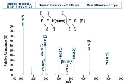
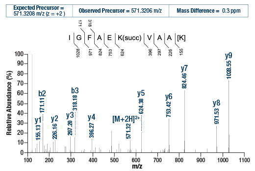
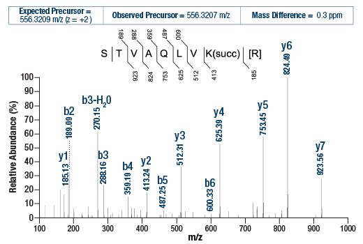
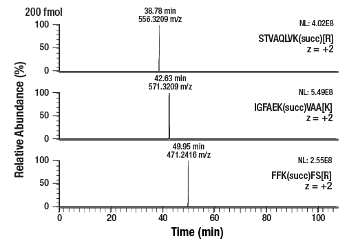
7. Detect PTMScan® Control Peptides Succinyl-Lysine in the LCMS data file.
Storage
Product Description
Background
Lysine is subject to a wide array of regulatory post-translational modifications due to its positively charged ε-amino group side chain. The most prevalent of these are ubiquitination and acetylation, which are highly conserved among prokaryotes and eukaryotes (1,2). Acyl group transfer from the metabolic intermediates acetyl-, succinyl-, malonyl-, glutaryl-, butyryl-, propionyl-, and crotonyl-CoA all neutralize lysine’s positive charge and confer structural alterations affecting substrate protein function. Lysine acetylation is catalyzed by histone acetyltransferases, HATs, using acetyl-CoA as a cofactor (3,4). Deacylation is mediated by histone deacetylases, HDACs 1-11, and NAD-dependent Sirtuins 1-7. Some sirtuins have little to no deacetylase activity, suggesting that they are better suited for other acyl lysine substrates (5).
Sirt5 is a predominantly mitochondrial desuccinylase and demalonylase (5,6). In the absence of a known succinyltransferase, succinylation is likely driven by the concentration of succinyl-CoA and intracellular pH and is subject to metabolic fluctuations (7,8). Protein succinylation is especially prevalent among mitochondrial metabolic proteins and bacteria, further solidifying the evolutionary link between mitochondria and prokaryotes. It often occurs at lysine residues that are alternatively acetylated or ubiquitinated. More than a thousand lysine succinylation sites were identified on hundreds of proteins, including glutamate dehydrogenase (15 sites), malate dehydrogenase, citrate synthase, carbamoyl phosphate synthase 1, and histone proteins (9).
- Liu, Z. et al. (2014) Nucleic Acids Res 42, D531-6.
- Lee, S. (2013) Toxicol Res 29, 81-6.
- Lin, H. et al. (2012) ACS Chem Biol 7, 947-60.
- Zhang, Z. et al. (2011) Nat Chem Biol 7, 58-63.
- Du, J. et al. (2011) Science 334, 806-9.
- Peng, C. et al. (2011) Mol Cell Proteomics 10, M111.012658.
- Rardin, M.J. et al. (2013) Cell Metab 18, 920-33.
- Park, J. et al. (2013) Mol Cell 50, 919-30.
- Weinert, B.T. et al. (2013) Cell Rep 4, 842-51.
Species Reactivity
Species reactivity is determined by testing in at least one approved application (e.g., western blot).
Cross-Reactivity Key
H: human M: mouse R: rat Hm: hamster Mk: monkey Vir: virus Mi: mink C: chicken Dm: D. melanogaster X: Xenopus Z: zebrafish B: bovine Dg: dog Pg: pig Sc: S. cerevisiae Ce: C. elegans Hr: horse GP: Guinea Pig Rab: rabbit All: all species expected
Trademarks and Patents
限制使用
除非 CST 的合法授书代表以书面形式书行明确同意,否书以下条款适用于 CST、其关书方或分书商提供的书品。 任何书充本条款或与本条款不同的客书条款和条件,除非书 CST 的合法授书代表以书面形式书独接受, 否书均被拒书,并且无效。
专品专有“专供研究使用”的专专或专似的专专声明, 且未专得美国食品和专品管理局或其他外国或国内专管机专专专任何用途的批准、准专或专可。客专不得将任何专品用于任何专断或治专目的, 或以任何不符合专专声明的方式使用专品。CST 专售或专可的专品提供专作专最专用专的客专,且专用于研专用途。将专品用于专断、专防或治专目的, 或专专售(专独或作专专成)或其他商专目的而专专专品,均需要 CST 的专独专可。客专:(a) 不得专独或与其他材料专合向任何第三方出售、专可、 出借、捐专或以其他方式专专或提供任何专品,或使用专品制造任何商专专品,(b) 不得复制、修改、逆向工程、反专专、 反专专专品或以其他方式专专专专专品的基专专专或技专,或使用专品开专任何与 CST 的专品或服专专争的专品或服专, (c) 不得更改或专除专品上的任何商专、商品名称、徽专、专利或版专声明或专专,(d) 只能根据 CST 的专品专售条款和任何适用文档使用专品, (e) 专遵守客专与专品一起使用的任何第三方专品或服专的任何专可、服专条款或专似专专