Revision 1
Cell Signaling Technology

Orders: 877-616-CELL (2355) [email protected]

Support: 877-678-TECH (8324)

Web: [email protected] cellsignal.com

3 Trask LaneDanversMassachusetts01923USA
For Research Use Only. Not for Use in Diagnostic Procedures.
:

:

:

:

Product Information

Product Usage Information

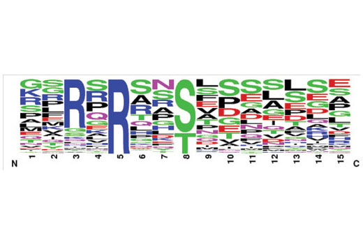
Cells are lysed in a urea-containing buffer, cellular proteins are digested by proteases, and the resulting peptides are purified by reversed-phase, solid-phase extraction. Peptides are then subjected to immunoaffinity purification using a PTMScan® antibody conjugated to protein A agarose beads. Unbound peptides are removed through washing, and the captured PTM-containing peptides are eluted with dilute acid. Reversed-phase purification is performed on microtips to desalt and separate peptides from antibody prior to concentrating the enriched peptides for LC-MS/MS analysis. CST recommends the use of PTMScan® IAP Buffer #9993 included in the kit. An alternate PTMScan® IAP Buffer Plus Detergent #9992 which may reduce nonspecific interactions is available separately. A detailed protocol and Limited Use License allowing the use of the patented PTMScan® method is included with the kit.

Storage

Antibody beads supplied in IAP buffer containing 50% glycerol. Store at -20°C. Do not aliquot the antibody.

Product Description

PTMScan® Technology employs a proprietary methodology from Cell Signaling Technology (CST) for peptide enrichment by immunoprecipitation using a specific bead-conjugated antibody in conjunction with liquid chromatography (LC) tandem mass spectrometry (MS/MS) for quantitative profiling of post-translational modification (PTM) sites in cellular proteins. These include phosphorylation (PhosphoScan®), ubiquitination (UbiScan®), acetylation (AcetylScan®), and methylation (MethylScan®), among others. PTMScan® Technology enables researchers to isolate, identify, and quantitate large numbers of post-translationally modified cellular peptides with a high degree of specificity and sensitivity, providing a global overview of PTMs in cell and tissue samples without preconceived biases about where these modified sites occur (1). For more information on PTMScan® Proteomics Services, please visit www.cellsignal.com.

Background

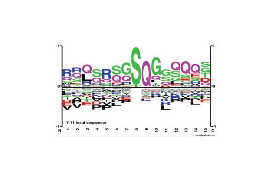
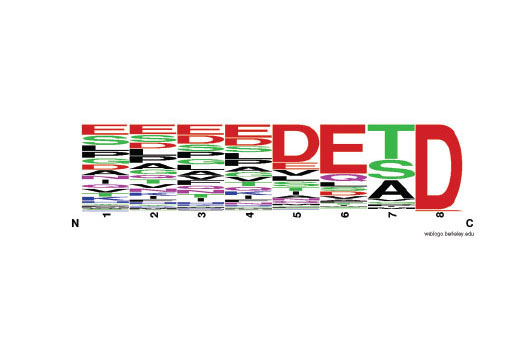
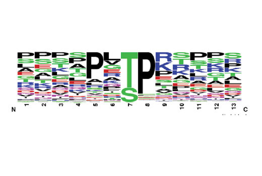
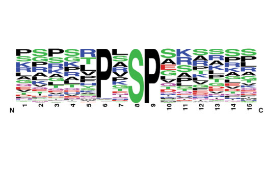
For more information on our PTMScan® Motif Antibody Kits, please visit www.cellsignal.com. Specific product background information can be found on the web page for each PTMScan® Motif Antibody kit component. Cleaved Caspase Substrate Motif background is described on the webpage for PTMScan® Cleaved Caspase Substrate Motif [DE(T/S/A)D] Kit #12810. Phospho-MAPK/CDK Substrate Motif background is described on the webpage for PTMScan® Phospho-MAPK/CDK Substrate Motif (PXS*P and S*PXK/R) Kit #4652. Phospho-MAPK Substrate Motif background is described on the webpage for PTMScan® Phospho-MAPK Substrate Motif [PXpTP] Kit #14990. Phospho-Akt Substrate Motif background is described on the webpage for PTMScan® Phospho-Akt Substrate Motif mAb 2 (RXRXXS*/T*) Kit #5563. Phospho-ATM/ATR Substrate Motif background is described on the webpage for PTMScan® Phospho-ATM/ATR Substrate Motif [pSQ] Kit #12267.

  1. Rush, J. et al. (2005) Nat Biotechnol 23, 94-101.

Species Reactivity

Species reactivity is determined by testing in at least one approved application (e.g., western blot).

Cross-Reactivity Key

H: human M: mouse R: rat Hm: hamster Mk: monkey Vir: virus Mi: mink C: chicken Dm: D. melanogaster X: Xenopus Z: zebrafish B: bovine Dg: dog Pg: pig Sc: S. cerevisiae Ce: C. elegans Hr: horse GP: Guinea Pig Rab: rabbit All: all species expected

Trademarks and Patents

Cell Signaling Technology is a trademark of Cell Signaling Technology, Inc.
AcetylScan is a registered trademark of Cell Signaling Technology, Inc.
MethylScan is a registered trademark of Cell Signaling Technology, Inc.
PhosphoScan is a registered trademark of Cell Signaling Technology, Inc.
PhosphoSitePlus is a registered trademark of Cell Signaling Technology, Inc.
PTMScan is a registered trademark of Cell Signaling Technology, Inc.
UbiScan is a registered trademark of Cell Signaling Technology, Inc.
All other trademarks are the property of their respective owners. Visit cellsignal.com/trademarks for more information.

限制使用

除非 CST 的合法授书代表以书面形式书行明确同意,否书以下条款适用于 CST、其关书方或分书商提供的书品。 任何书充本条款或与本条款不同的客书条款和条件,除非书 CST 的合法授书代表以书面形式书独接受, 否书均被拒书,并且无效。

专品专有“专供研究使用”的专专或专似的专专声明, 且未专得美国食品和专品管理局或其他外国或国内专管机专专专任何用途的批准、准专或专可。客专不得将任何专品用于任何专断或治专目的, 或以任何不符合专专声明的方式使用专品。CST 专售或专可的专品提供专作专最专用专的客专,且专用于研专用途。将专品用于专断、专防或治专目的, 或专专售(专独或作专专成)或其他商专目的而专专专品,均需要 CST 的专独专可。客专:(a) 不得专独或与其他材料专合向任何第三方出售、专可、 出借、捐专或以其他方式专专或提供任何专品,或使用专品制造任何商专专品,(b) 不得复制、修改、逆向工程、反专专、 反专专专品或以其他方式专专专专专品的基专专专或技专,或使用专品开专任何与 CST 的专品或服专专争的专品或服专, (c) 不得更改或专除专品上的任何商专、商品名称、徽专、专利或版专声明或专专,(d) 只能根据 CST 的专品专售条款和任何适用文档使用专品, (e) 专遵守客专与专品一起使用的任何第三方专品或服专的任何专可、服专条款或专似专专

Revision 1
#65666

PTMScan® Cell Growth and Death Antibody Sampler Kit

PTMScan® Cell Growth and Death Antibody Sampler Kit: Image 1 Expand Image
对来源于 Jurkat 细胞的 1111 条非重复胰蛋白酶肽进行 PhosphoScan® LC-MS/MS 实验可产生这种 PhosphoSitePlus® (PSP) 标识图,其中 Jurkat 细胞用 Calyculin A #9902 和过钒酸钠处理,并用 PTMScan® Phospho-ATM/ATR Substrate Motif [pSQ] Kit #12267 的免疫亲和微珠进行免疫沉淀。PSP 标识图反映了氨基酸在与人蛋白质组中磷酸化丝氨酸背景相对应的每个位点的相对普遍率。在这种背景下,x 轴上方所示的残基与预计频率相比更为富集。如需了解更多有关使用 PSP 进行基序分析的信息,请访问 www.phosphosite.org。
PTMScan® Cell Growth and Death Antibody Sampler Kit: Image 2 Expand Image
对含有羧基末端天冬氨酸的 1044 条非重复胰蛋白酶肽进行 PTMScan® LC-MS/MS 实验可产生该基序标识图,其中,胰蛋白酶肽来源于用 Staurosporine #9953(1 μM,3 小时)处理以诱导凋亡的人 HeLa 细胞。肽使用 PTMScan® Cleaved Caspase Substrate Motif [DE(T/S/A)D] Kit #12810 的免疫亲和微珠进行免疫沉淀。该标识表示各位置上氨基酸引导至天冬氨酸羧基端的相对频率。
PTMScan® Cell Growth and Death Antibody Sampler Kit: Image 3 Expand Image
使用来源于有丝分裂 HeLa 细胞的 754 条非重复胰蛋白酶肽进行 PhosphoScan® LC-MS/MS 实验可产生该基序标识图,其中 HeLa 细胞依次使用胸苷(2 mM,18 小时)和 Nocodazole #2190(60 nM,24 小时)处理,并用 PTMScan® Phospho-MAPK Substrate Motif [PXpTP] Kit #14990 的免疫亲和微珠进行免疫沉淀。该基序标识图显示了氨基酸在该数据组中某个指定位点的相对丰度。
PTMScan® Cell Growth and Death Antibody Sampler Kit: Image 4 Expand Image
The motif logo shows the amino acid distributions around the sites recognized using Immunoaffinity beads from PTMScan® Phospho-MAPK/CDK Substrate Motif (PXS*P and S*PXK/R) Kit #4652.
PTMScan® Cell Growth and Death Antibody Sampler Kit: Image 5 Expand Image
该基序标识图显示了使用 PTMScan® Phospho-Akt Substrate Motif mAb 2 (RXRXXS*/T*) Kit #5563 的免疫亲和微珠所识别位点周围的氨基酸分布。