Revision 1
#9765
Store at -20C
Vesicle Trafficking Antibody Sampler Kit
1 Kit
(8 x 20 microliters)
877-616-CELL (2355)
877-678-TECH (8324)
3 Trask Lane | Danvers | Massachusetts | 01923 | USA
For Research Use Only. Not for Use in Diagnostic Procedures.
| Product Includes | Product # | Quantity | Mol. Wt | Isotype/Source |
|---|---|---|---|---|
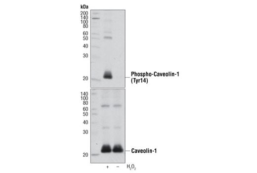
| Phospho-Caveolin-1 (Tyr14) Antibody | 3251 | 20 µl | 23, 25 kDa | Rabbit |
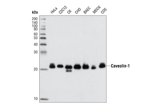
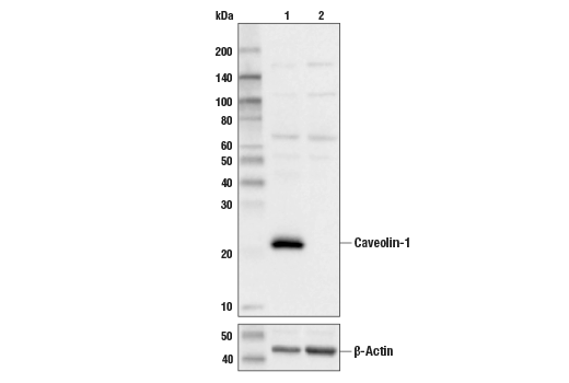
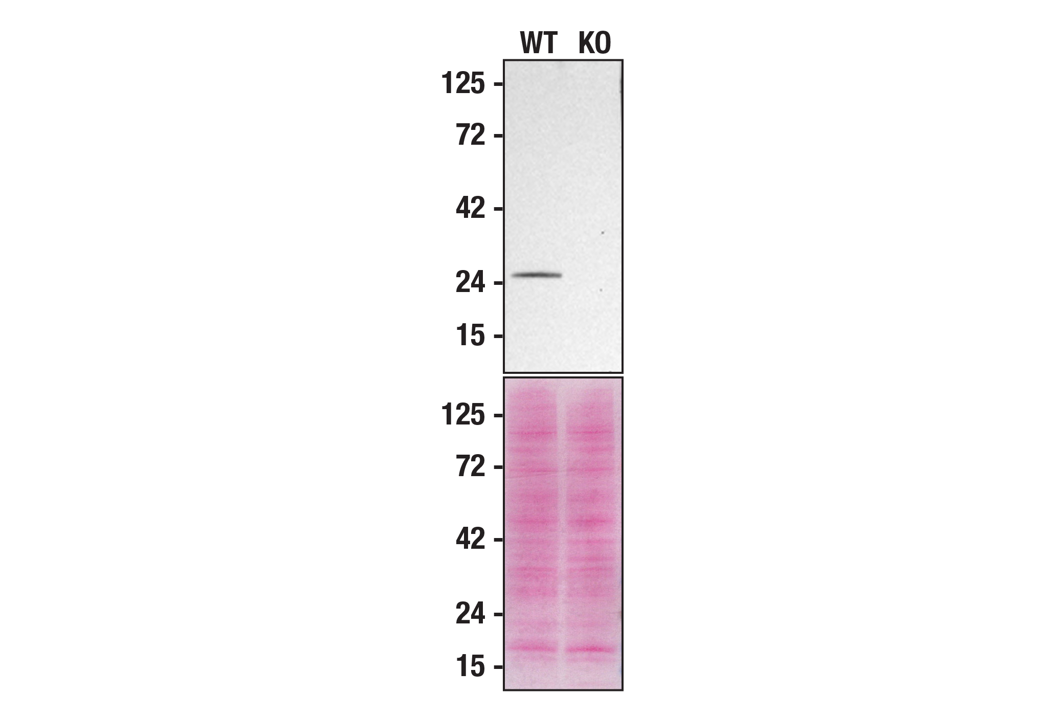
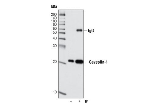
| Caveolin-1 (D46G3) Rabbit Monoclonal Antibody | 3267 | 20 µl | 21, 24 kDa | Rabbit IgG |
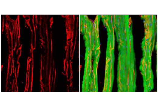
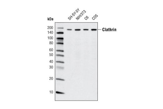
| Clathrin Heavy Chain (D3C6) Rabbit Monoclonal Antibody | 4796 | 20 µl | 190 kDa | Rabbit IgG |
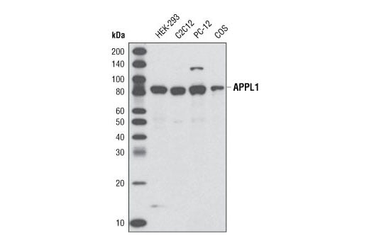
| APPL1 (D83H4) Rabbit Monoclonal Antibody | 3858 | 20 µl | 82 kDa | Rabbit IgG |
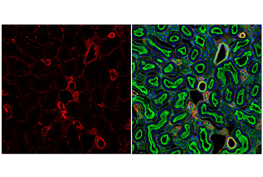
| EEA1 (C45B10) Rabbit Monoclonal Antibody | 3288 | 20 µl | 170 kDa | Rabbit IgG |
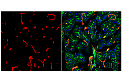
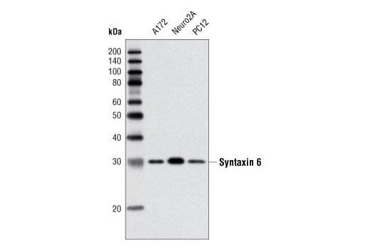
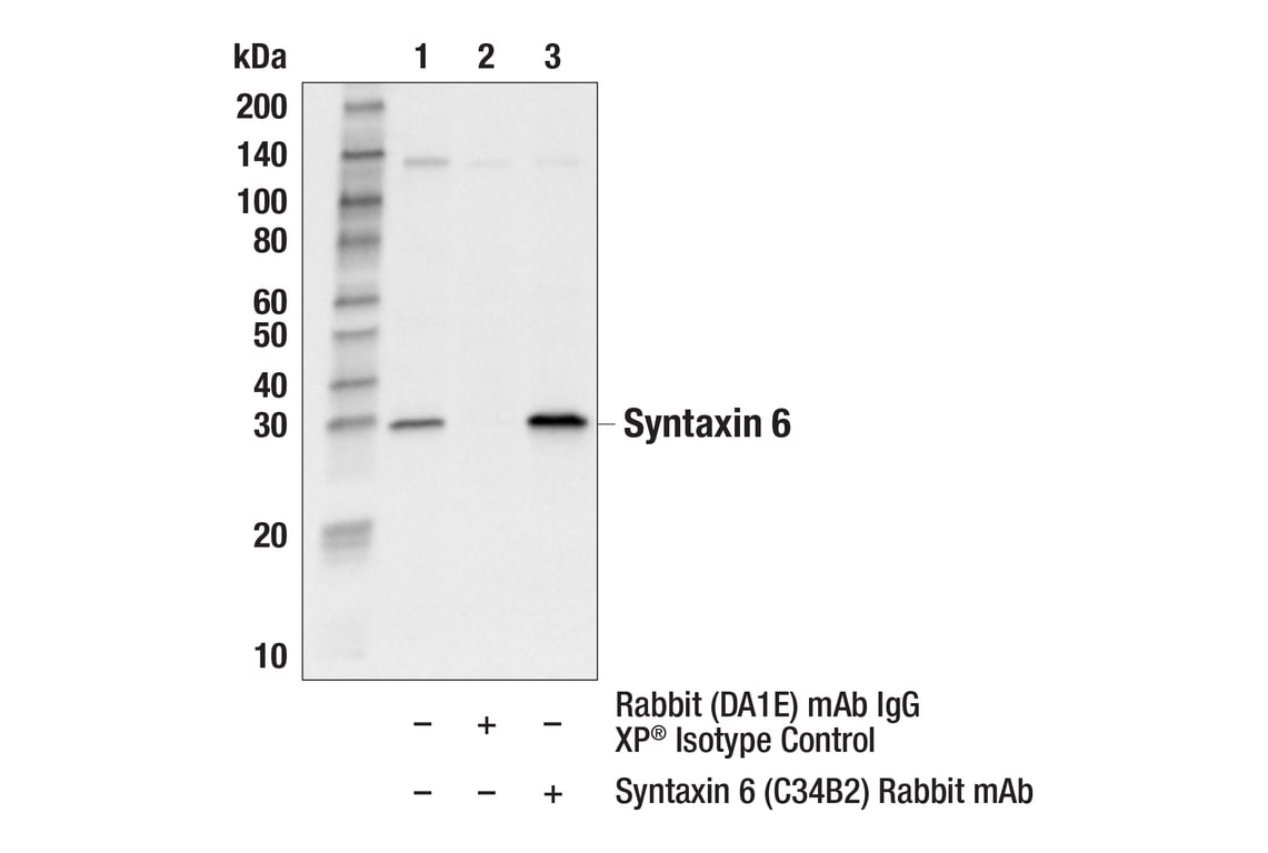
| Syntaxin 6 (C34B2) Rabbit Monoclonal Antibody | 2869 | 20 µl | 32 kDa | Rabbit IgG |
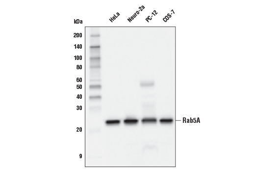
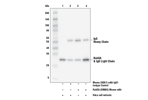
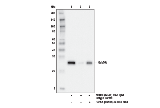
| Rab5A (E6N8S) Mouse Monoclonal Antibody | 46449 | 20 µl | 25 kDa | Mouse IgG1 |
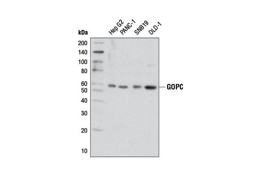
| GOPC (D10A12) Rabbit Monoclonal Antibody | 8576 | 20 µl | 59 kDa | Rabbit IgG |
| Anti-rabbit IgG, HRP-linked Antibody | 7074 | 100 µl | Goat | |
| Anti-mouse IgG, HRP-linked Antibody | 7076 | 100 µl | Horse |
Please visit cellsignal.com for individual component applications, species cross-reactivity, dilutions, protocols, and additional product information.
Description
Storage
Background
Clathrin-coated vesicles provide for the intracellular transport of proteins following endocytosis and during multiple vesicle trafficking pathways. Vesicles form at specialized areas of the cell membrane where clathrin and associated proteins form clathrin-coated pits. Invagination of these cell membrane-associated pits internalizes proteins and forms an intracellular clathrin-coated vesicle (5,6). Clathrin is the most abundant protein in these vesicles and is present as a basic assembly unit called a triskelion. Each clathrin triskelion is composed of three clathrin heavy chains and three clathrin light chains. Clathrin heavy chain proteins are composed of several functional domains that associate with other vesicle proteins (6).
The APPL1 multidomain adaptor protein is a BAR-domain protein family member that is involved in membrane trafficking within a number of signal transduction pathways (7).
EEA1 is an early endosomal marker and a Rab5 effector protein essential for early endosomal membrane fusion and trafficking (8,9). Syntaxin 6 is a ubiquitously expressed S25C family member of the SNARE proteins (10,11). Syntaxin 6 protein is localized to the trans-Golgi and within endosomes and regulates membrane trafficking by partnering with a variety of other SNARE proteins (12-14). It has two coiled-coil domains (CC1 and CC2) located in the amino-terminal region and a PDZ domain in the carboxy-terminal region (15). The CC2 domain and its adjacent linker region mediate the association of GOPC with the Golgi protein golgin-160 and the Q-SNARE protein syntaxin 6 (15,16). The PDZ domain of GOPC interacts with the carboxy terminus of target proteins to mediate target protein vesicular trafficking and surface expression (17-20).
Rab5 is a member of the Ras superfamily of small Rab GTPases. Rab5 is localized at the plasma membrane and early endosomes and functions as a key regulator of vesicular trafficking during early endocytosis (21).
Background References
- Smart, E.J. et al. (1999) Mol Cell Biol 19, 7289-304.
- Nomura, R. and Fujimoto, T. (1999) Mol Biol Cell 10, 975-86.
- Volonté, D. et al. (2001) J Biol Chem 276, 8094-103.
- Lee, H. et al. (2000) Mol Endocrinol 14, 1750-75.
- Rodriguez-Boulan, E. et al. (2005) Nat Rev Mol Cell Biol 6, 233-47.
- Mousavi, S.A. et al. (2004) Biochem J 377, 1-16.
- Habermann, B. (2004) EMBO Rep 5, 250-5.
- Mu, F.T. et al. (1995) J Biol Chem 270, 13503-11.
- Christoforidis, S. et al. (1999) Nature 397, 621-5.
- Bock, J.B. et al. (2001) Nature 409, 839-41.
- Bock, J.B. et al. (1996) J Biol Chem 271, 17961-5.
- Wendler, F. and Tooze, S. (2001) Traffic 2, 606-11.
- Bock, J.B. et al. (1997) Mol Biol Cell 8, 1261-71.
- Mallard, F. et al. (2002) J Cell Biol 156, 653-64.
- Charest, A. et al. (2001) J Biol Chem 276, 29456-65.
- Hicks, S.W. and Machamer, C.E. (2005) J Biol Chem 280, 28944-51.
- Cheng, J. et al. (2002) J Biol Chem 277, 3520-9.
- He, J. et al. (2004) J Biol Chem 279, 50190-6.
- Wente, W. et al. (2005) J Biol Chem 280, 32419-25.
- Ito, H. et al. (2006) Biochem J 397, 389-98.
- Zerial, M. and McBride, H. (2001) Nat Rev Mol Cell Biol 2, 107-17.
Trademarks and Patents
Cell Signaling Technology is a trademark of Cell Signaling Technology, Inc.
U.S. Patent No. 7,429,487, foreign equivalents, and child patents deriving therefrom.
All other trademarks are the property of their respective owners. Visit cellsignal.com/trademarks for more information.
Limited Uses
Except as otherwise expressly agreed in a writing signed by a legally authorized representative of CST, the following terms apply to Products provided by CST, its affiliates or its distributors. Any Customer's terms and conditions that are in addition to, or different from, those contained herein, unless separately accepted in writing by a legally authorized representative of CST, are rejected and are of no force or effect.
Products are labeled with For Research Use Only or a similar labeling statement and have not been approved, cleared, or licensed by the FDA or other regulatory foreign or domestic entity, for any purpose. Customer shall not use any Product for any diagnostic or therapeutic purpose, or otherwise in any manner that conflicts with its labeling statement. Products sold or licensed by CST are provided for Customer as the end-user and solely for research and development uses. Any use of Product for diagnostic, prophylactic or therapeutic purposes, or any purchase of Product for resale (alone or as a component) or other commercial purpose, requires a separate license from CST. Customer shall (a) not sell, license, loan, donate or otherwise transfer or make available any Product to any third party, whether alone or in combination with other materials, or use the Products to manufacture any commercial products, (b) not copy, modify, reverse engineer, decompile, disassemble or otherwise attempt to discover the underlying structure or technology of the Products, or use the Products for the purpose of developing any products or services that would compete with CST products or services, (c) not alter or remove from the Products any trademarks, trade names, logos, patent or copyright notices or markings, (d) use the Products solely in accordance with CST Product Terms of Sale and any applicable documentation, and (e) comply with any license, terms of service or similar agreement with respect to any third party products or services used by Customer in connection with the Products.
Revision 1
Revision 1

Revision 1
Revision 1


Revision 1

Revision 1
Revision 1
Revision 1
Revision 1
Revision 1
Revision 1
Revision 1