| Product Includes | Product # | Quantity | Mol. Wt | Isotype/Source |
|---|---|---|---|---|
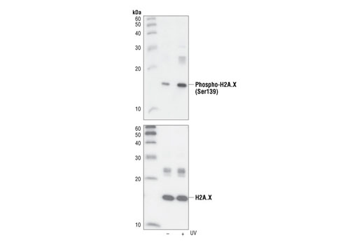
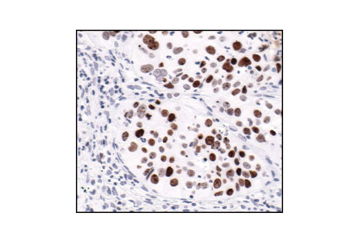
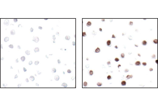
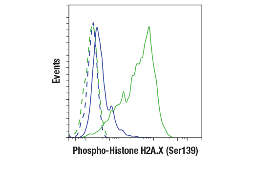
| Phospho-Histone H2A.X (Ser139) (20E3) Rabbit mAb | 9718 | 20 µl | 15 kDa | Rabbit IgG |
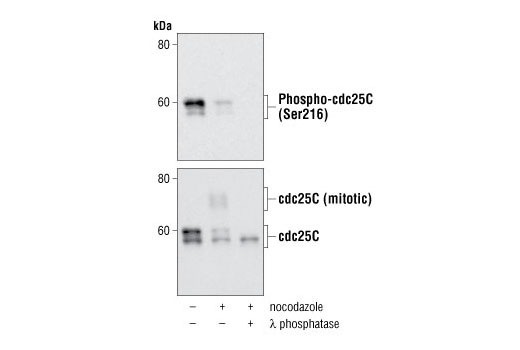
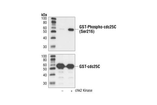
| Phospho-cdc25C (Ser216) (63F9) Rabbit mAb | 4901 | 20 µl | 60 kDa | Rabbit IgG |
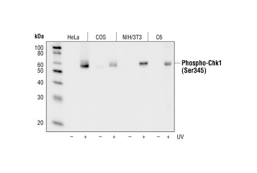
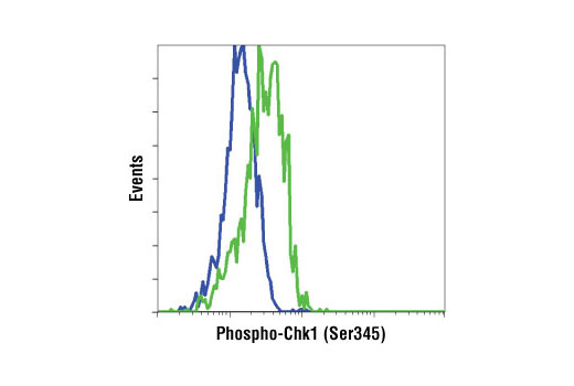
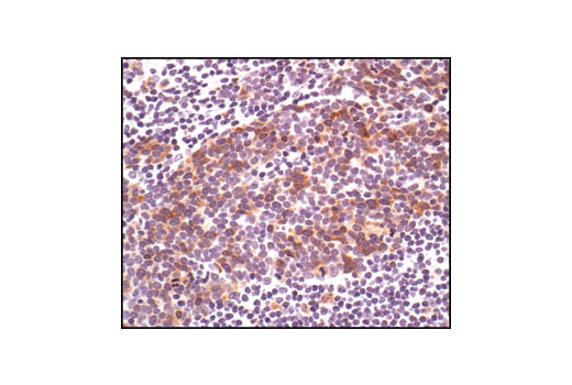
| Phospho-Chk1 (Ser345) (133D3) Rabbit mAb | 2348 | 20 µl | 56 kDa | Rabbit IgG |
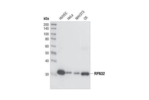
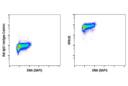
| RPA32/RPA2 (4E4) Rat mAb | 2208 | 20 µl | 32 kDa | Rat IgG1 |
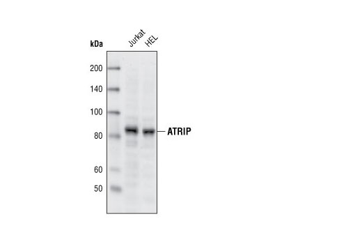
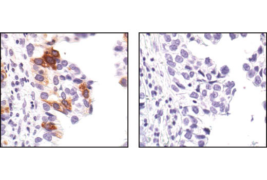
| ATRIP Antibody | 2737 | 20 µl | 82 kDa | Rabbit |
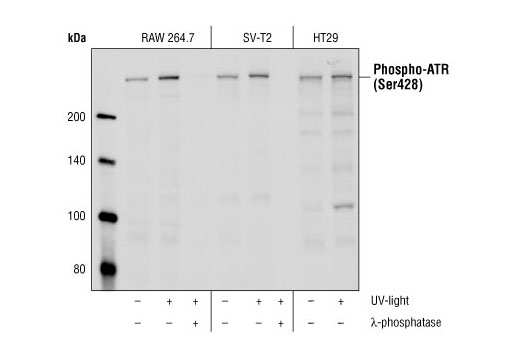
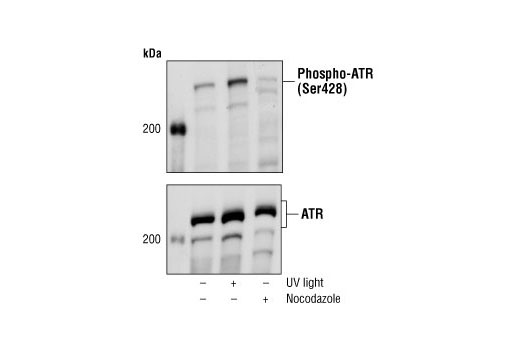
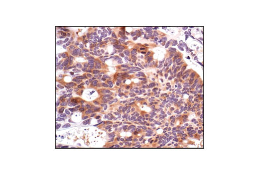
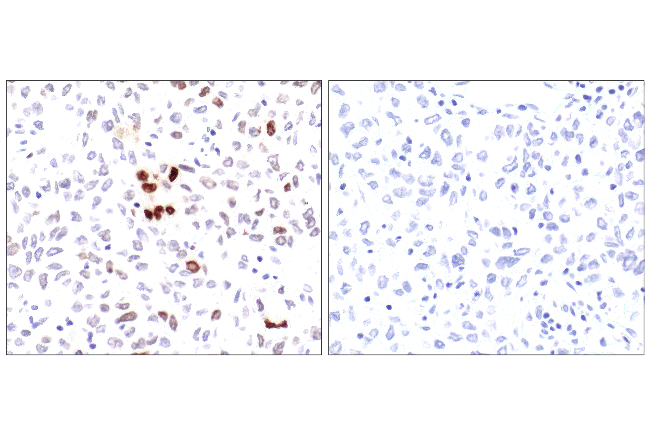
| Phospho-ATR (Ser428) Antibody | 2853 | 20 µl | 300 kDa | Rabbit |
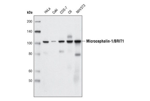
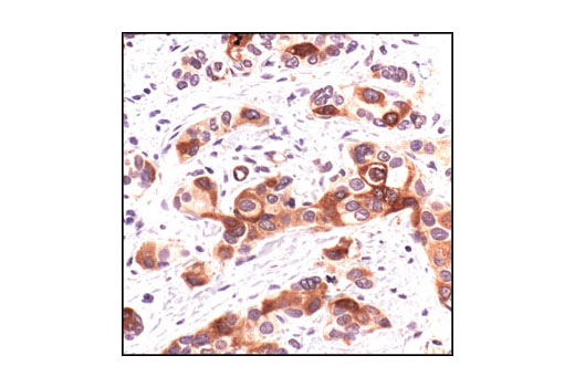
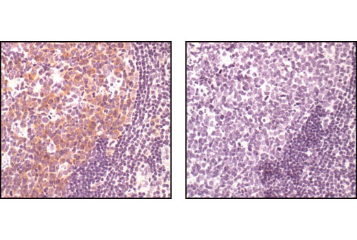
| Microcephalin-1/BRIT1 (D38G5) Rabbit mAb | 4120 | 20 µl | 100 kDa | Rabbit IgG |
| Anti-rabbit IgG, HRP-linked Antibody | 7074 | 100 µl | Goat | |
| Anti-rat IgG, HRP-linked Antibody | 7077 | 100 µl | Goat |
Please visit cellsignal.com for individual component applications, species cross-reactivity, dilutions, protocols, and additional product information.
Description
The UV Induced DNA Damage Response Antibody Sampler Kit offers an economical means of investigating proteins involved in the cellular response to UV-induced DNA damage. The kit contains enough primary and secondary antibody to perform two western blot experiments per primary.
Storage
Background
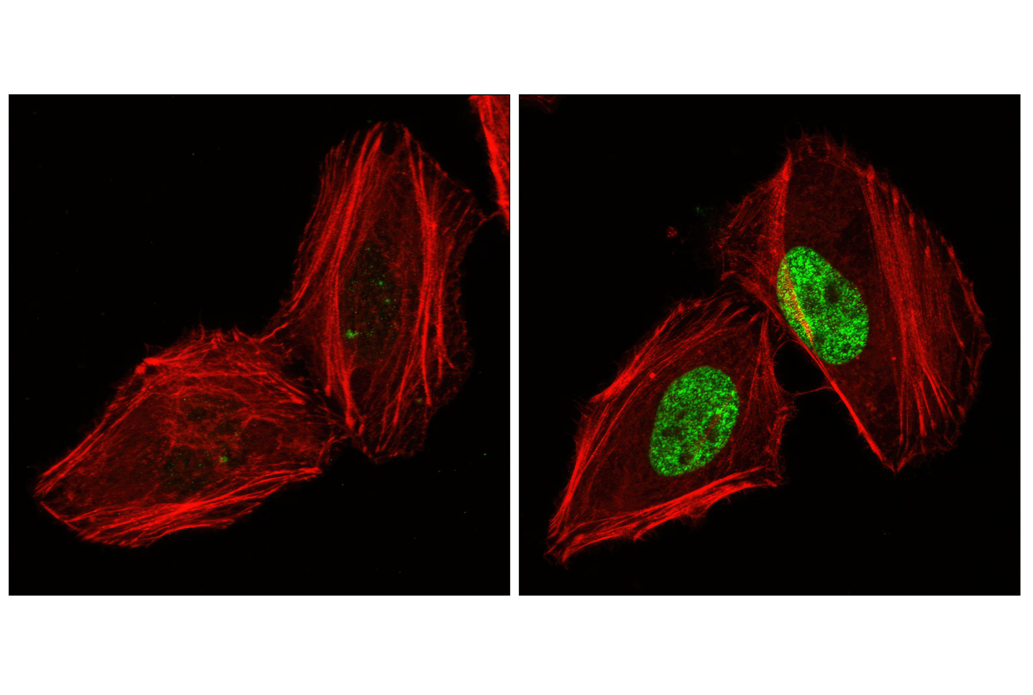
Exposure to ultraviolet radiation (UV) has a profound impact on human health and disease (1). Low level UV exposure induces the production of vitamin D and is a key regulator of calcium metabolism. Conversely, overexposure to UV is associated with an increased risk of cancer, immunosuppression, and many eye disorders, such as cataracts. Photons of UV light can directly damage DNA causing thymine dimers and other pyrimidine dimers between adjacent bases (2). Free radicals and reactive oxygen species induced by UV exposure also result in DNA lesions and have been linked to malignant melanoma (3). DNA damage from replicative stress and genotoxic agents like UV activate the ATR-mediated checkpoint pathway and stimulate DNA repair, cell cycle arrest, and apoptosis (4). ATR recruitment to sites of DNA damage and activation depends, at least in part, on interaction with the complex of single-stranded DNA, Replication Protein A (RPA), and direct binding to the ATR-associated adapter protein, ATRIP (5). In addition, the Rad17-RFC and Rad9-Rad1-Hus1 (9-1-1) protein complexes are independently recruited with TopBP1 to fully activate the checkpoint response (6,7). BRIT1 (MCPH1) is required for UV-induced formation of ATR, RPA, and p-Rad17 foci at sites of DNA damage (8-10) and may regulate the expression of several DNA damage response proteins (11). Once activated, ATR phosphorylates a number of mediators, including histone H2AX Ser139 and Chk1 kinase at Ser345. H2AX phosphorylation is a marker of DNA damage. Complete loss of H2AX results in reduced Chk1 activation and impaired survival of cells after UV exposure (12). Chk1 and Chk2 kinase activation is essential for checkpoint-mediated control of cell cycle progression (4). Checkpoint kinases stimulate cell cycle arrest by phosphorylation of a group of tyrosine phosphatases known as Cdc25A, Cdc25B, and Cdc25C (13 -15). Both Chk1 and Chk2 kinases phosphorylate Cdc25C at Ser216 in response to DNA damage and stimulate arrest (16-17).
- von Thaler, A.K. et al. (2010) Exp Dermatol 19, 81-8.
- Rastogi, R.P. et al. (2010) J Nucleic Acids 2010, 592980.
- Narayanan, D.L. et al. (2010) Int J Dermatol 49, 978-86.
- Zhou, B.B. and Elledge, S.J. (2000) Nature 408, 433-9.
- Zou, L. and Elledge, S.J. (2003) Science 300, 1542-8.
- Zou, L. et al. (2002) Genes Dev 16, 198-208.
- Mordes, D.A. and Cortez, D. (2008) Cell Cycle 7, 2809-12.
- Rai, R. et al. (2006) Cancer Cell 10, 145-57.
- Peng, G. et al. (2009) Nat Cell Biol 11, 865-72.
- Lin, S.Y. et al. (2010) Yonsei Med J 51, 295-301.
- Lin, S.Y. et al. (2005) Proc Natl Acad Sci U S A 102, 15105-9.
- Revet, I. et al. (2011) Proc Natl Acad Sci U S A 108, 8663-7.
- Mailand, N. et al. (2000) Science 288, 1425-9.
- Sanchez, Y. et al. (1997) Science 277, 1497-501.
- Matsuoka, S. et al. (1998) Science 282, 1893-7.
- Blasina, A. et al. (1999) Curr Biol 9, 1-10.
- Furnari, B. et al. (1999) Mol Biol Cell 10, 833-45.
Background References
Trademarks and Patents
限制使用
除非 CST 的合法授书代表以书面形式书行明确同意,否书以下条款适用于 CST、其关书方或分书商提供的书品。 任何书充本条款或与本条款不同的客书条款和条件,除非书 CST 的合法授书代表以书面形式书独接受, 否书均被拒书,并且无效。
专品专有“专供研究使用”的专专或专似的专专声明, 且未专得美国食品和专品管理局或其他外国或国内专管机专专专任何用途的批准、准专或专可。客专不得将任何专品用于任何专断或治专目的, 或以任何不符合专专声明的方式使用专品。CST 专售或专可的专品提供专作专最专用专的客专,且专用于研专用途。将专品用于专断、专防或治专目的, 或专专售(专独或作专专成)或其他商专目的而专专专品,均需要 CST 的专独专可。客专:(a) 不得专独或与其他材料专合向任何第三方出售、专可、 出借、捐专或以其他方式专专或提供任何专品,或使用专品制造任何商专专品,(b) 不得复制、修改、逆向工程、反专专、 反专专专品或以其他方式专专专专专品的基专专专或技专,或使用专品开专任何与 CST 的专品或服专专争的专品或服专, (c) 不得更改或专除专品上的任何商专、商品名称、徽专、专利或版专声明或专专,(d) 只能根据 CST 的专品专售条款和任何适用文档使用专品, (e) 专遵守客专与专品一起使用的任何第三方专品或服专的任何专可、服专条款或专似专专