| Product Includes | Product # | Quantity | Mol. Wt | Isotype/Source |
|---|---|---|---|---|
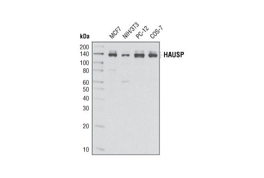
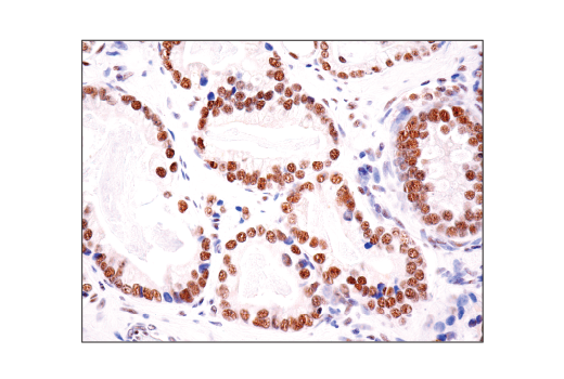
| HAUSP (D17C6) XP® Rabbit mAb | 4833 | 20 µl | 135, 140 kDa | Rabbit IgG |
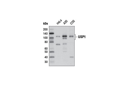
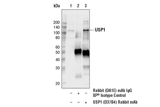
| USP1 (D37B4) Rabbit mAb | 8033 | 20 µl | 110 kDa | Rabbit IgG |
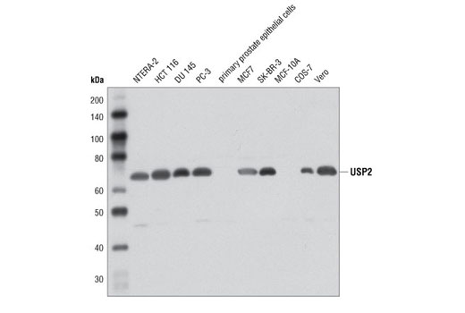
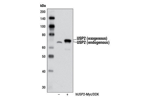
| USP2 Antibody | 8036 | 20 µl | 68 kDa | Rabbit |
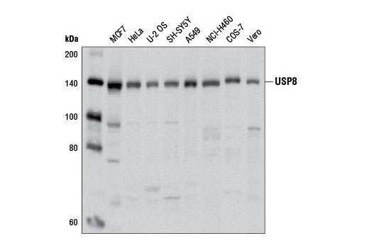
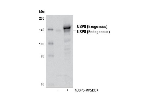
| USP8 Antibody | 8728 | 20 µl | 130 kDa | Rabbit |
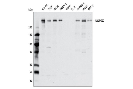
| USP9X Antibody | 5751 | 20 µl | 270 kDa | Rabbit |
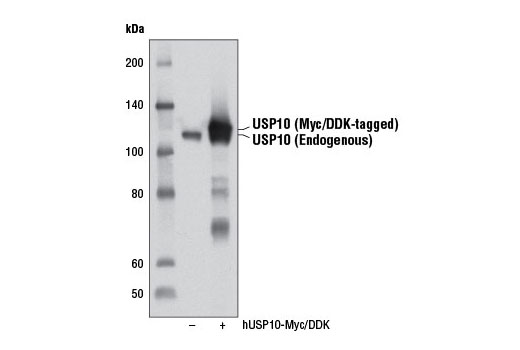
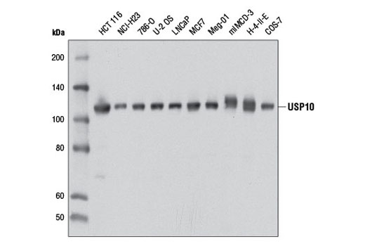
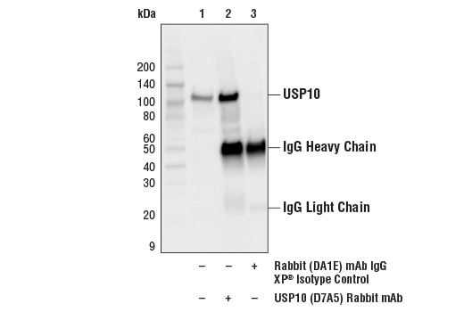
| USP10 (D7A5) Rabbit mAb | 8501 | 20 µl | 110 kDa | Rabbit IgG |
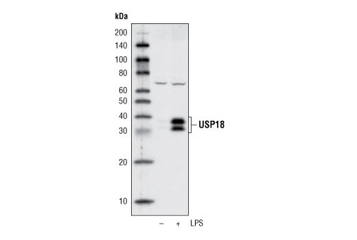
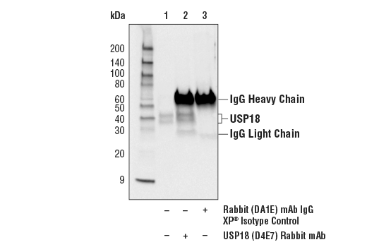
| USP18 (D4E7) Rabbit mAb | 4813 | 20 µl | 34, 39 kDa | Rabbit IgG |
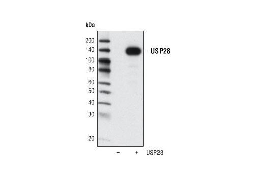
| USP28 Antibody | 4217 | 20 µl | 135 kDa | Rabbit |
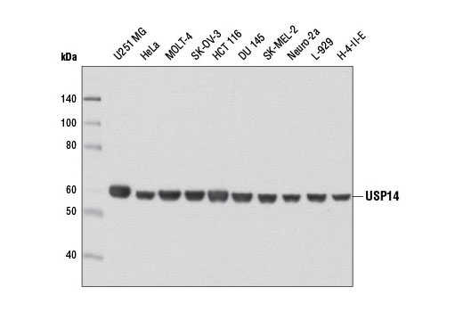
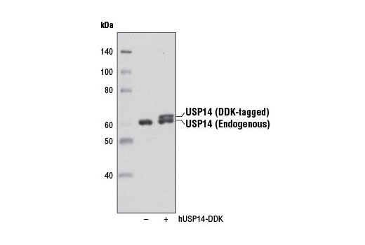
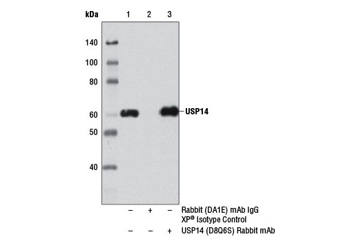
| USP14 (D8Q6S) Rabbit mAb | 11931 | 20 µl | 60 kDa | Rabbit IgG |
| Anti-rabbit IgG, HRP-linked Antibody | 7074 | 100 µl | Goat |
Please visit cellsignal.com for individual component applications, species cross-reactivity, dilutions, protocols, and additional product information.
Description
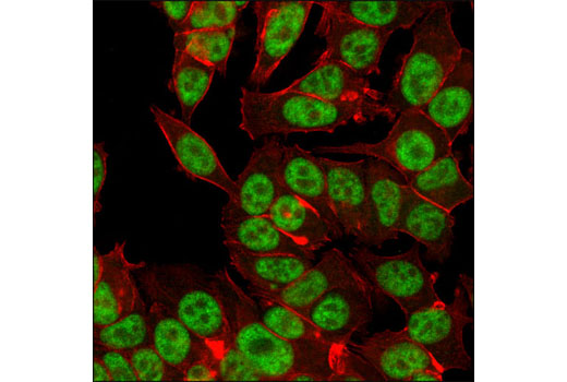
The USP Antibody Sampler Kit provides an economical means of detecting members of the ubiquitin-specific protease (USP) family. The kit includes enough primary antibody to perform two western blot experiments per primary antibody.
Storage
Background
Ubiquitinating enzymes (UBEs) catalyze protein ubiquitination, a reversible process countered by deubiquitinating enzyme (DUB) action (1,2). The ubiquitin-specific protease (USP) subfamily is one of five distinct groups of DUB enzymes. Ubiquitin-specific-processing protease 1 (USP1) is regulated in a cell cycle dependent manner by both transcriptional and ubiquitin-proteasomal mechanisms (3). Nuclear USP1 localizes to chromatin where it deubiquitinates monoubiquitinated FANCD2 and plays an important role in DNA damage repair and Chk1 protein stability (3,4). Ubiquitin-specific-processing protease 2 (USP2) contains C19 peptidase activity and is involved in ubiquitin recycling and disassembly of polymeric ubiquitin and ubiquitin-like protein complexes (5). USP2 is a putative oncoprotein that is highly over expressed in prostate cancer and drives tumor growth by binding and stabilizing fatty acid synthase through deubiquitination (6,7).
Herpesvirus-associated ubiquitin-specific protease (HAUSP, USP7) binds and deubiquitinates transcription factor p53 and regulator protein Mdm2, stabilizing both proteins (8,9). HAUSP modifies other ubiquitinated proteins, including FoxO family forkhead transcription factors and the mitotic stress checkpoint protein CHFR (10,11). Ubiquitin-specific protease 8 (USP8, UBPy) is a cysteine protease and growth-regulated enzyme that is essential for cell proliferation and survival (12,13). The catalytic domain of USP9X possesses cysteine peptidase activity that cleaves ubiquitin and polyubiquitin conjugates. USP9X may help stabilize adherens and tight junction molecules during epithelial cell polarization (14,15). USP10 is regulated at the posttranslational level through protein-protein interactions and phosphorylation. Interaction of USP10 with the Ras-GAP SH3 domain binding protein (G3BP) inhibits the ability of USP10 to catalyze ubiquitin chain disassembly (16). ATM-mediated phosphorylation of USP10 at Thr42 and Ser337 promotes USP10 stabilization and relocation from the cytoplasm to the nucleus, where it functions in p53 deubiquitination, stabilization, and activation in response to genotoxic stress (17).
USP14 is recruited to the proteasome through association with the PSMD2 (S2/hRPN1) subunit of the 19S regulatory particle, where it may antagonize substrate degradation (18,19). USP14 trims ubiquitin residues from distal polyubiquitin chain ends, decreasing chain affinity for proteasome ubiquitin receptors and allowing for enhanced substrate stability (20,21). USP18 (UBP43) catalyzes the removal of the interferon-regulated, ubiquitin-like protein ISG15 from conjugated proteins (22). Removal of ISG15 from target proteins maintains a critical balance of cellular ISG15-conjugated proteins, which is important for normal development and brain function (23,24). USP28 can bind, deubiquitinate and stabilize several DNA-damage pathway proteins, including p53BP1 and Chk2 (25). USP28 plays an important role in Myc-related signaling as it catalyzes Myc deubiquitination and promotes Myc stabilization, which contributes to tumor-cell growth (26).
- Nijman, S.M. et al. (2005) Cell 123, 773-86.
- Nalepa, G. et al. (2006) Nat Rev Drug Discov 5, 596-613.
- Nijman, S.M. et al. (2005) Mol Cell 17, 331-9.
- Guervilly, J.H. et al. (2011) Hum Mol Genet 20, 2171-81.
- Wilkinson, K.D. (1997) FASEB J 11, 1245-56.
- Graner, E. et al. (2004) Cancer Cell 5, 253-61.
- Priolo, C. et al. (2006) Cancer Res 66, 8625-32.
- Li, M. et al. (2002) Nature 416, 648-53.
- Brooks, C.L. et al. (2007) Oncogene 26, 7262-6.
- van der Horst, A. et al. (2006) Nat Cell Biol 8, 1064-73.
- Oh, Y.M. et al. (2007) Biochem Biophys Res Commun 357, 615-9.
- Naviglio, S. et al. (1998) EMBO J 17, 3241-50.
- Niendorf, S. et al. (2007) Mol Cell Biol 27, 5029-39.
- Murray, R.Z. et al. (2004) Mol Biol Cell 15, 1591-9.
- Théard, D. et al. (2010) EMBO J 29, 1499-509.
- Soncini, C. et al. (2001) Oncogene 20, 3869-79.
- Yuan, J. et al. (2010) Cell 140, 384-96.
- Lee, B.H. et al. (2010) Nature 467, 179-84.
- Koulich, E. et al. (2008) Mol Biol Cell 19, 1072-82.
- Hanna, J. et al. (2006) Cell 127, 99-111.
- Thrower, J.S. et al. (2000) EMBO J 19, 94-102.
- Malakhov, M.P. et al. (2002) J Biol Chem 277, 9976-81.
- Rempel, L.A. et al. (2007) Reprod Biol Endocrinol 5, 13.
- Ritchie, K.J. et al. (2002) Genes Dev 16, 2207-12.
- Zhang, D. et al. (2006) Cell 126, 529-42.
- Popov, N. et al. (2007) Nat Cell Biol 9, 765-74.
Background References
Trademarks and Patents
限制使用
除非 CST 的合法授书代表以书面形式书行明确同意,否书以下条款适用于 CST、其关书方或分书商提供的书品。 任何书充本条款或与本条款不同的客书条款和条件,除非书 CST 的合法授书代表以书面形式书独接受, 否书均被拒书,并且无效。
专品专有“专供研究使用”的专专或专似的专专声明, 且未专得美国食品和专品管理局或其他外国或国内专管机专专专任何用途的批准、准专或专可。客专不得将任何专品用于任何专断或治专目的, 或以任何不符合专专声明的方式使用专品。CST 专售或专可的专品提供专作专最专用专的客专,且专用于研专用途。将专品用于专断、专防或治专目的, 或专专售(专独或作专专成)或其他商专目的而专专专品,均需要 CST 的专独专可。客专:(a) 不得专独或与其他材料专合向任何第三方出售、专可、 出借、捐专或以其他方式专专或提供任何专品,或使用专品制造任何商专专品,(b) 不得复制、修改、逆向工程、反专专、 反专专专品或以其他方式专专专专专品的基专专专或技专,或使用专品开专任何与 CST 的专品或服专专争的专品或服专, (c) 不得更改或专除专品上的任何商专、商品名称、徽专、专利或版专声明或专专,(d) 只能根据 CST 的专品专售条款和任何适用文档使用专品, (e) 专遵守客专与专品一起使用的任何第三方专品或服专的任何专可、服专条款或专似专专