Revision 4
#85493
Store at -20C
ULK1 Substrate Antibody Sampler Kit
1 Kit
(9 x 20 microliters)
877-616-CELL (2355)
877-678-TECH (8324)
3 Trask Lane | Danvers | Massachusetts | 01923 | USA
For Research Use Only. Not for Use in Diagnostic Procedures.
| Product Includes | Product # | Quantity | Mol. Wt | Isotype/Source |
|---|---|---|---|---|
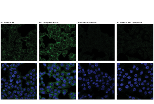
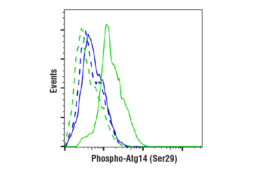
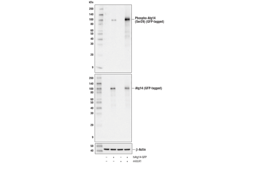
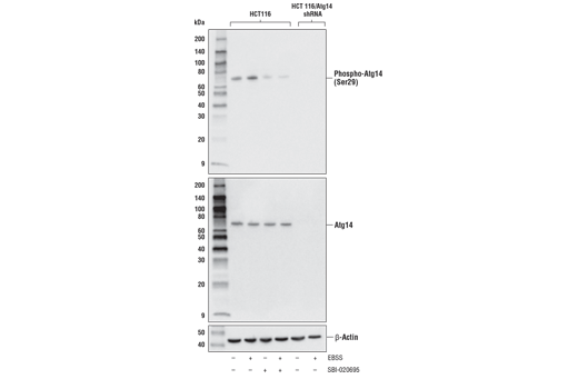
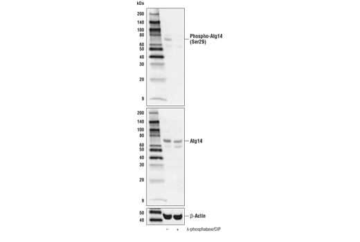
| Phospho-Atg14 (Ser29) (D4B8M) Rabbit mAb | 92340 | 20 µl | 65 kDa | Rabbit IgG |
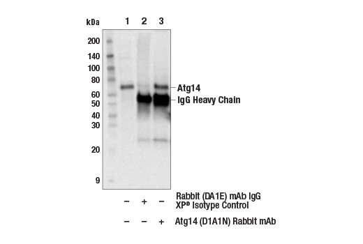
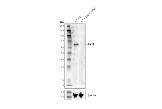
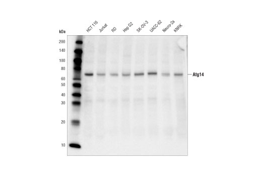
| Atg14 (D1A1N) Rabbit mAb | 96752 | 20 µl | 65 kDa | Rabbit IgG |
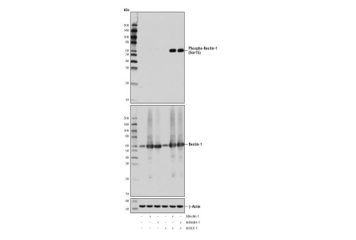
| Phospho-Beclin-1 (Ser15) (D4B7R) Rabbit mAb | 84966 | 20 µl | 60 kDa | Rabbit IgG |
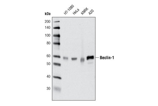
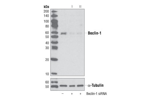
| Beclin-1 (D40C5) Rabbit mAb | 3495 | 20 µl | 60 kDa | Rabbit IgG |
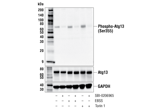
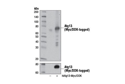
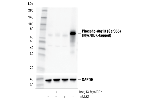
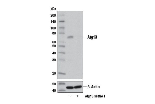
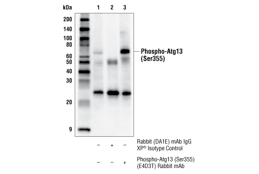
| Phospho-Atg13 (Ser355) (E4D3T) Rabbit mAb | 46329 | 20 µl | 72 kDa | Rabbit IgG |
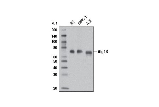
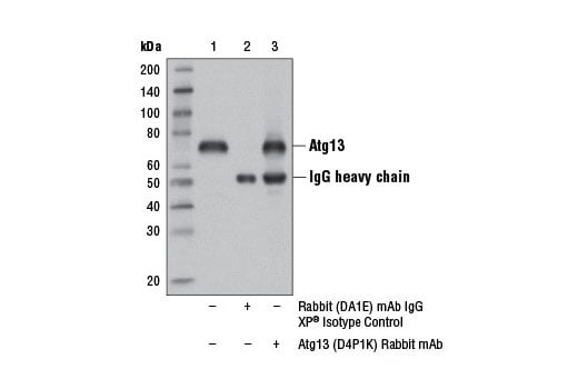
| Atg13 (D4P1K) Rabbit mAb | 13273 | 20 µl | 72 kDa | Rabbit IgG |
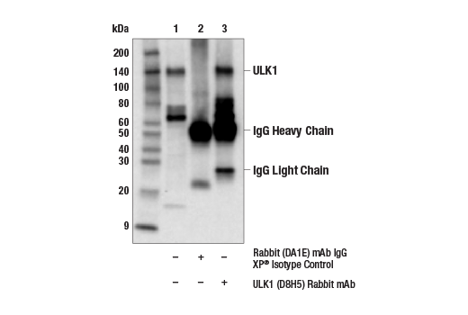
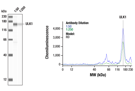
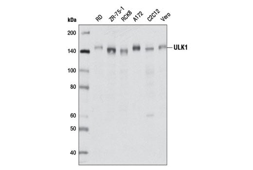
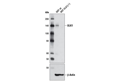
| ULK1 (D8H5) Rabbit mAb | 8054 | 20 µl | 150 kDa | Rabbit IgG |
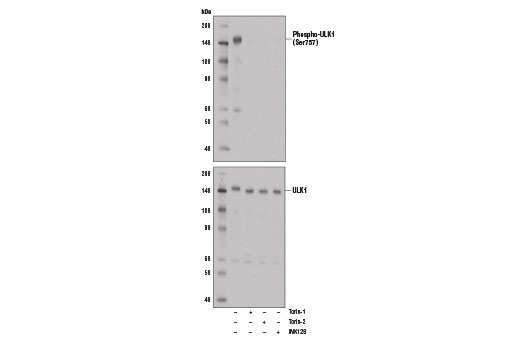
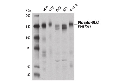
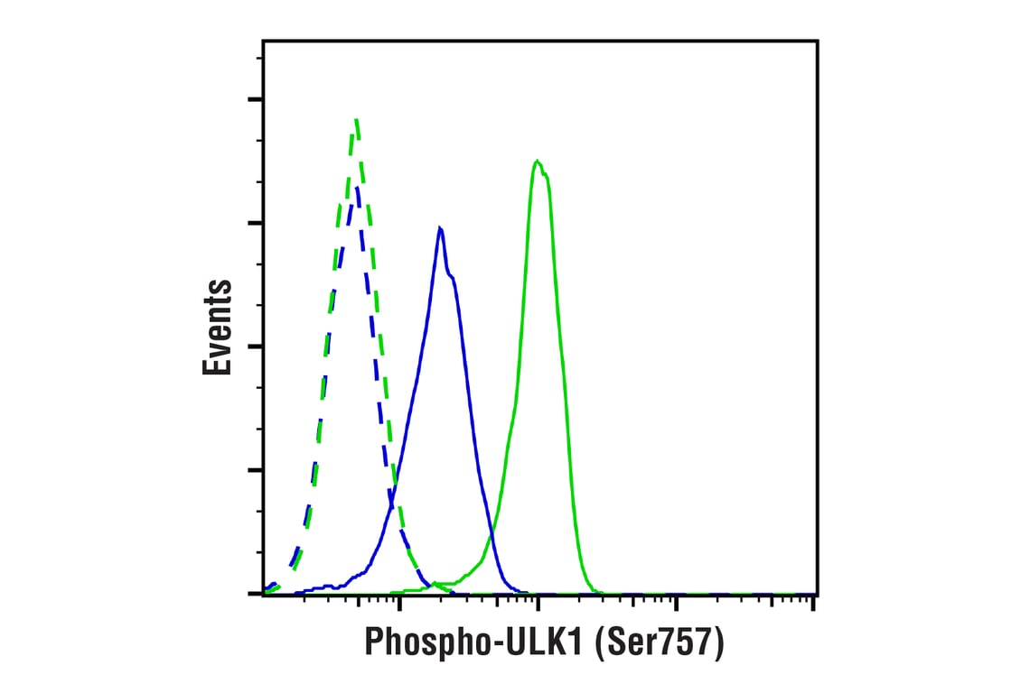
| Phospho-ULK1 (Ser757) (D7O6U) Rabbit mAb | 14202 | 20 µl | 140-150 kDa | Rabbit IgG |
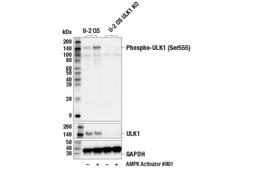
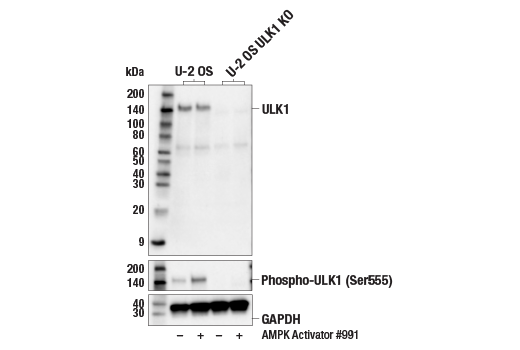
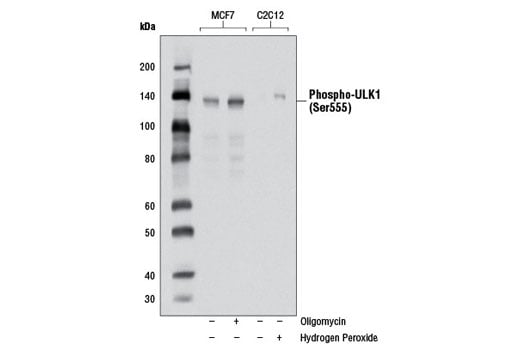
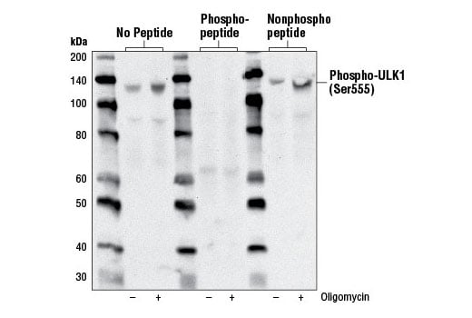
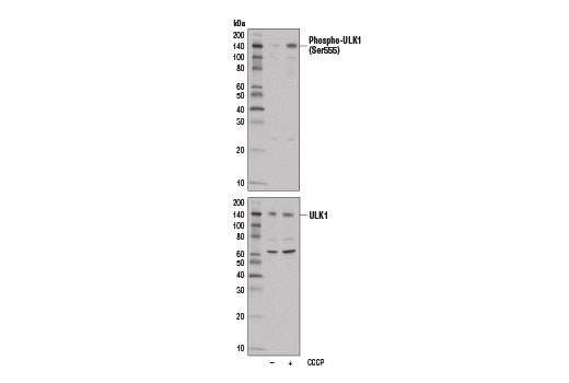
| Phospho-ULK1 (Ser555) (D1H4) Rabbit mAb | 5869 | 20 µl | 140-150 kDa | Rabbit IgG |
| Anti-rabbit IgG, HRP-linked Antibody | 7074 | 100 µl | Goat |
Please visit cellsignal.com for individual component applications, species cross-reactivity, dilutions, protocols, and additional product information.
Description
Storage
Background
AMPK, activated during low nutrient conditions, directly phosphorylates ULK1 at multiple sites, including Ser317, Ser555, and Ser777 (17,18). Conversely, mTOR, which is a regulator of cell growth and is an inhibitor of autophagy, phosphorylates ULK1 at Ser757 and disrupts the interaction between ULK1 and AMPK (17). ULK1 has been shown to phoshorylate several targets in the autophagy pathway, including Ser29 of Atg14, Ser15 of Beclin-1, and Ser355 of Atg13 (19-22).
Background References
- Ogura, K. et al. (1994) Genes Dev 8, 2389-400.
- Kuroyanagi, H. et al. (1998) Genomics 51, 76-85.
- Yan, J. et al. (1998) Biochem Biophys Res Commun 246, 222-7.
- Yan, J. et al. (1999) Oncogene 18, 5850-9.
- Zhou, X. et al. (2007) Proc Natl Acad Sci USA 104, 5842-7.
- Tomoda, T. et al. (2004) Genes Dev 18, 541-58.
- Matsuura, A. et al. (1997) Gene 192, 245-50.
- Chan, E.Y. et al. (2007) J Biol Chem 282, 25464-74.
- Reggiori, F. and Klionsky, D.J. (2002) Eukaryot Cell 1, 11-21.
- Codogno, P. and Meijer, A.J. (2005) Cell Death Differ 12 Suppl 2, 1509-18.
- Stephan, J.S. and Herman, P.K. (2006) Autophagy 2, 146-8.
- Okazaki, N. et al. (2000) Brain Res Mol Brain Res 85, 1-12.
- Young, A.R. et al. (2006) J Cell Sci 119, 3888-900.
- Kamada, Y. et al. (2000) J Cell Biol 150, 1507-13.
- Lee, S.B. et al. (2007) EMBO Rep 8, 360-5.
- Hara, T. et al. (2008) J Cell Biol 181, 497-510.
- Kim, J. et al. (2011) Nat Cell Biol 13, 132-41.
- Egan, D.F. et al. (2011) Science 331, 456-61.
- Park, J.M. et al. (2016) Autophagy 12, 547-64.
- Russell, R.C. et al. (2013) Nat Cell Biol 15, 741-50.
- Joo, J.H. et al. (2011) Mol Cell 43, 572-85.
- Egan, D.F. et al. (2015) Mol Cell 59, 285-97.
Trademarks and Patents
Cell Signaling Technology is a trademark of Cell Signaling Technology, Inc.
All other trademarks are the property of their respective owners. Visit cellsignal.com/trademarks for more information.
限制使用
除非 CST 的合法授书代表以书面形式书行明确同意,否书以下条款适用于 CST、其关书方或分书商提供的书品。 任何书充本条款或与本条款不同的客书条款和条件,除非书 CST 的合法授书代表以书面形式书独接受, 否书均被拒书,并且无效。
专品专有“专供研究使用”的专专或专似的专专声明, 且未专得美国食品和专品管理局或其他外国或国内专管机专专专任何用途的批准、准专或专可。客专不得将任何专品用于任何专断或治专目的, 或以任何不符合专专声明的方式使用专品。CST 专售或专可的专品提供专作专最专用专的客专,且专用于研专用途。将专品用于专断、专防或治专目的, 或专专售(专独或作专专成)或其他商专目的而专专专品,均需要 CST 的专独专可。客专:(a) 不得专独或与其他材料专合向任何第三方出售、专可、 出借、捐专或以其他方式专专或提供任何专品,或使用专品制造任何商专专品,(b) 不得复制、修改、逆向工程、反专专、 反专专专品或以其他方式专专专专专品的基专专专或技专,或使用专品开专任何与 CST 的专品或服专专争的专品或服专, (c) 不得更改或专除专品上的任何商专、商品名称、徽专、专利或版专声明或专专,(d) 只能根据 CST 的专品专售条款和任何适用文档使用专品 , (e) 专遵守客专与专品一起使用的任何第三方专品或服专的任何专可、服专条款或专似专专
Revision 4
Revision 4

Revision 4
Revision 4
Revision 4
Revision 4
Revision 4
Revision 4
Revision 4
Revision 4

Revision 4