#O75385
8408
| Product Includes | Quantity | Reactivity | MW(kDa) | Isotype | |
|---|---|---|---|---|---|
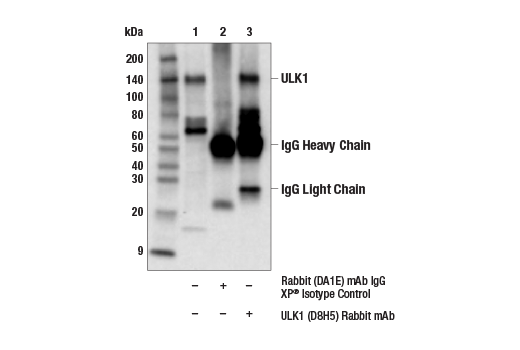
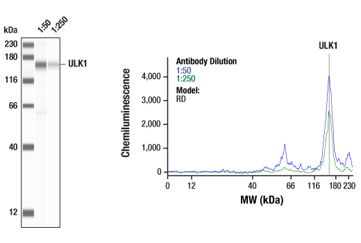
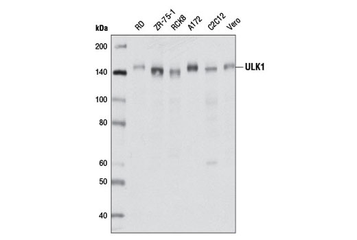
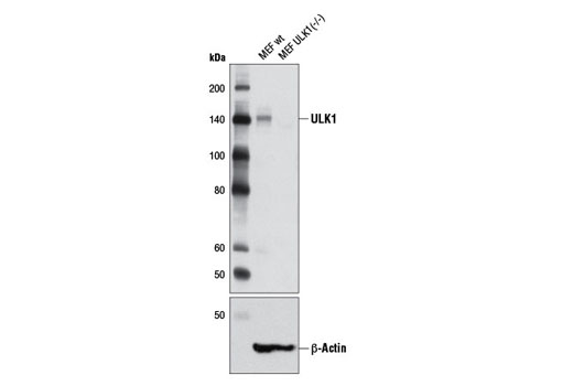
| ULK1 (D8H5) Rabbit mAb 8054 | 100 µl | H M R Mk | 150 | Rabbit IgG | |
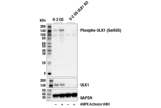
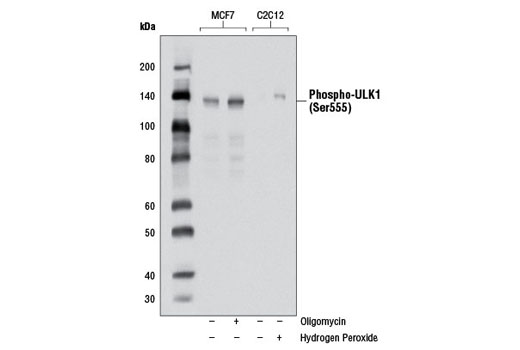
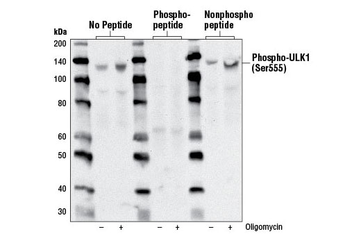
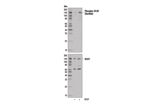
| Phospho-ULK1 (Ser555) (D1H4) Rabbit mAb 5869 | 100 µl | H M | 140-150 | Rabbit IgG |
Please visit cellsignal.com for individual component applications, species cross-reactivity, dilutions, protocols, and additional product information.
Description
PhosphoPlus® Duets from Cell Signaling Technology (CST) provide a means to assess protein activation status. Each Duet contains an activation-state and total protein antibody to your target of interest. These antibodies have been selected from CST's product offering based upon superior performance in specified applications.
Storage
Background
Two related serine/threonine kinases, UNC-51-like kinase 1 and 2 (ULK1, ULK2), were discovered as mammalian homologs of the C. elegans gene unc-51 in which mutants exhibited abnormal axonal extension and growth (1-4). Both proteins are widely expressed and contain an amino-terminal kinase domain followed by a central proline/serine rich domain and a highly conserved carboxy-terminal domain. The roles of ULK1 and ULK2 in axon growth have been linked to studies showing that the kinases are localized to neuronal growth cones and are involved in endocytosis of critical growth factors, such as NGF (5). Yeast two-hybrid studies found ULK1/2 associated with modulators of the endocytic pathway, SynGAP, and syntenin (6). Structural similarity of ULK1/2 has also been recognized with the yeast autophagy protein Atg1/Apg1 (7). Knockdown experiments using siRNA demonstrated that ULK1 is essential for autophagy (8), a catabolic process for the degradation of bulk cytoplasmic contents (9,10). It appears that Atg1/ULK1 can act as a convergence point for multiple signals that control autophagy (11), and can bind to several autophagy-related (Atg) proteins, regulating phosphorylation states and protein trafficking (12-16).
Phosphorylation of ULK1 by AMPK at Ser555 is critical for starvation-induced autophagy, cell survival under conditions of low nutrients and energy, and mitochondrial homeostasis (17).
- Ogura, K. et al. (1994) Genes Dev 8, 2389-400.
- Kuroyanagi, H. et al. (1998) Genomics 51, 76-85.
- Yan, J. et al. (1998) Biochem Biophys Res Commun 246, 222-7.
- Yan, J. et al. (1999) Oncogene 18, 5850-9.
- Zhou, X. et al. (2007) Proc Natl Acad Sci USA 104, 5842-7.
- Tomoda, T. et al. (2004) Genes Dev 18, 541-58.
- Matsuura, A. et al. (1997) Gene 192, 245-50.
- Chan, E.Y. et al. (2007) J Biol Chem 282, 25464-74.
- Reggiori, F. and Klionsky, D.J. (2002) Eukaryot Cell 1, 11-21.
- Codogno, P. and Meijer, A.J. (2005) Cell Death Differ 12 Suppl 2, 1509-18.
- Stephan, J.S. and Herman, P.K. (2006) Autophagy 2, 146-8.
- Okazaki, N. et al. (2000) Brain Res Mol Brain Res 85, 1-12.
- Young, A.R. et al. (2006) J Cell Sci 119, 3888-900.
- Kamada, Y. et al. (2000) J Cell Biol 150, 1507-13.
- Lee, S.B. et al. (2007) EMBO Rep 8, 360-5.
- Hara, T. et al. (2008) J Cell Biol 181, 497-510.
- Egan, D.F. et al. (2011) Science 331, 456-61.
Background References
Trademarks and Patents
限制使用
除非 CST 的合法授书代表以书面形式书行明确同意,否书以下条款适用于 CST、其关书方或分书商提供的书品。 任何书充本条款或与本条款不同的客书条款和条件,除非书 CST 的合法授书代表以书面形式书独接受, 否书均被拒书,并且无效。
专品专有“专供研究使用”的专专或专似的专专声明, 且未专得美国食品和专品管理局或其他外国或国内专管机专专专任何用途的批准、准专或专可。客专不得将任何专品用于任何专断或治专目的, 或以任何不符合专专声明的方式使用专品。CST 专售或专可的专品提供专作专最专用专的客专,且专用于研专用途。将专品用于专断、专防或治专目的, 或专专售(专独或作专专成)或其他商专目的而专专专品,均需要 CST 的专独专可。客专:(a) 不得专独或与其他材料专合向任何第三方出售、专可、 出借、捐专或以其他方式专专或提供任何专品,或使用专品制造任何商专专品,(b) 不得复制、修改、逆向工程、反专专、 反专专专品或以其他方式专专专专专品的基专专专或技专,或使用专品开专任何与 CST 的专品或服专专争的专品或服专, (c) 不得更改或专除专品上的任何商专、商品名称、徽专、专利或版专声明或专专,(d) 只能根据 CST 的专品专售条款和任何适用文档使用专品, (e) 专遵守客专与专品一起使用的任何第三方专品或服专的任何专可、服专条款或专似专专