WB, IP, IHC-P, IF-IC
H
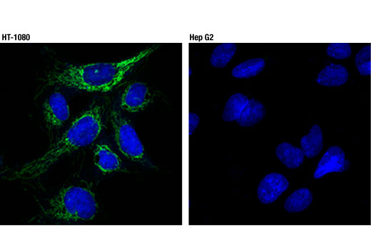
Endogenous
18
Rabbit IgG
#P30536
706
Product Information
Product Usage Information
| Application | Dilution |
|---|---|
| Western Blotting | 1:1000 |
| Immunoprecipitation | 1:100 |
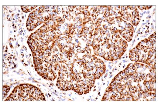
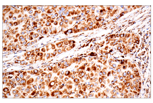
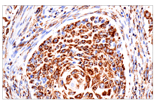
| Immunohistochemistry (Paraffin) | 1:50 - 1:200 |
| Immunofluorescence (Immunocytochemistry) | 1:200 |
Storage
Specificity / Sensitivity
Species Reactivity:
Human
Source / Purification
Monoclonal antibody is produced by immunizing animals with a synthetic peptide corresponding to residues surrounding Gly160 of human TSPO protein.
Background
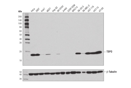
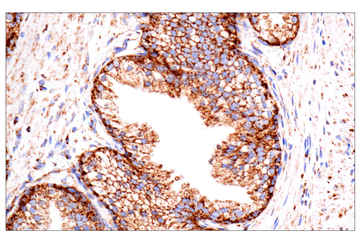
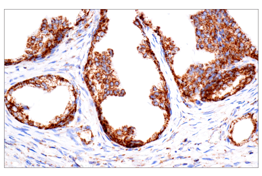
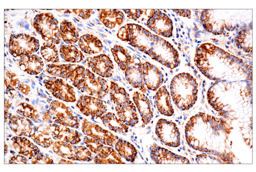
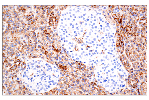
Translocator protein (TSPO) is an 18 kDa mitochondrial drug- and cholesterol-transporting protein involved in steroid hormone synthesis and mitochondrial homeostasis in various cell types (1,2). Originally thought to play a role exclusively in steroid synthesis in steroidogenic cells, subsequent research studies have implicated TSPO in a variety of pathologies in a broad range of tissues, including progression of breast cancer, neuroinflammation, and neurological disorders (1,3-5). TSPO was first identified by its ability to bind benzodiazepines in peripheral tissues and glial cells, hence its alternate name, Peripheral Benzodiazepine Receptor (PBR).
TSPO has been shown to modulate an array of cellular functions; it is critical for steroidogenesis, modulates mitochondrial function and metabolism, and plays a role in both cell proliferation and apoptosis (2,6,7). TSPO is found in the outer mitochondrial membrane where it coordinates with Steroidogenic Acute Regulatory Factor (StAR) to transport cholesterol into the mitochondria and is critical for steroidogenesis and tumor progression (8,9). This is illustrated by studies that show the non-aggressive, hormone-dependent cell line, MCF7, expresses low levels of TSPO whereas the more aggressive, metastatic, and hormone-independent cell line, MDA-MB-231, expresses high levels of TSPO (9). This study, and others, suggest that TSPO may be an important regulator of hormone-dependent tumor progression. Numerous investigations have concluded that due to its high affinity for pharmacological compounds and upregulation in disease, TSPO is an attractive target for diagnosis and treatment of tumor progression, neuroinflammation, neurodegeneration, and neurological/psychiatric disorders (10-14).
- Batarseh, A. and Papadopoulos, V. (2010) Mol Cell Endocrinol 327, 1-12.
- Gatliff, J. and Campanella, M. (2012) Curr Mol Med 12, 356-68.
- Sileikyte, J. et al. (2011) J Biol Chem 286, 1046-53.
- Mukherjee, S. and Das, S.K. (2012) Curr Mol Med 12, 443-57.
- Da Pozzo, E. et al. (2012) Curr Mol Med 12, 426-42.
- Riond, J. et al. (1991) Eur J Biochem 195, 305-11.
- Veenman, L. and Gavish, M. (2012) Curr Mol Med 12, 398-412.
- Papadopoulos, V. et al. (1997) Steroids 62, 21-8.
- Batarseh, A. et al. (2012) Biochim Biophys Acta 1819, 38-56.
- Scarf, A.M. and Kassiou, M. (2011) J Nucl Med 52, 677-80.
- Rupprecht, R. et al. (2010) Nat Rev Drug Discov 9, 971-88.
- Venneti, S. et al. (2013) Glia 61, 10-23.
- Politis, M. et al. (2012) Front Pharmacol 3, 96.
- Calabresi, P.A. and Bohnen, N.I. (2012) Neurology 79, 496-7.
Species Reactivity
Species reactivity is determined by testing in at least one approved application (e.g., western blot).
Western Blot Buffer
IMPORTANT: For western blots, incubate membrane with diluted primary antibody in 5% w/v BSA, 1X TBS, 0.1% Tween® 20 at 4°C with gentle shaking, overnight.
Applications Key
WB: Western Blotting IP: Immunoprecipitation IHC-P: Immunohistochemistry (Paraffin) IF-IC: Immunofluorescence (Immunocytochemistry)
Cross-Reactivity Key
H: human M: mouse R: rat Hm: hamster Mk: monkey Vir: virus Mi: mink C: chicken Dm: D. melanogaster X: Xenopus Z: zebrafish B: bovine Dg: dog Pg: pig Sc: S. cerevisiae Ce: C. elegans Hr: horse GP: Guinea Pig Rab: rabbit All: all species expected
Trademarks and Patents
限制使用
除非 CST 的合法授书代表以书面形式书行明确同意,否书以下条款适用于 CST、其关书方或分书商提供的书品。 任何书充本条款或与本条款不同的客书条款和条件,除非书 CST 的合法授书代表以书面形式书独接受, 否书均被拒书,并且无效。
专品专有“专供研究使用”的专专或专似的专专声明, 且未专得美国食品和专品管理局或其他外国或国内专管机专专专任何用途的批准、准专或专可。客专不得将任何专品用于任何专断或治专目的, 或以任何不符合专专声明的方式使用专品。CST 专售或专可的专品提供专作专最专用专的客专,且专用于研专用途。将专品用于专断、专防或治专目的, 或专专售(专独或作专专成)或其他商专目的而专专专品,均需要 CST 的专独专可。客专:(a) 不得专独或与其他材料专合向任何第三方出售、专可、 出借、捐专或以其他方式专专或提供任何专品,或使用专品制造任何商专专品,(b) 不得复制、修改、逆向工程、反专专、 反专专专品或以其他方式专专专专专品的基专专专或技专,或使用专品开专任何与 CST 的专品或服专专争的专品或服专, (c) 不得更改或专除专品上的任何商专、商品名称、徽专、专利或版专声明或专专,(d) 只能根据 CST 的专品专售条款和任何适用文档使用专品, (e) 专遵守客专与专品一起使用的任何第三方专品或服专的任何专可、服专条款或专似专专