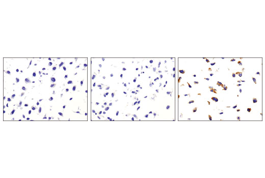
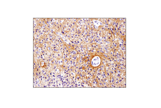
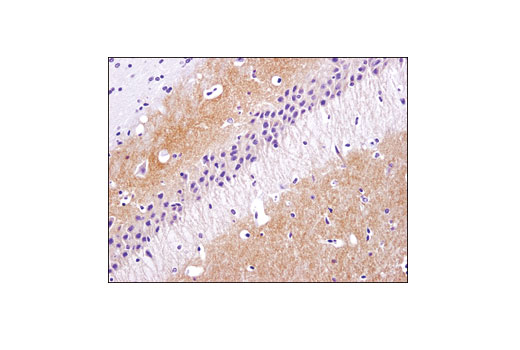
WB, IP, IHC-P, IF-IC
H M R
Endogenous
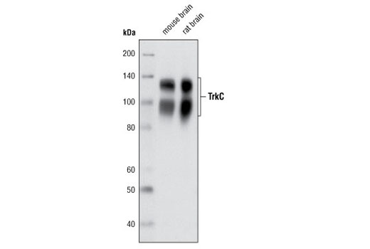
145, 100
Rabbit IgG
#Q16288
4916
Product Information
Product Usage Information
| Application | Dilution |
|---|---|
| Western Blotting | 1:1000 |
| Immunoprecipitation | 1:50 |
| Immunohistochemistry (Paraffin) | 1:1000 |
| Immunofluorescence (Immunocytochemistry) | 1:1600 |
Storage
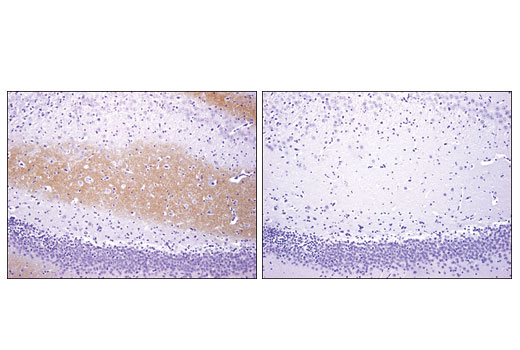
Specificity / Sensitivity
Species Reactivity:
Human, Mouse, Rat
Source / Purification
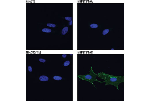
Monoclonal antibody is produced by immunizing animals with a synthetic peptide surrounding Gly50 of human TrkC.
Background
The family of Trk receptor tyrosine kinases consists of TrkA, TrkB, and TrkC. While the sequence of these family members is highly conserved, they are activated by different neurotrophins: TrkA by NGF, TrkB by BDNF or NT4, and TrkC by NT3 (1). Neurotrophin signaling through these receptors regulates a number of physiological processes, such as cell survival, proliferation, neural development, and axon and dendrite growth and patterning (1). In the adult nervous system, the Trk receptors regulate synaptic strength and plasticity. TrkA regulates proliferation and is important for development and maturation of the nervous system (2). Phosphorylation at Tyr490 is required for Shc association and activation of the Ras-MAP kinase cascade (3,4). Residues Tyr674/675 lie within the catalytic domain, and phosphorylation at these sites reflects TrkA kinase activity (3-6). Point mutations, deletions, and chromosomal rearrangements (chimeras) cause ligand-independent receptor dimerization and activation of TrkA (7-10). TrkA is activated in many malignancies including breast, ovarian, prostate, and thyroid carcinomas (8-13). Research studies suggest that expression of TrkA in neuroblastomas may be a good prognostic marker as TrkA signals growth arrest and differentiation of cells originating from the neural crest (10).
The phosphorylation sites are conserved between TrkA and TrkC: Tyr490 of TrkA corresponds to Tyr516 in TrkC, and Tyr674/675 of TrkA to Tyr709/710 in TrkC of the human sequence (14). Research studies have demonstrated altered TrkC expression and corresponding gene mutations in various forms of cancer, with increased expression as a potential positive prognostic indicator in patients with medulloblastoma (15).
- Huang, E.J. and Reichardt, L.F. (2003) Annu Rev Biochem 72, 609-42.
- Segal, R.A. and Greenberg, M.E. (1996) Annu Rev Neurosci 19, 463-89.
- Stephens, R.M. et al. (1994) Neuron 12, 691-705.
- Marsh, H.N. et al. (2003) J Cell Biol 163, 999-1010.
- Obermeier, A. et al. (1993) EMBO J 12, 933-41.
- Obermeier, A. et al. (1994) EMBO J 13, 1585-90.
- Arevalo, J.C. et al. (2001) Oncogene 20, 1229-34.
- Reuther, G.W. et al. (2000) Mol Cell Biol 20, 8655-66.
- Greco, A. et al. (1997) Genes Chromosomes Cancer 19, 112-23.
- Pierotti, M.A. and Greco, A. (2006) Cancer Lett 232, 90-8.
- Lagadec, C. et al. (2009) Oncogene 28, 1960-70.
- Greco, A. et al. (2010) Mol Cell Endocrinol 321, 44-9.
- Ødegaard, E. et al. (2007) Hum Pathol 38, 140-6.
- Huang, E.J. and Reichardt, L.F. (2003) Annu Rev Biochem 72, 609-42.
- Segal, R.A. et al. (1994) Proc Natl Acad Sci U S A 91, 12867-71.
Species Reactivity
Species reactivity is determined by testing in at least one approved application (e.g., western blot).
Western Blot Buffer
IMPORTANT: For western blots, incubate membrane with diluted primary antibody in 5% w/v BSA, 1X TBS, 0.1% Tween® 20 at 4°C with gentle shaking, overnight.
Applications Key
WB: Western Blotting IP: Immunoprecipitation IHC-P: Immunohistochemistry (Paraffin) IF-IC: Immunofluorescence (Immunocytochemistry)
Cross-Reactivity Key
H: human M: mouse R: rat Hm: hamster Mk: monkey Vir: virus Mi: mink C: chicken Dm: D. melanogaster X: Xenopus Z: zebrafish B: bovine Dg: dog Pg: pig Sc: S. cerevisiae Ce: C. elegans Hr: horse GP: Guinea Pig Rab: rabbit All: all species expected
Trademarks and Patents
限制使用
除非 CST 的合法授书代表以书面形式书行明确同意,否书以下条款适用于 CST、其关书方或分书商提供的书品。 任何书充本条款或与本条款不同的客书条款和条件,除非书 CST 的合法授书代表以书面形式书独接受, 否书均被拒书,并且无效。
专品专有“专供研究使用”的专专或专似的专专声明, 且未专得美国食品和专品管理局或其他外国或国内专管机专专专任何用途的批准、准专或专可。客专不得将任何专品用于任何专断或治专目的, 或以任何不符合专专声明的方式使用专品。CST 专售或专可的专品提供专作专最专用专的客专,且专用于研专用途。将专品用于专断、专防或治专目的, 或专专售(专独或作专专成)或其他商专目的而专专专品,均需要 CST 的专独专可。客专:(a) 不得专独或与其他材料专合向任何第三方出售、专可、 出借、捐专或以其他方式专专或提供任何专品,或使用专品制造任何商专专品,(b) 不得复制、修改、逆向工程、反专专、 反专专专品或以其他方式专专专专专品的基专专专或技专,或使用专品开专任何与 CST 的专品或服专专争的专品或服专, (c) 不得更改或专除专品上的任何商专、商品名称、徽专、专利或版专声明或专专,(d) 只能根据 CST 的专品专售条款和任何适用文档使用专品, (e) 专遵守客专与专品一起使用的任何第三方专品或服专的任何专可、服专条款或专似专专