| Product Includes | Product # | Quantity | Mol. Wt | Isotype/Source |
|---|---|---|---|---|
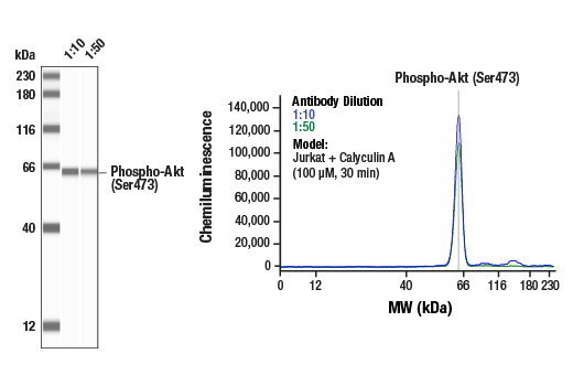
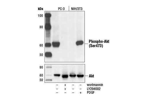
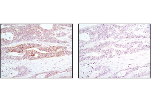
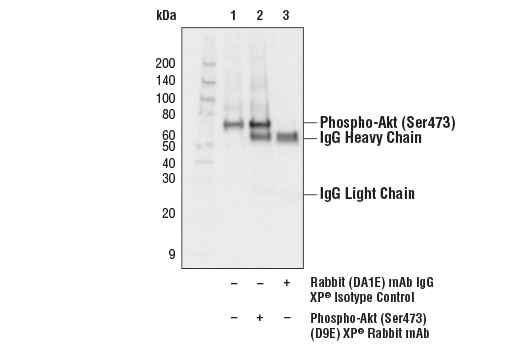
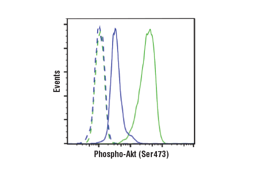
| Phospho-Akt (Ser473) (D9E) XP® Rabbit mAb | 4060 | 20 µl | 60 kDa | Rabbit IgG |
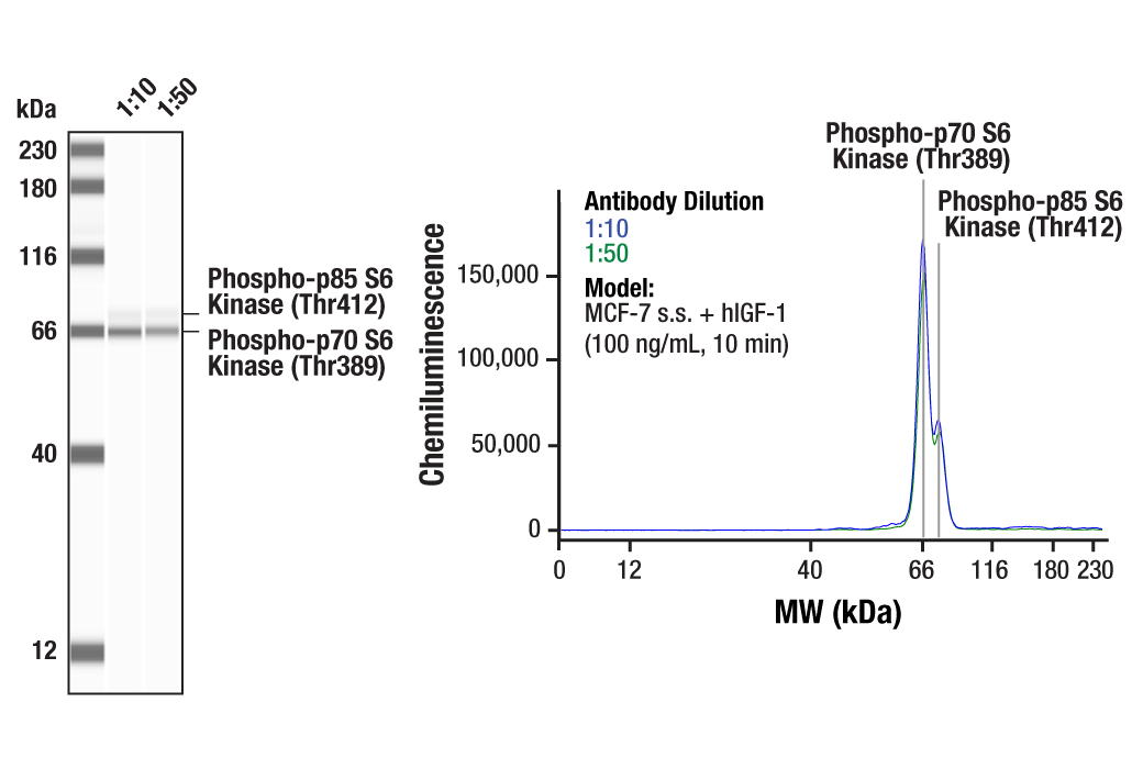
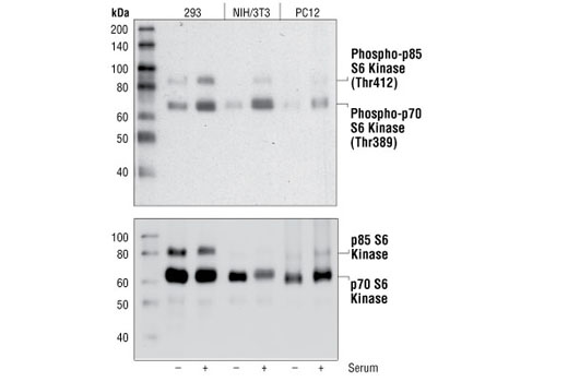
| Phospho-p70 S6 Kinase (Thr389) (108D2) Rabbit mAb | 9234 | 20 µl | 70, 85 kDa | Rabbit IgG |
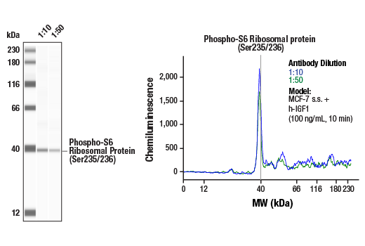
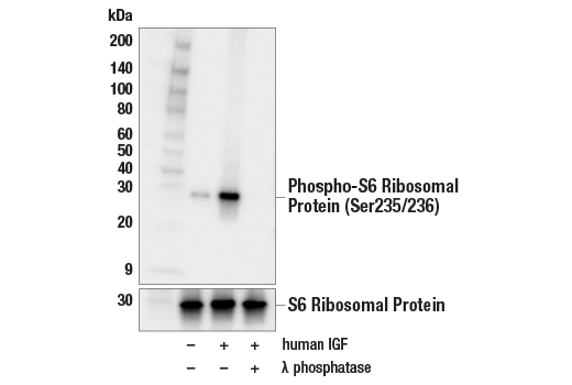
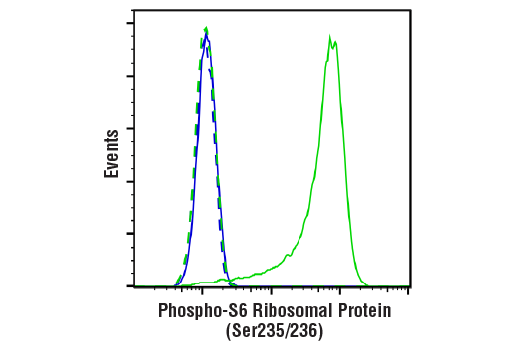
| Phospho-S6 Ribosomal Protein (Ser235/236) (D57.2.2E) XP® Rabbit mAb | 4858 | 20 µl | 32 kDa | Rabbit IgG |
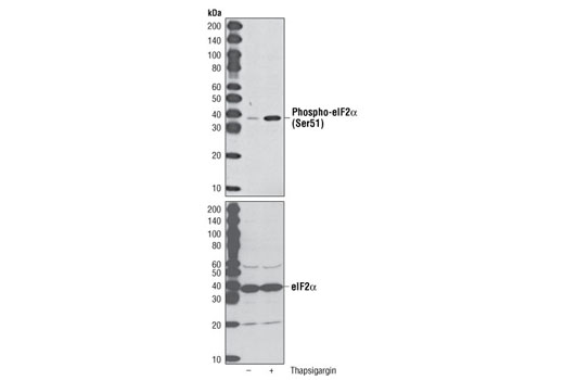
| Phospho-eIF2α (Ser51) (D9G8) XP® Rabbit mAb | 3398 | 20 µl | 38 kDa | Rabbit IgG |
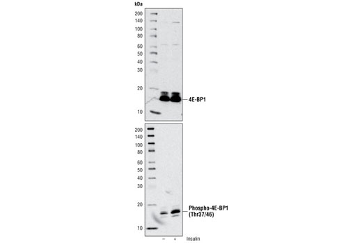
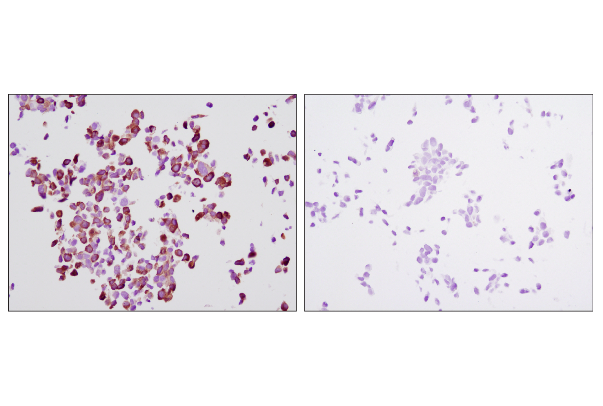
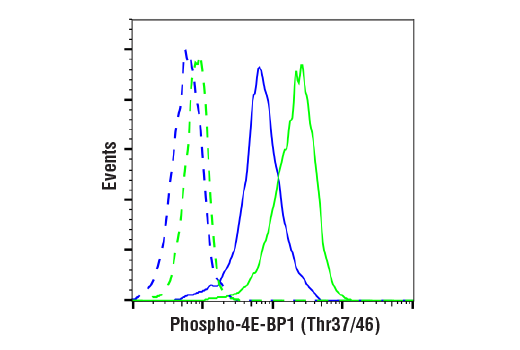
| Phospho-4E-BP1 (Thr37/46) (236B4) Rabbit mAb | 2855 | 20 µl | 15 to 20 kDa | Rabbit IgG |
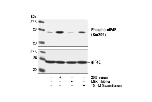
| Phospho-eIF4E (Ser209) Antibody | 9741 | 20 µl | 25 kDa | Rabbit |
| Anti-rabbit IgG, HRP-linked Antibody | 7074 | 100 µl | Goat |
Please visit cellsignal.com for individual component applications, species cross-reactivity, dilutions, protocols, and additional product information.
Description
The Translational Control Antibody Sampler Kit provides a fast and economical means of evaluating multiple proteins involved in translational control. The kit contains enough primary and secondary antibody to perform two Western blot experiments.
Storage
Background
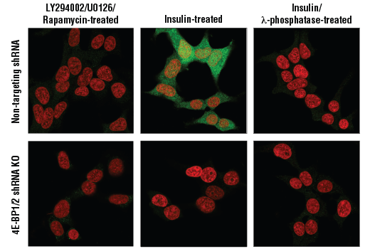
Key steps in translational control occur at the level of eukaryotic initiation factor 4F (eIF4F) and p70 S6 kinase regulation. eIF4F is a complex whose functions include the recognition of the mRNA 5' cap structure. Several stimuli, such as insulin and various growth and survival factors, regulate the eIF4F complex and p70 S6 kinase primarily by triggering a signaling cascade dependent on sequential activation of PI3K, Akt/PKB and mTOR/FRAP kinases. Akt is activated by phosphorylation within the C-terminus at Ser473 and within the activation loop at Thr308 by phospholipid-dependent kinases. Inactivation in vivo of PI3K by the highly selective inhibitor LY294002 inhibits Akt and downstream elements of this cascade. Direct phosphorylation of mTOR/FRAP at Ser2448 by Akt is a key regulatory event controlling its kinase activity. mTOR/FRAP activity can be effectively blocked by Rapamycin, leading to inactivation of eukaryotic initiation factor 4E binding protein 1 (4E-BP1), an inhibitor of translation initiation, and activation of p70 S6 kinases. Inactivation of 4E-BP1 by sequential phosphorylation causes the release of eIF4E, which, together with eIF4G and other factors, forms a functional eIF4F cap binding complex. p70 S6 kinases phosphorylates the 40S ribosomal subunit protein S6 and stimulates the translation of 5' oligopyrimidine tract containing mRNAs. The Erk pathway is also involved in regulation at this level by regulating the eIF4E kinase, Mnk1, and activating p70 S6 kinase. Tuberin, a product of the tumor supressor gene TSG2, is directly phosphorylated atThr1462 by Akt/PKB. Tuberin inhibits the mammalian target of rapamycin, mTOR, which results in inhibition of p70 S6 kinase and activation of 4E-BP1 and, therefore, inhibition of translation.
- Gingras, A. et al. (1999) Annu. Rev. Biochem. 68, 913-963.
- Gingras, A. C. et al. (2001) Genes and Develop. 15, 807-826.
- Dennis, P. B. et al. (1999) Curr. Opin. Genet. Dev. 9, 49-54.
- Volarevic, S. and Thomas, G. (2000) Prog. Nucleic Acid Res. Mol. Biol. 65, 101-127.
- Pyronnet, S. et al. (2000) Biochem. Pharmacol. 60, 1237-1243.
- Dever, T.E. (2002) Cell 108, 545-56.
- Goncharova, E.A. et al. (2002) J Biol Chem 277, 30958-67.
Background References
Trademarks and Patents
限制使用
除非 CST 的合法授书代表以书面形式书行明确同意,否书以下条款适用于 CST、其关书方或分书商提供的书品。 任何书充本条款或与本条款不同的客书条款和条件,除非书 CST 的合法授书代表以书面形式书独接受, 否书均被拒书,并且无效。
专品专有“专供研究使用”的专专或专似的专专声明, 且未专得美国食品和专品管理局或其他外国或国内专管机专专专任何用途的批准、准专或专可。客专不得将任何专品用于任何专断或治专目的, 或以任何不符合专专声明的方式使用专品。CST 专售或专可的专品提供专作专最专用专的客专,且专用于研专用途。将专品用于专断、专防或治专目的, 或专专售(专独或作专专成)或其他商专目的而专专专品,均需要 CST 的专独专可。客专:(a) 不得专独或与其他材料专合向任何第三方出售、专可、 出借、捐专或以其他方式专专或提供任何专品,或使用专品制造任何商专专品,(b) 不得复制、修改、逆向工程、反专专、 反专专专品或以其他方式专专专专专品的基专专专或技专,或使用专品开专任何与 CST 的专品或服专专争的专品或服专, (c) 不得更改或专除专品上的任何商专、商品名称、徽专、专利或版专声明或专专,(d) 只能根据 CST 的专品专售条款和任何适用文档使用专品, (e) 专遵守客专与专品一起使用的任何第三方专品或服专的任何专可、服专条款或专似专专