Revision 3
Cell Signaling Technology

Orders: 877-616-CELL (2355) [email protected]

Support: 877-678-TECH (8324)

Web: [email protected] cellsignal.com

3 Trask LaneDanversMassachusetts01923USA
For Research Use Only. Not for Use in Diagnostic Procedures.
Applications:

WB, IHC-Bond, IHC-P

REACTIVITY:

H M Mk

SENSITIVITY:

Endogenous

MW (kDa):

90, 120

Source/Isotype:

Rabbit IgG

UniProt ID:

#P28908

Entrez-Gene Id:

943

Product Information

Product Usage Information

This product is the carrier-free version of product #54535. All data were generated using the same antibody clone in the standard formulation which contains BSA and glycerol.

This formulation is ideal for use with technologies requiring specialized or custom antibody labeling, including fluorophores, metals, lanthanides, and oligonucleotides. It is not recommended for ChIP, ChIP-seq, CUT&RUN, or CUT&Tag assays. If you require a carrier-free formulation for chromatin profiling, please contact us. Optimal dilutions/concentrations should be determined by the end user.

Formulation

Supplied in 1X PBS, BSA and Azide Free.

For standard formulation of this product see product #54535.

Storage

Store at -20°C. This product will freeze at -20°C so it is recommended to aliquot into single-use vials to avoid multiple freeze/thaw cycles. A slight precipitate may be present and can be dissolved by gently vortexing. This will not interfere with antibody performance.

Specificity / Sensitivity

TNFRSF8/CD30 (E4L4I) XP® Rabbit mAb (BSA and Azide Free) recognizes endogenous levels of total TNFRSF8/CD30 protein. This antibody is predicted to detect CD30V, which is a truncated isoform that consists of the CD30 cytoplasmic domain. Non-specific staining was observed in the epithelium of mouse prostate tissue.

Species Reactivity:

Human, Mouse, Monkey

Source / Purification

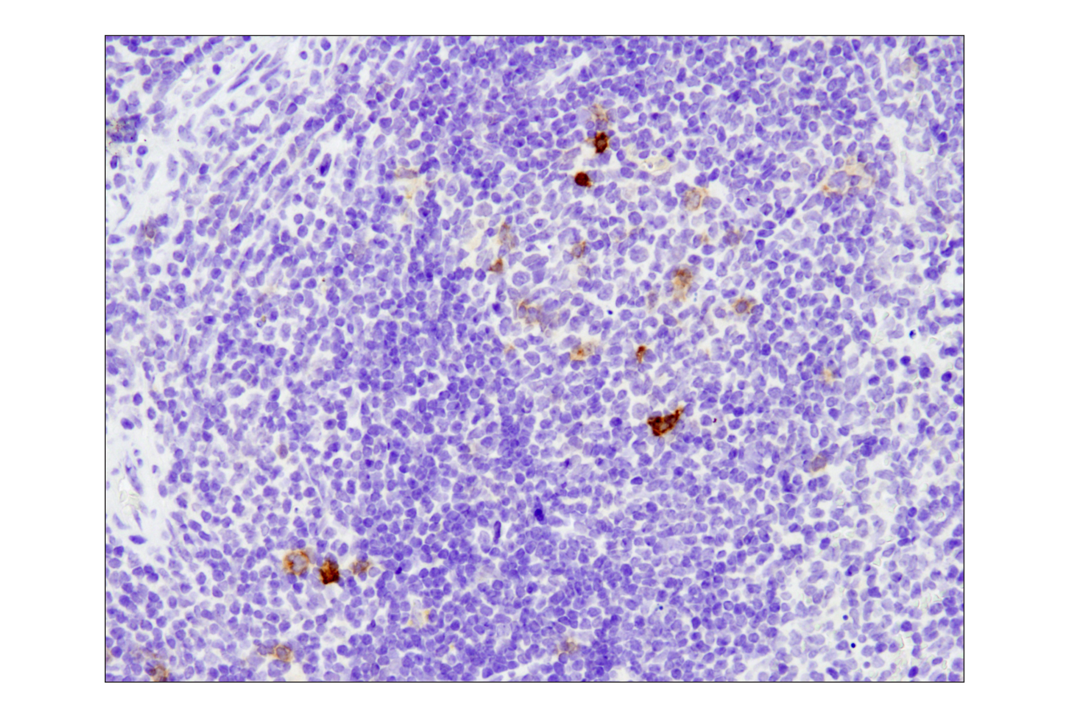
Monoclonal antibody is produced by immunizing animals with a synthetic peptide corresponding to residues surrounding Leu500 of human TNFRSF8/CD30 protein.

Background

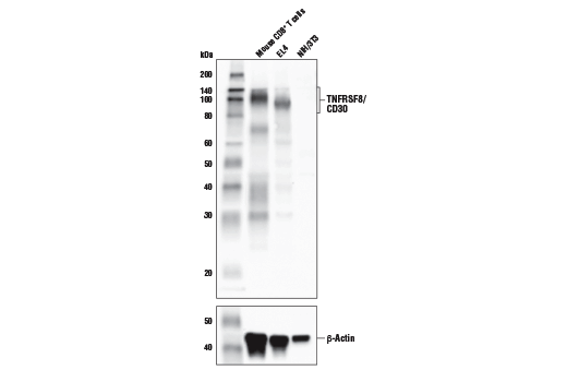
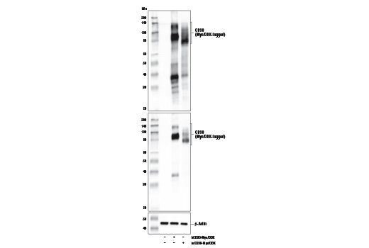
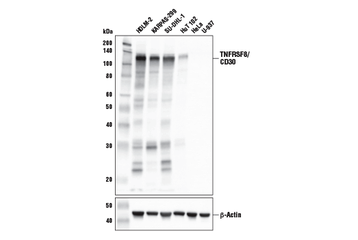
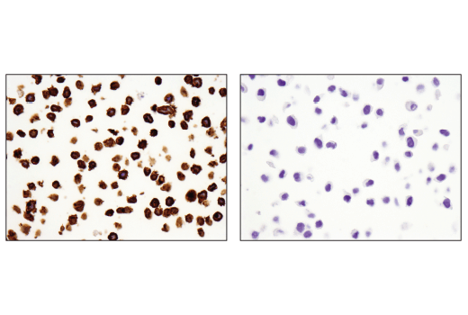
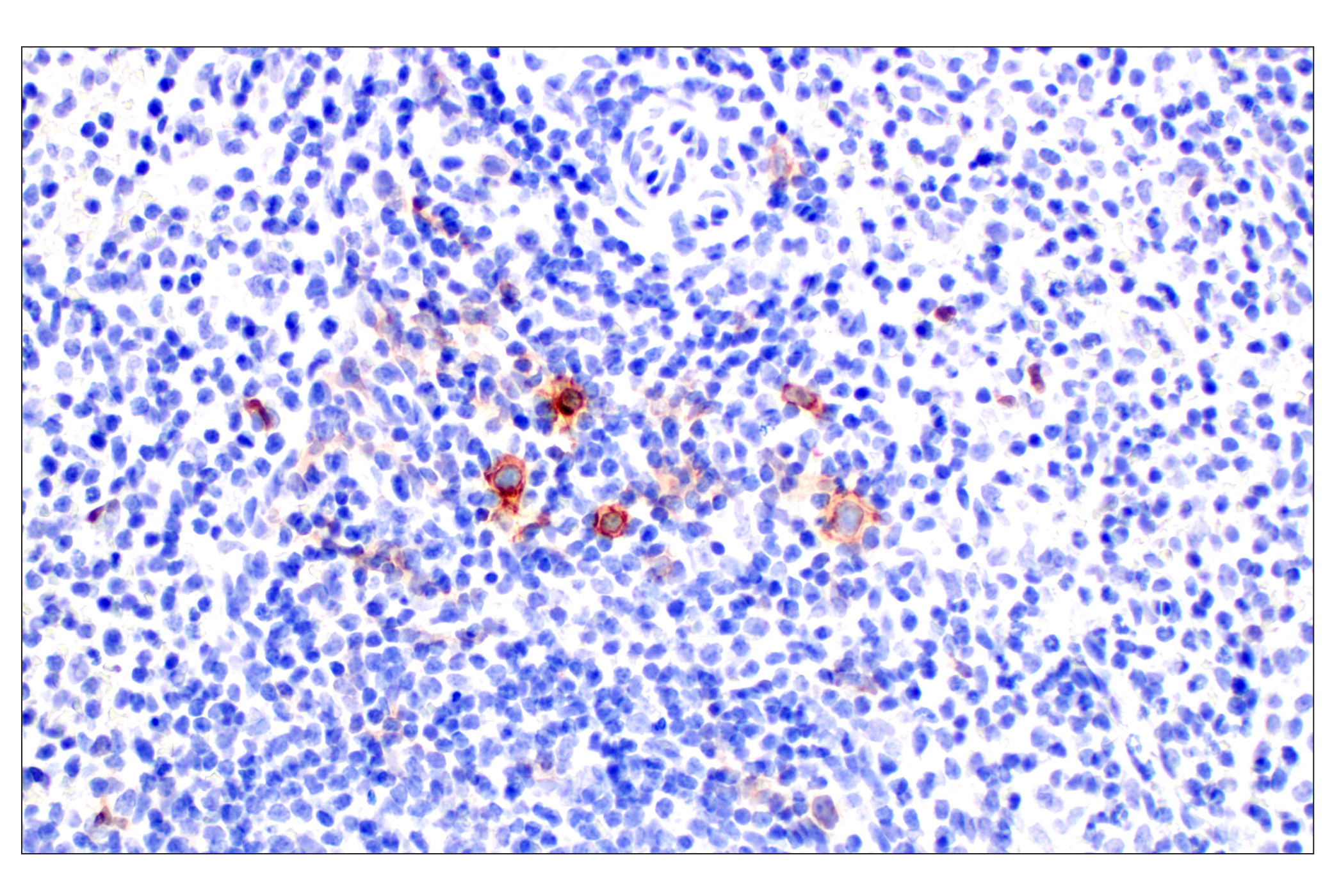
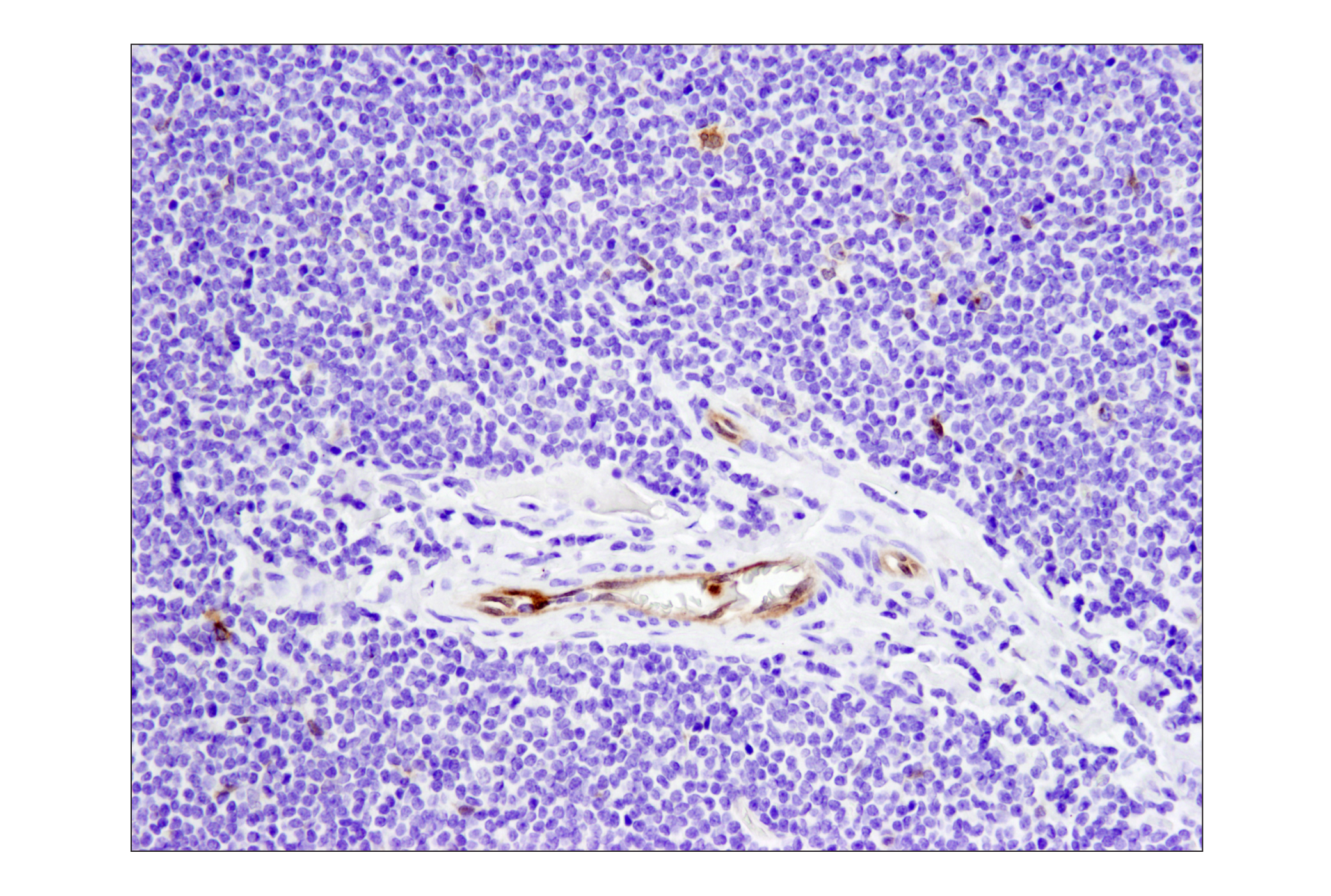
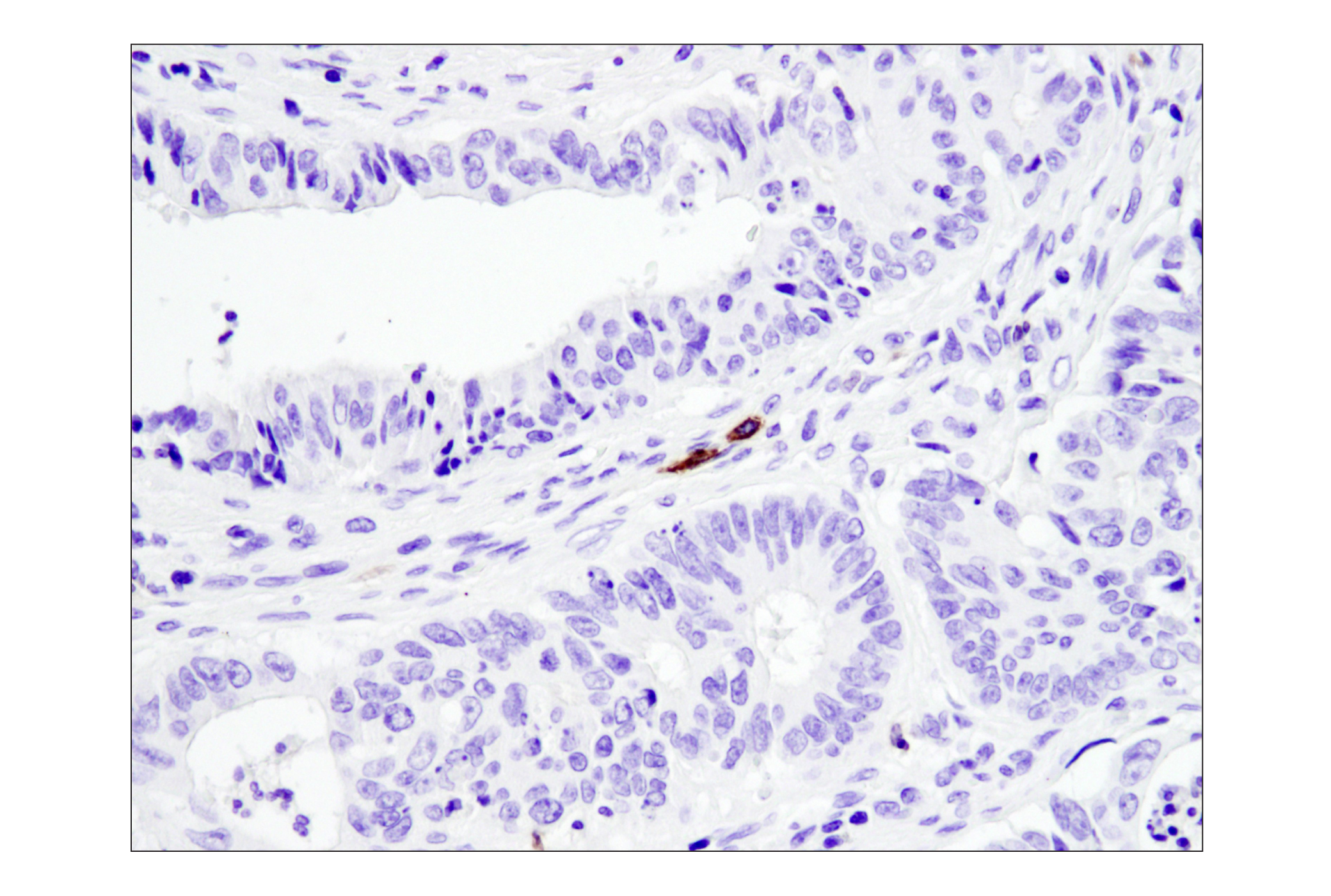
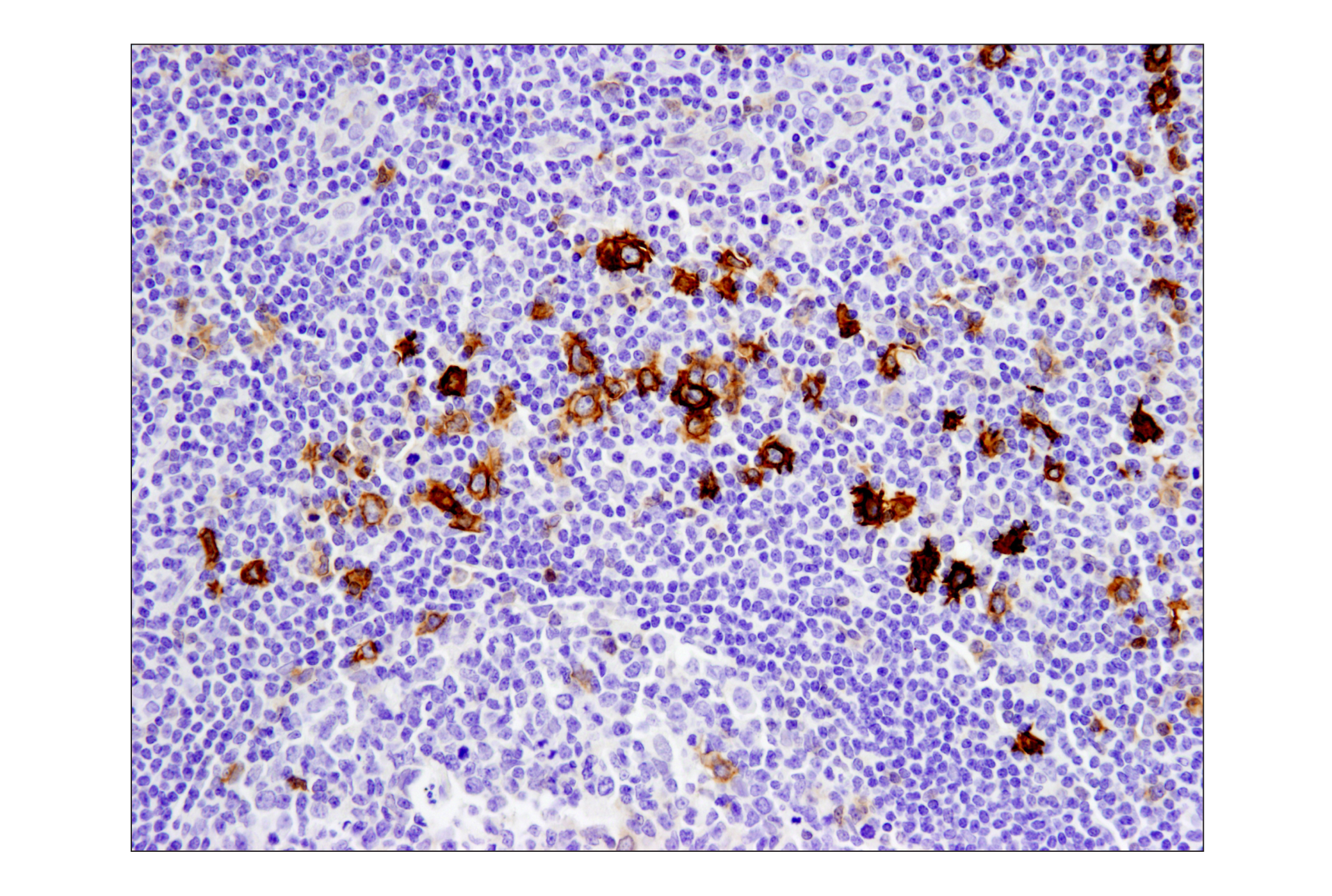
TNFRSF8/CD30 is a type-I transmembrane glycoprotein that is a member of the TNFR superfamily. CD30 is synthesized as a precursor protein that undergoes extensive post-translational modification before becoming embedded in the plasma membrane as a 120-kDa transmembrane protein (1,2). The expression of CD30 is upregulated in activated T cells and may trigger costimulatory signaling pathways upon its engagement (3,4). While its expression is normally restricted to subsets of activated T cells and B cells, CD30 expression is robustly upregulated in hematologic malignancies, such as Hodgkin lymphoma (HL), anaplastic large cell lymphoma (ALCL), and adult T-cell leukemia, thus making it an attractive target for therapeutic intervention (5,6). Research studies have suggested that in certain disease contexts, CD30 recruits TRAF2 and TRAF5 adaptor proteins to drive NF-kappa B activation, aberrant cell growth, and cytokine production (7-9). CD30 signaling is also regulated by TACE-dependent proteolytic cleavage of its ectodomain, which results in reduced CD30L-dependent activation of CD30+ cells (10,11).

  1. Froese, P. et al. (1987) J Immunol 139, 2081-7.
  2. Nawrocki, J.F. et al. (1988) J Immunol 141, 672-80.
  3. Del Prete, G. et al. (1995) J Exp Med 182, 1655-61.
  4. Gilfillan, M.C. et al. (1998) J Immunol 160, 2180-7.
  5. Stein, H. et al. (1985) Blood 66, 848-58.
  6. Chiarle, R. et al. (1999) Clin Immunol 90, 157-64.
  7. Horie, R. et al. (2002) Am J Pathol 160, 1647-54.
  8. Horie, R. et al. (2002) Oncogene 21, 2493-503.
  9. Horie, R. et al. (2004) Cancer Cell 5, 353-64.
  10. Hansen, H.P. et al. (2000) J Immunol 165, 6703-9.
  11. Gruss, H.J. et al. (1997) Immunol Today 18, 156-63.

Species Reactivity

Species reactivity is determined by testing in at least one approved application (e.g., western blot).

Applications Key

WB: Western Blotting IHC-Bond: IHC Leica Bond IHC-P: Immunohistochemistry (Paraffin)

Cross-Reactivity Key

H: human M: mouse R: rat Hm: hamster Mk: monkey Vir: virus Mi: mink C: chicken Dm: D. melanogaster X: Xenopus Z: zebrafish B: bovine Dg: dog Pg: pig Sc: S. cerevisiae Ce: C. elegans Hr: horse GP: Guinea Pig Rab: rabbit All: all species expected

Trademarks and Patents

Cell Signaling Technology is a trademark of Cell Signaling Technology, Inc.
SignalStain is a registered trademark of Cell Signaling Technology, Inc.
XP is a registered trademark of Cell Signaling Technology, Inc.
KARPAS cell line source: Dr. Abraham Karpas at the University of Cambridge.
All other trademarks are the property of their respective owners. Visit cellsignal.com/trademarks for more information.

限制使用

除非 CST 的合法授书代表以书面形式书行明确同意,否书以下条款适用于 CST、其关书方或分书商提供的书品。 任何书充本条款或与本条款不同的客书条款和条件,除非书 CST 的合法授书代表以书面形式书独接受, 否书均被拒书,并且无效。

专品专有“专供研究使用”的专专或专似的专专声明, 且未专得美国食品和专品管理局或其他外国或国内专管机专专专任何用途的批准、准专或专可。客专不得将任何专品用于任何专断或治专目的, 或以任何不符合专专声明的方式使用专品。CST 专售或专可的专品提供专作专最专用专的客专,且专用于研专用途。将专品用于专断、专防或治专目的, 或专专售(专独或作专专成)或其他商专目的而专专专品,均需要 CST 的专独专可。客专:(a) 不得专独或与其他材料专合向任何第三方出售、专可、 出借、捐专或以其他方式专专或提供任何专品,或使用专品制造任何商专专品,(b) 不得复制、修改、逆向工程、反专专、 反专专专品或以其他方式专专专专专品的基专专专或技专,或使用专品开专任何与 CST 的专品或服专专争的专品或服专, (c) 不得更改或专除专品上的任何商专、商品名称、徽专、专利或版专声明或专专,(d) 只能根据 CST 的专品专售条款和任何适用文档使用专品, (e) 专遵守客专与专品一起使用的任何第三方专品或服专的任何专可、服专条款或专似专专

Revision 3
#47448

TNFRSF8/CD30 (E4L4I) XP® Rabbit mAb (BSA and Azide Free)

Western Blotting Image 1: TNFRSF8/CD30 (E4L4I) XP® Rabbit mAb (BSA and Azide Free) Expand Image
使用 TNFRSF8/CD30 (E4L4I) XP® Rabbit mAb(上图)或 β-Actin (D6A8) Rabbit mAb #8457(下图),对来自小鼠原代 CD8+ T 细胞、EL4 细胞和 NIH/3T3 细胞的提取物进行蛋白质印迹分析。正如预计的一样,未在 NIH/3T3 细胞中检测到 TNFRSF8/CD30 蛋白。CD8+ T 细胞用小鼠脾细胞纯化而来,并在添加小鼠 interleukin-2 (20 ng/mL) 的情况下,使用覆有 CD3 和 CD28 抗体的珠子刺激 7 天。数据的生成使用的是该产品的常规组分产品。
Western Blotting Image 2: TNFRSF8/CD30 (E4L4I) XP® Rabbit mAb (BSA and Azide Free) Expand Image
使用 NFRSF8/CD30 (E4L4I) XP® Rabbit mAb(上图)、DYKDDDDK Tag Antibody #2368(中间图)或 β-Actin (D6A8) Rabbit mAb #8457(下图),对空载转染 (-) 或经表达 Myc/DDK 标记的全长人 TNFRSF8/CD30 蛋白(hCD30-Myc/DDK;+)的构建体转染或经表达 Myc/DDK 标记的全长小鼠 TNFRSF8/CD30 蛋白(mCD30-Myc/DDK;+)的构建体转染的 293T 细胞的提取物进行蛋白质印迹分析。数据的生成使用的是该产品的常规组分产品。
Western Blotting Image 3: TNFRSF8/CD30 (E4L4I) XP® Rabbit mAb (BSA and Azide Free) Expand Image
使用 TNFRSF8/CD30 (E4L4I) XP® Rabbit mAb(上图)或 β-Actin (D6A8) Rabbit mAb #8457(下图)对各种细胞系的提取物进行蛋白质印迹分析。有如预期,TNFRSF8/CD30 蛋白在 U-937 或 HeLa 细胞中均不表达。KARPAS-299 细胞系来源:剑桥大学 Abraham Karpas 博士。数据的生成使用的是该产品的常规组分产品。
Immunohistochemistry Image 1: TNFRSF8/CD30 (E4L4I) XP® Rabbit mAb (BSA and Azide Free) Expand Image
在 Leica BOND Rx 上使用 TNFRSF8/CD30 (E4L4I) XP® Rabbit mAb 对石蜡包埋的人霍奇金氏淋巴瘤进行免疫组织化学分析。注意 Reed-Sternberg 细胞的深染。数据的生成使用的是该产品的常规组分产品。
Immunohistochemistry Image 1: TNFRSF8/CD30 (E4L4I) XP® Rabbit mAb (BSA and Azide Free) Expand Image
使用 TNFRSF8/CD30 (E4L4I) XP® Rabbit mAb 对石蜡包埋的 KARPAS-299 细胞沉淀物(左图,阳性)或 HeLa 细胞沉淀物(右图,阴性)进行免疫组织化学分析。数据的生成使用的是该产品的常规组分产品。
Immunohistochemistry Image 2: TNFRSF8/CD30 (E4L4I) XP® Rabbit mAb (BSA and Azide Free) Expand Image
Immunohistochemical analysis of paraffin-embedded rhesus monkey spleen using TNFRSF8/CD30 (E4L4I) XP® Rabbit mAb. 数据的生成使用的是该产品的常规组分产品。
Immunohistochemistry Image 3: TNFRSF8/CD30 (E4L4I) XP® Rabbit mAb (BSA and Azide Free) Expand Image
使用 TNFRSF8/CD30 (E4L4I) XP® Rabbit mAb 对石蜡包埋的人霍奇金氏淋巴瘤进行免疫组织化学分析。注意 Reed-Sternberg 细胞的深染。数据的生成使用的是该产品的常规组分产品。
Immunohistochemistry Image 4: TNFRSF8/CD30 (E4L4I) XP® Rabbit mAb (BSA and Azide Free) Expand Image
使用 TNFRSF8/CD30 (E4L4I) XP® Rabbit mAb 对石蜡包埋的人非霍奇金氏淋巴瘤进行免疫组织化学分析。数据的生成使用的是该产品的常规组分产品。
Immunohistochemistry Image 5: TNFRSF8/CD30 (E4L4I) XP® Rabbit mAb (BSA and Azide Free) Expand Image
使用 TNFRSF8/CD30 (E4L4I) XP® Rabbit mAb 对石蜡包埋的人结肠癌细胞进行免疫组织化学分析。数据的生成使用的是该产品的常规组分产品。
Immunohistochemistry Image 6: TNFRSF8/CD30 (E4L4I) XP® Rabbit mAb (BSA and Azide Free) Expand Image
使用 TNFRSF8/CD30 (E4L4I) XP® Rabbit mAb 对石蜡包埋的人扁桃体细胞进行免疫组织化学分析。数据的生成使用的是该产品的常规组分产品。
Immunohistochemistry Image 7: TNFRSF8/CD30 (E4L4I) XP® Rabbit mAb (BSA and Azide Free) Expand Image
使用 TNFRSF8/CD30 (E4L4I) XP® Rabbit mAb 对石蜡包埋的小鼠脾细胞进行免疫组织化学分析。数据的生成使用的是该产品的常规组分产品。