WB, IHC-Bond, IHC-P, FC-L
H
Endogenous (IHC-P), Transfected (W, F)
40-60
Rabbit IgG
#Q96H15
91937
Product Information
Product Usage Information
| Application | Dilution |
|---|---|
| Western Blotting | 1:1000 |
| IHC Leica Bond | 1:100 |
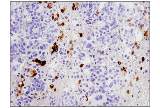
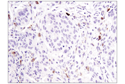
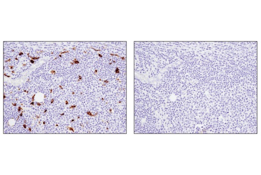
| Immunohistochemistry (Paraffin) | 1:100 |
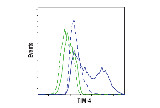
| Flow Cytometry (Live) | 1:800 |
Storage
For a carrier-free (BSA and azide free) version of this product see product #27337.
Specificity / Sensitivity
Species Reactivity:
Human
Source / Purification
Monoclonal antibody is produced by immunizing animals with a synthetic peptide corresponding to residues surrounding Pro215 of human TIM-4 protein.
Background
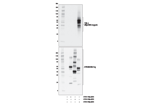
T cell Ig- and mucin-domain-containing molecules (TIMs) are a family of transmembrane proteins expressed by various immune cells. TIM-4 is expressed by dendritic cells and macrophages (1). Like TIM-1, TIM-4 is a receptor for phosphatidylserine (2). Interaction of TIM-4 with phosphatidylserine exposed by cells undergoing apoptosis leads to uptake of apoptotic cells (2,3). In this way, TIM-4 regulates adaptive immunity by removing apoptotic antigen-specific T cells following clearance of infection. Peritoneal macrophages from mice deficient in TIM-4 fail to effectively clear apoptotic cells leading to hyperactive T and B cells and autoimmunity (4). In addition, TIM-4 expressed by tumor-associated macrophages and dendritic cells leads to autophagy-mediated degradation of dying tumor cells, resulting in reduced antigen presentation and T cell responses (5). TIM-4 was also identified as a ligand for TIM-1, and interaction between these proteins regulates T cell proliferation (1). Finally, TIM-4 is expressed by a subset of proinflammatory B cells that produce IFN-γ and promote Th1 cell differentiation (6).
- Meyers, J.H. et al. (2005) Nat Immunol 6, 455-64.
- Miyanishi, M. et al. (2007) Nature 450, 435-9.
- Kobayashi, N. et al. (2007) Immunity 27, 927-40.
- Rodriguez-Manzanet, R. et al. (2010) Proc Natl Acad Sci U S A 107, 8706-11.
- Baghdadi, M. et al. (2013) Immunity 39, 1070-81.
- Ding, Q. et al. (2017) J Immunol 199, 2585-2595.
Species Reactivity
Species reactivity is determined by testing in at least one approved application (e.g., western blot).
Western Blot Buffer
IMPORTANT: For western blots, incubate membrane with diluted primary antibody in 5% w/v BSA, 1X TBS, 0.1% Tween® 20 at 4°C with gentle shaking, overnight.
Applications Key
WB: Western Blotting IHC-Bond: IHC Leica Bond IHC-P: Immunohistochemistry (Paraffin) FC-L: Flow Cytometry (Live)
Cross-Reactivity Key
H: human M: mouse R: rat Hm: hamster Mk: monkey Vir: virus Mi: mink C: chicken Dm: D. melanogaster X: Xenopus Z: zebrafish B: bovine Dg: dog Pg: pig Sc: S. cerevisiae Ce: C. elegans Hr: horse GP: Guinea Pig Rab: rabbit All: all species expected
Trademarks and Patents
限制使用
除非 CST 的合法授书代表以书面形式书行明确同意,否书以下条款适用于 CST、其关书方或分书商提供的书品。 任何书充本条款或与本条款不同的客书条款和条件,除非书 CST 的合法授书代表以书面形式书独接受, 否书均被拒书,并且无效。
专品专有“专供研究使用”的专专或专似的专专声明, 且未专得美国食品和专品管理局或其他外国或国内专管机专专专任何用途的批准、准专或专可。客专不得将任何专品用于任何专断或治专目的, 或以任何不符合专专声明的方式使用专品。CST 专售或专可的专品提供专作专最专用专的客专,且专用于研专用途。将专品用于专断、专防或治专目的, 或专专售(专独或作专专成)或其他商专目的而专专专品,均需要 CST 的专独专可。客专:(a) 不得专独或与其他材料专合向任何第三方出售、专可、 出借、捐专或以其他方式专专或提供任何专品,或使用专品制造任何商专专品,(b) 不得复制、修改、逆向工程、反专专、 反专专专品或以其他方式专专专专专品的基专专专或技专,或使用专品开专任何与 CST 的专品或服专专争的专品或服专, (c) 不得更改或专除专品上的任何商专、商品名称、徽专、专利或版专声明或专专,(d) 只能根据 CST 的专品专售条款和任何适用文档使用专品, (e) 专遵守客专与专品一起使用的任何第三方专品或服专的任何专可、服专条款或专似专专