WB, IHC-P, IF-F, IF-IC
H M R Mk
Endogenous
Rabbit
#P63165
7341
Product Information
Product Usage Information
| Application | Dilution |
|---|---|
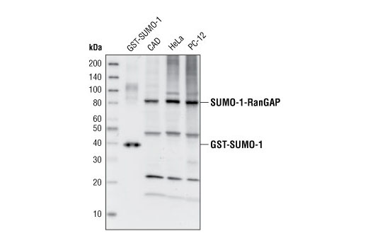
| Western Blotting | 1:1000 |
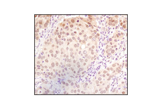
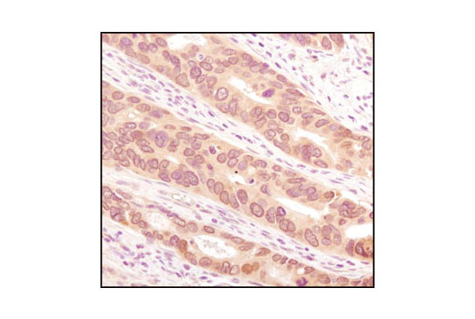
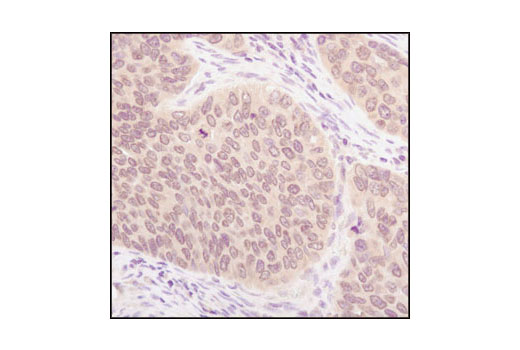
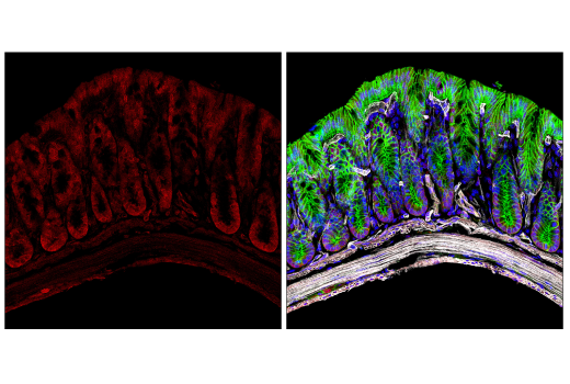
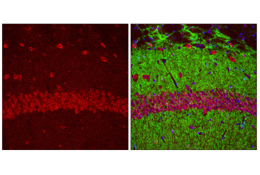
| Immunohistochemistry (Paraffin) | 1:50 |
| Immunofluorescence (Frozen) | 1:200 |
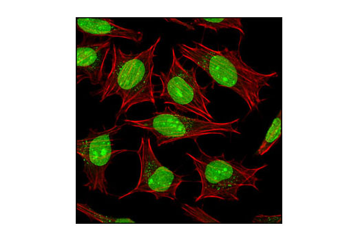
| Immunofluorescence (Immunocytochemistry) | 1:100 - 1:400 |
Storage
Specificity / Sensitivity
Species Reactivity:
Human, Mouse, Rat, Monkey
Source / Purification
Polyclonal antibodies are produced by immunizing animals with a synthetic peptide corresponding to a sequence within human SUMO-1 that does not correspond to SUMO-2/3. Antibodies are purified by protein A and peptide affinity chromatography.
Background
Small ubiquitin-related modifier 1, 2 and 3 (SUMO-1, -2 and -3) are members of the ubiquitin-like protein family (1). The covalent attachment of the SUMO-1, -2 or -3 (SUMOylation) to target proteins is analogous to ubiquitination. This post-translational modification is a reversible, multi-step process that is initiated by cleaving a precursor protein to a mature protein. Mature SUMO-1, -2 or -3 is then linked to the activating enzyme E1, conjugated to E2 and in conjunction with E3, SUMO-1, -2 or -3 is ligated to the target protein (2). Ubiquitin and the individual SUMO family members are all targeted to different proteins with diverse biological functions. Ubiquitin predominantly regulates degradation of its target (1). In contrast, SUMO-1 is conjugated to RanGAP, PML, p53 and IκB-α to regulate nuclear trafficking, formation of subnuclear structures, regulation of transcriptional activity and protein stability (3-7). SUMO-2/-3 forms poly-(SUMO) chains, is conjugated to topoisomerase II and APP, regulates chromosomal segregation and cellular responses to environmental stress, and plays a role in the progression of Alzheimer disease (8-11).
- Schwartz, D.C. and Hochstrasser, M. (2003) Trends Biochem. Sci. 28, 321-8.
- Kim, K.I. et al. (2002) J. Cell Physiol. 191, 257-68.
- Matunis, M.J. et al. (1996) J. Cell Biol. 135, 1457-70.
- Duprez, E. et al. (1999) J. Cell Sci. 112, 381-93.
- Gostissa, M. et al. (1999) EMBO J. 18, 6462-74.
- Rodriguez, M.S. et al. (1999) EMBO J. 18, 6455-61.
- Desterro, J.M. et al. (1998) Mol. Cell 2, 233-9.
- Tatham, M.H. et al. (2001) J. Biol. Chem. 276, 35368-74.
- Azuma, Y. et al. (2003) J. Cell Biol. 163, 477-87.
- Li, Y. et al. (2003) Proc. Natl. Acad. Sci. USA 100, 259-64.
- Saitoh, H. and Hinchey, J. (2000) J. Biol. Chem. 275, 6252-8.
Species Reactivity
Species reactivity is determined by testing in at least one approved application (e.g., western blot).
Western Blot Buffer
IMPORTANT: For western blots, incubate membrane with diluted primary antibody in 5% w/v BSA, 1X TBS, 0.1% Tween® 20 at 4°C with gentle shaking, overnight.
Applications Key
WB: Western Blotting IHC-P: Immunohistochemistry (Paraffin) IF-F: Immunofluorescence (Frozen) IF-IC: Immunofluorescence (Immunocytochemistry)
Cross-Reactivity Key
H: human M: mouse R: rat Hm: hamster Mk: monkey Vir: virus Mi: mink C: chicken Dm: D. melanogaster X: Xenopus Z: zebrafish B: bovine Dg: dog Pg: pig Sc: S. cerevisiae Ce: C. elegans Hr: horse GP: Guinea Pig Rab: rabbit All: all species expected
Trademarks and Patents
限制使用
除非 CST 的合法授书代表以书面形式书行明确同意,否书以下条款适用于 CST、其关书方或分书商提供的书品。 任何书充本条款或与本条款不同的客书条款和条件,除非书 CST 的合法授书代表以书面形式书独接受, 否书均被拒书,并且无效。
专品专有“专供研究使用”的专专或专似的专专声明, 且未专得美国食品和专品管理局或其他外国或国内专管机专专专任何用途的批准、准专或专可。客专不得将任何专品用于任何专断或治专目的, 或以任何不符合专专声明的方式使用专品。CST 专售或专可的专品提供专作专最专用专的客专,且专用于研专用途。将专品用于专断、专防或治专目的, 或专专售(专独或作专专成)或其他商专目的而专专专品,均需要 CST 的专独专可。客专:(a) 不得专独或与其他材料专合向任何第三方出售、专可、 出借、捐专或以其他方式专专或提供任何专品,或使用专品制造任何商专专品,(b) 不得复制、修改、逆向工程、反专专、 反专专专品或以其他方式专专专专专品的基专专专或技专,或使用专品开专任何与 CST 的专品或服专专争的专品或服专, (c) 不得更改或专除专品上的任何商专、商品名称、徽专、专利或版专声明或专专,(d) 只能根据 CST 的专品专售条款和任何适用文档使用专品, (e) 专遵守客专与专品一起使用的任何第三方专品或服专的任何专可、服专条款或专似专专