| Product Includes | Product # | Quantity | Mol. Wt | Isotype/Source |
|---|---|---|---|---|
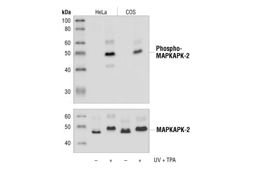
| Phospho-MAPKAPK-2 (Thr334) (27B7) Rabbit mAb | 3007 | 20 µl | 49 kDa | Rabbit IgG |
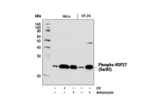
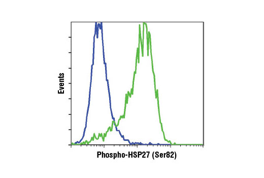
| Phospho-HSP27 (Ser82) (D1H2F6) XP® Rabbit mAb | 9709 | 20 µl | 27 kDa | Rabbit IgG |
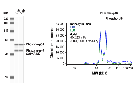
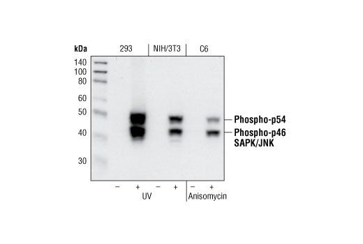
| Phospho-SAPK/JNK (Thr183/Tyr185) (81E11) Rabbit mAb | 4668 | 20 µl | 46, 54 kDa | Rabbit IgG |
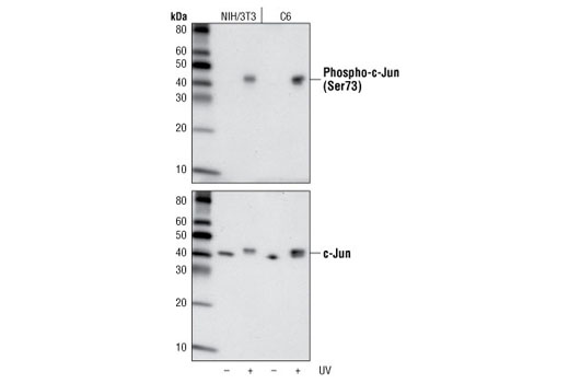
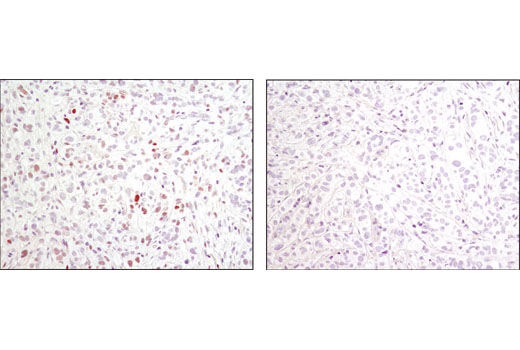
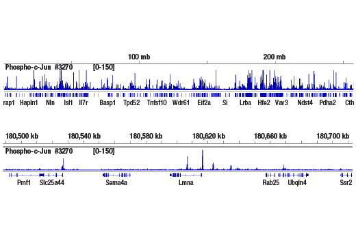
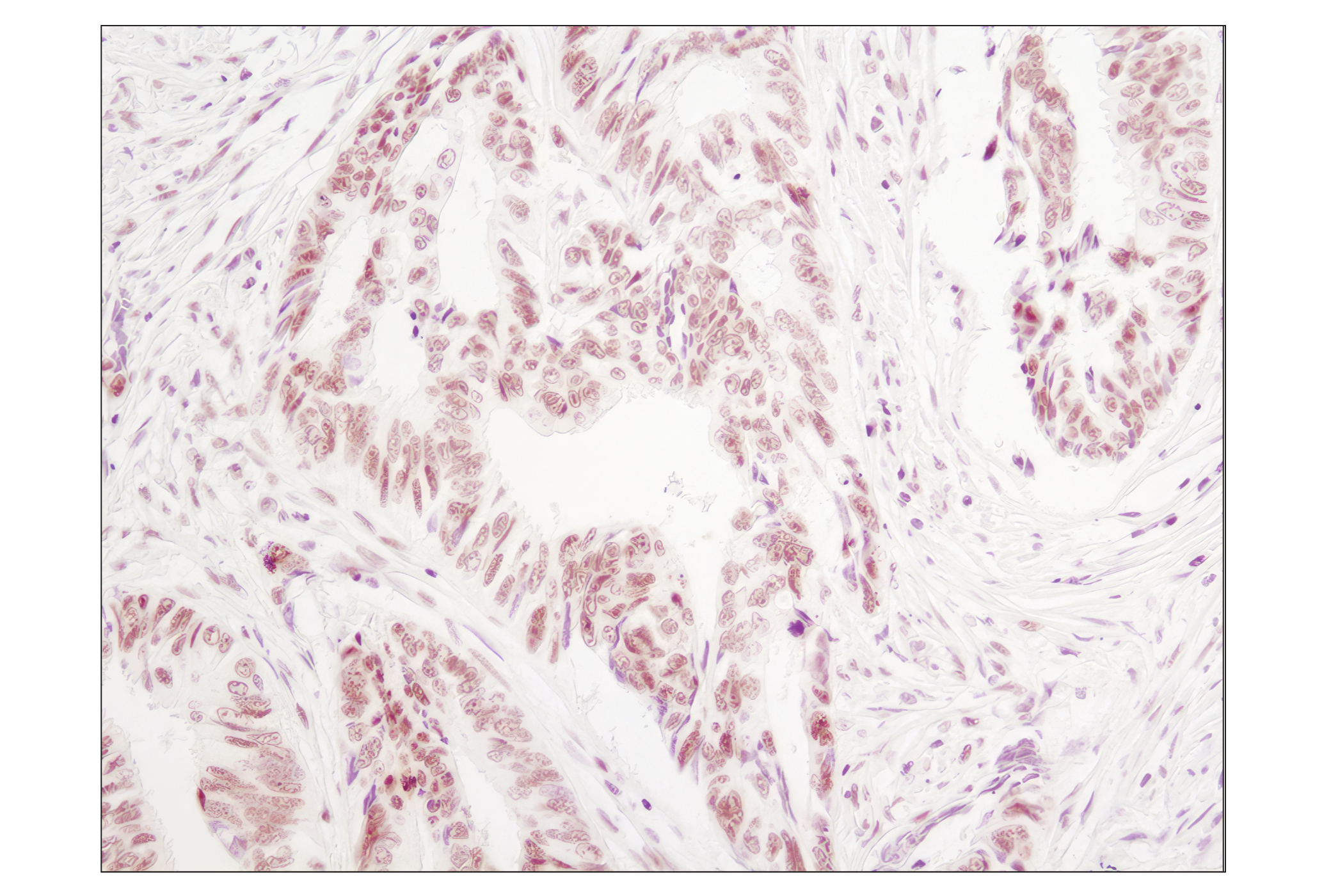
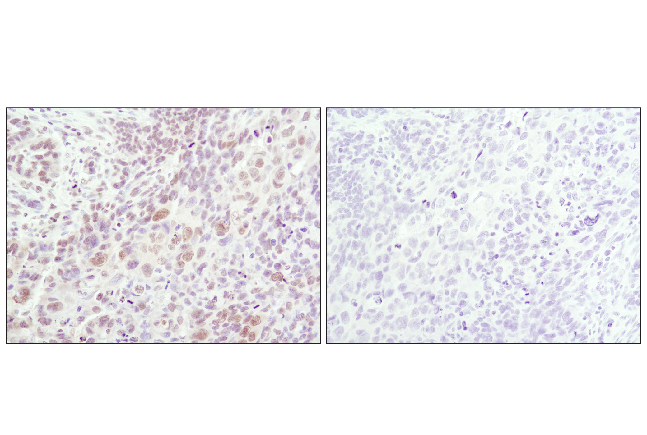
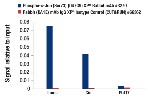
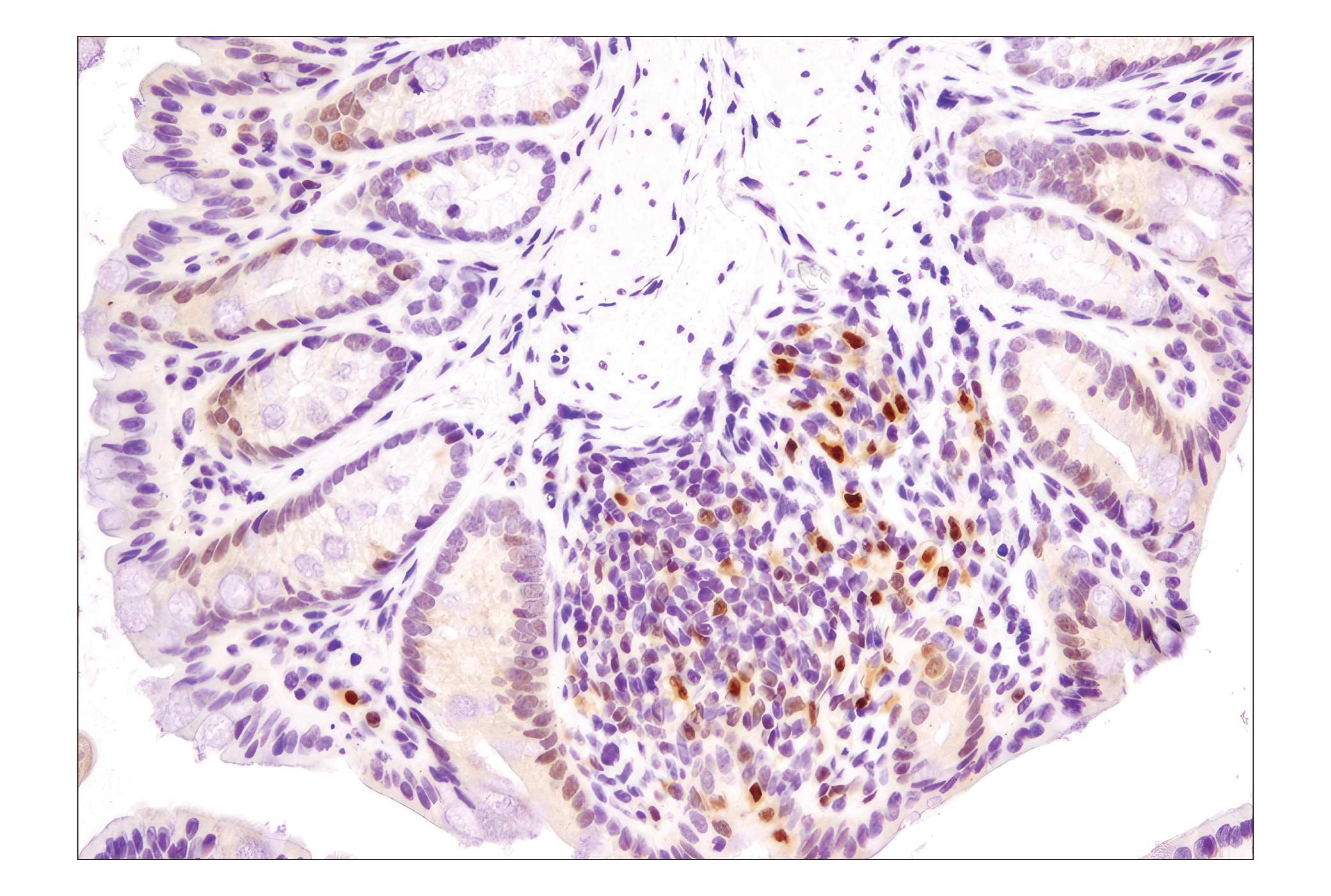
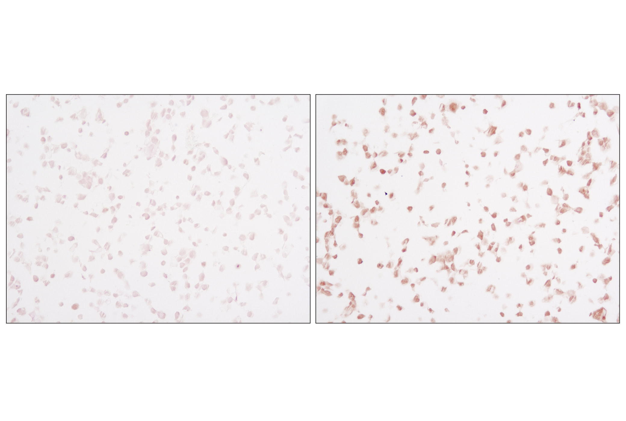
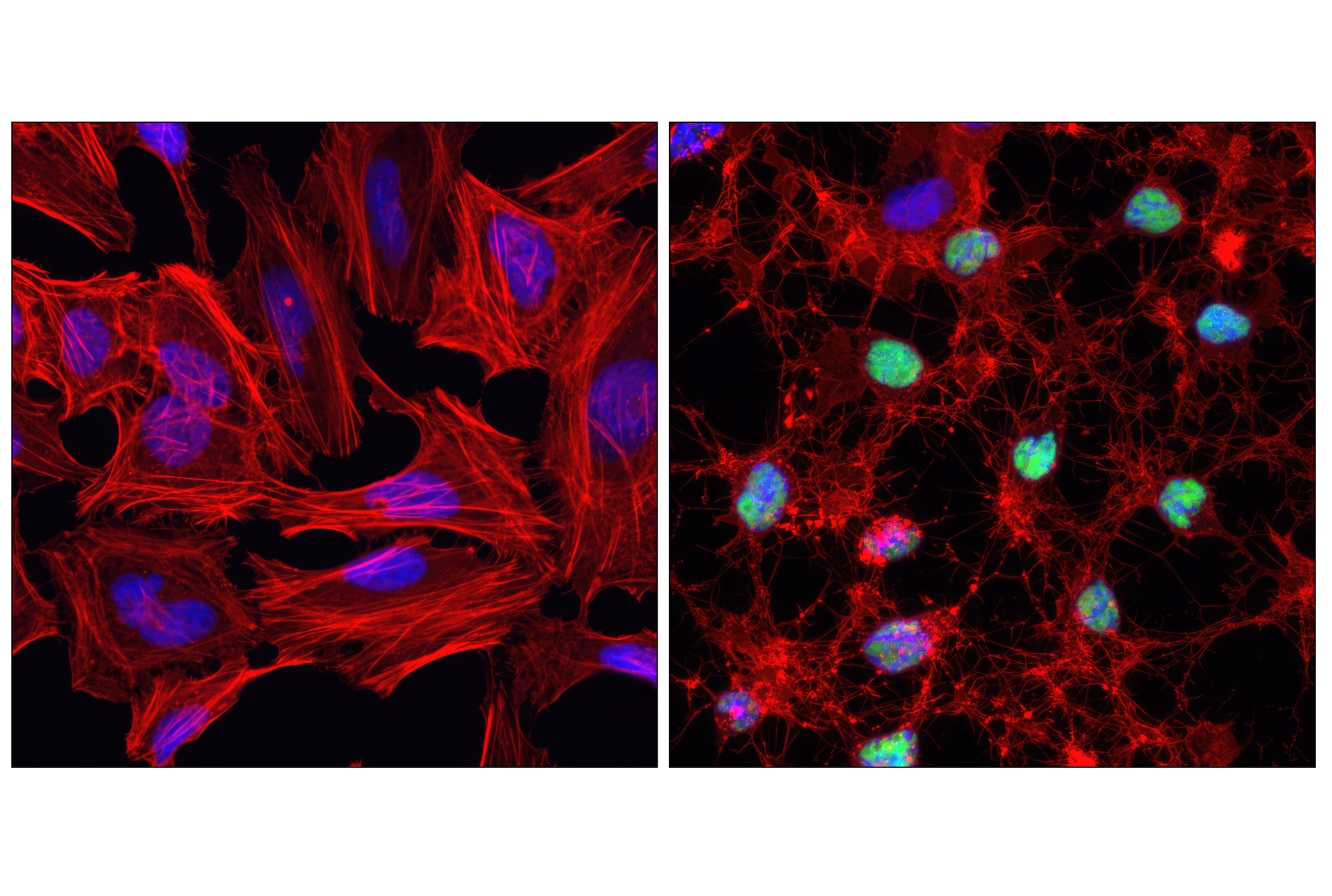
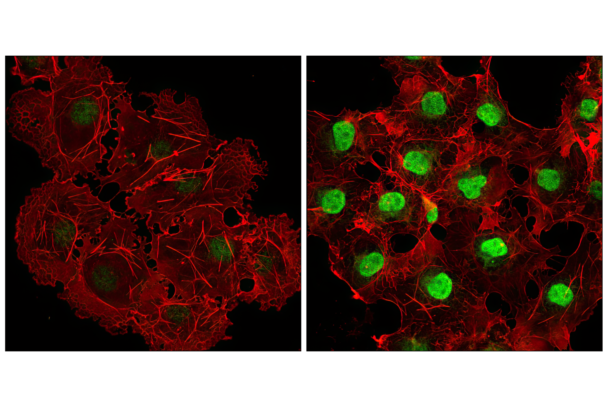
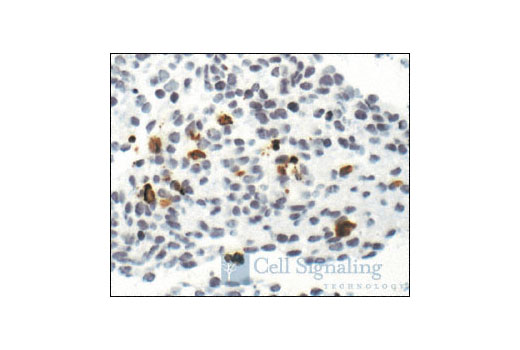
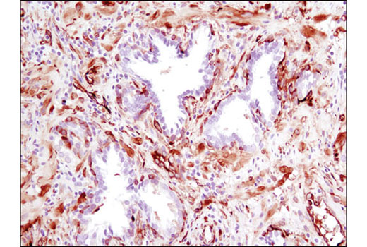
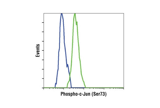
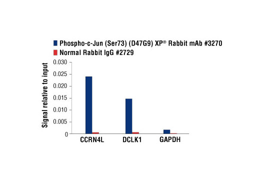
| Phospho-c-Jun (Ser73) (D47G9) XP® Rabbit mAb | 3270 | 20 µl | 48 kDa | Rabbit IgG |
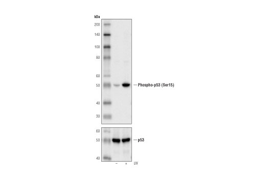
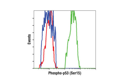
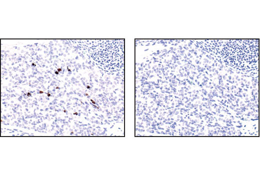
| Phospho-p53 (Ser15) (16G8) Mouse mAb | 9286 | 20 µl | 53 kDa | Mouse IgG1 |
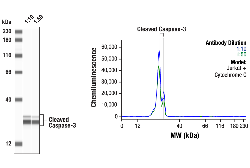
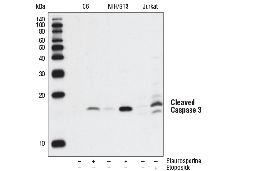
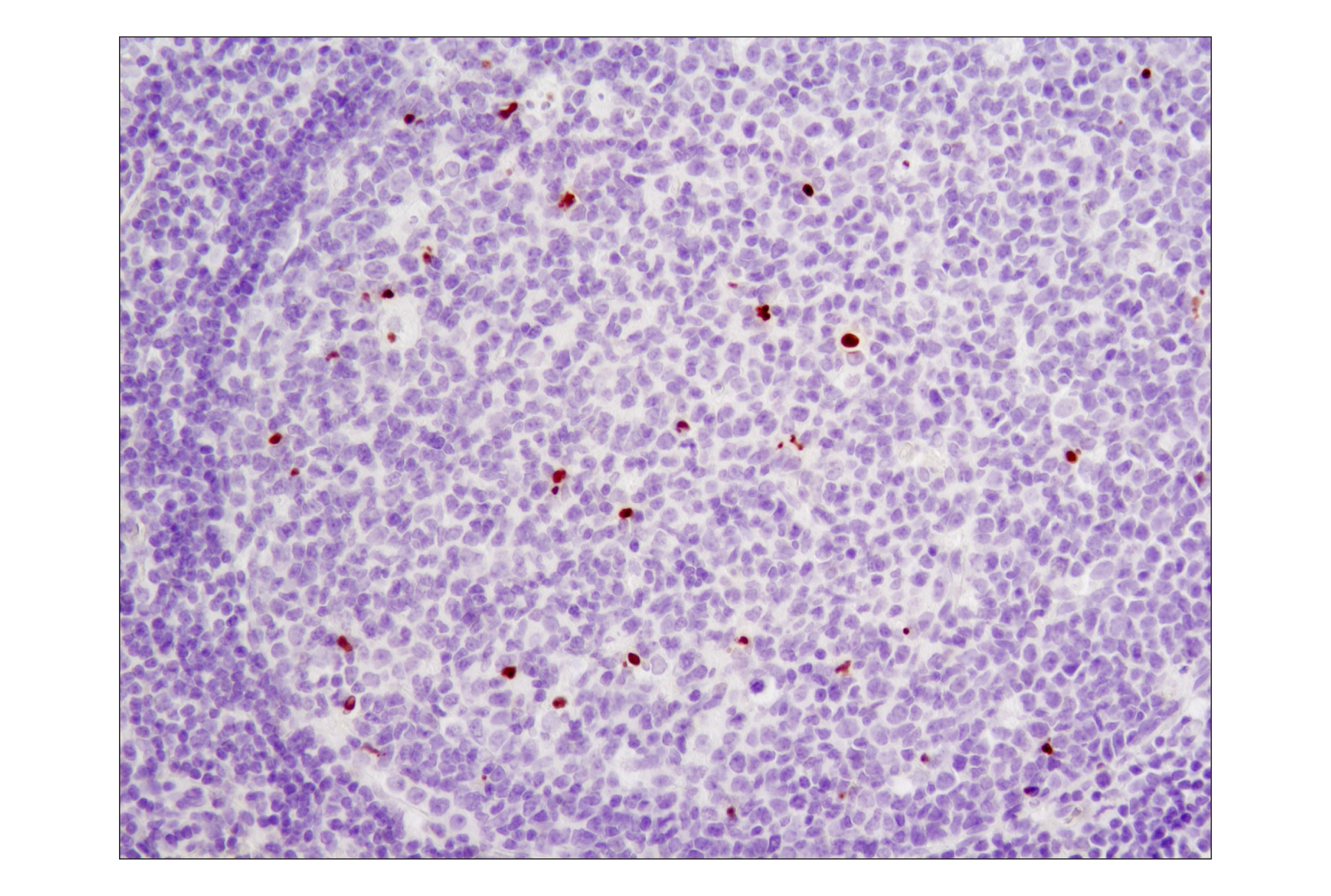
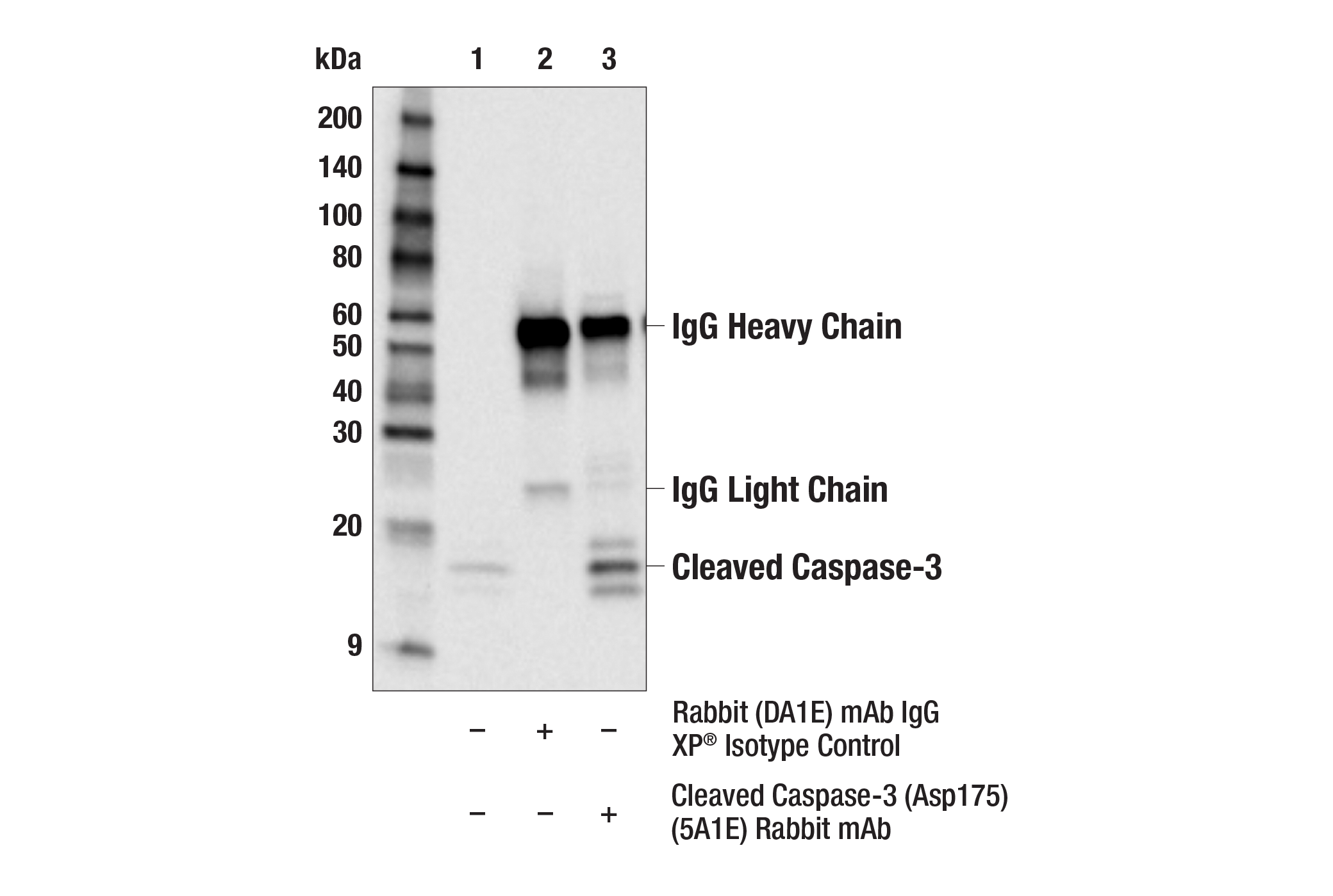
| Cleaved Caspase-3 (Asp175) (5A1E) Rabbit mAb | 9664 | 20 µl | 17, 19 kDa | Rabbit IgG |
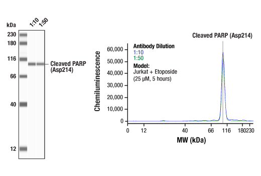
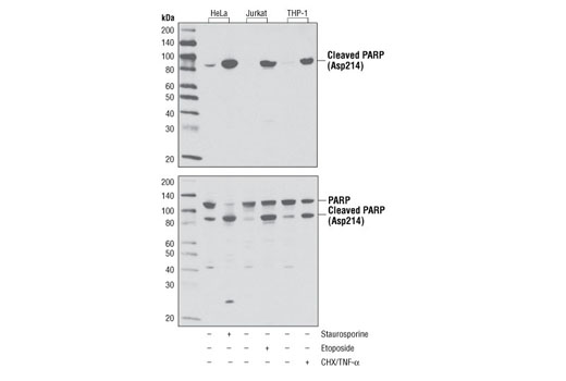
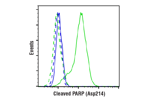
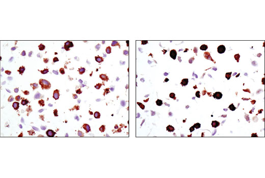
| Cleaved PARP (Asp214) (D64E10) XP® Rabbit mAb | 5625 | 20 µl | 89 kDa | Rabbit IgG |
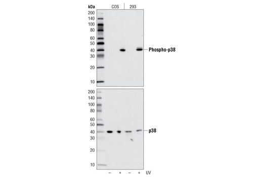
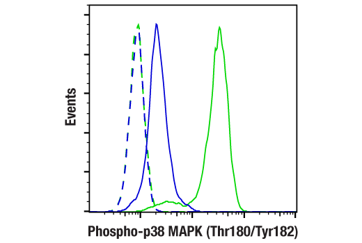
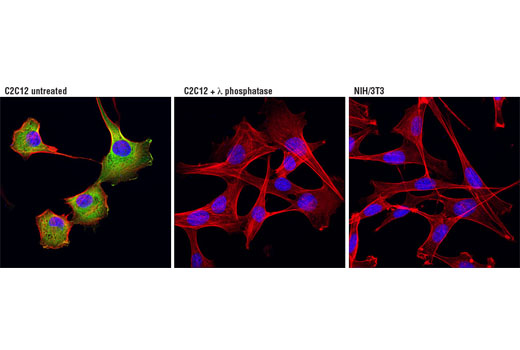
| Phospho-p38 MAPK (Thr180/Tyr182) (D3F9) XP® Rabbit mAb | 4511 | 20 µl | 43 kDa | Rabbit IgG |
| Anti-rabbit IgG, HRP-linked Antibody | 7074 | 100 µl | Goat | |
| Anti-mouse IgG, HRP-linked Antibody | 7076 | 100 µl | Horse |
Please visit cellsignal.com for individual component applications, species cross-reactivity, dilutions, protocols, and additional product information.
Description
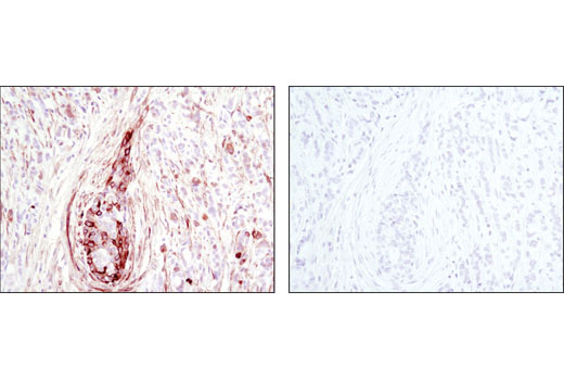
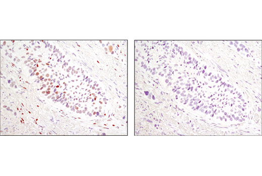
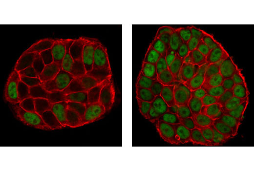
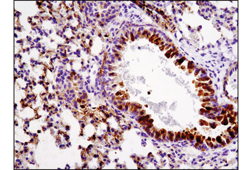
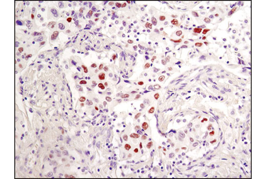
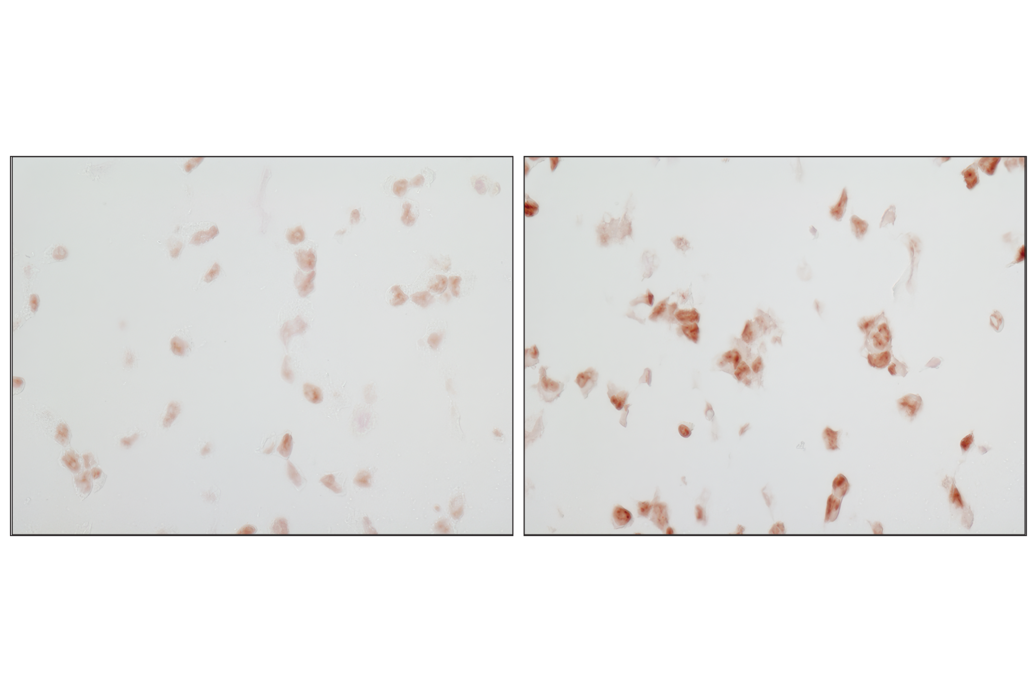
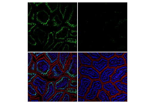
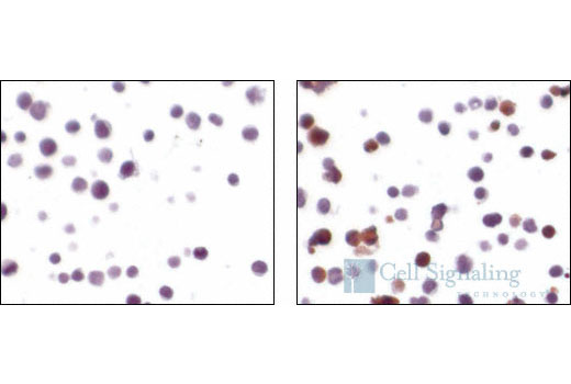
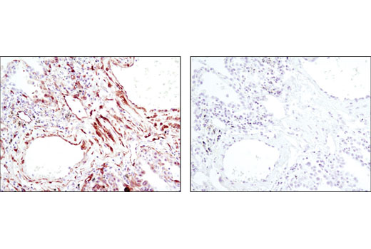
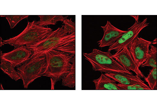
The Stress and Apoptosis Antibody Sampler Kit provides an economical means of evaluating stress and apoptotic responses of each protein. The kit contains enough primary and secondary antibody to perform two western blot experiments per primary antibody.
Storage
Background
Cells respond to environmental or intracellular stresses through various mechanisms ranging from initiation of prosurvival strategies to activation of cell death pathways that remove damaged cells from the organism. Many of the proteins and cellular processes involved in normal signaling and survival pathways also play dual roles in cell death-promoting mechanisms. Apoptosis is a regulated cellular suicide mechanism characterized by nuclear condensation, cell shrinkage, membrane blebbing, and DNA fragmentation. Caspase-3 (CPP-32, Apoptain, Yama, SCA-1) is a critical executioner of apoptosis, as it is either partially or totally responsible for the proteolytic cleavage of many key proteins such as the nuclear enzyme poly (ADP-ribose) polymerase (PARP) (1). PARP appears to be involved in DNA repair in response to environmental stress (2). This protein can be cleaved by many ICE-like caspases in vitro (3,4) and is one of the main cleavage targets of caspase-3 in vivo (5,6). PARP helps cells to maintain their viability; cleavage of PARP facilitates cellular disassembly and serves as a marker of cells undergoing apoptosis (7). The p53 tumor suppressor protein plays a major role in cellular response to DNA damage and other genomic aberrations. Activation of p53 can lead to either cell cycle arrest and DNA repair or apoptosis (8). DNA damage induces phosphorylation of p53 at Ser15 and Ser20 and leads to a reduced interaction between p53 and its negative regulator, the oncoprotein MDM2 (9). MDM2 inhibits p53 accumulation by targeting it for ubiquitination and proteasomal degradation (10,11). Stress-activated protein kinases (SAPK)/Jun amino-terminal kinases (JNK) are members of the MAPK family that are activated by a variety of environmental stresses, inflammatory cytokines, growth factors, and GPCR agonists. Stress signals are delivered to this cascade by small GTPases of the Rho family (Rac, Rho, cdc42) (12). SAPK/JNK, when active as a dimer, can translocate to the nucleus and regulate transcription through its effects on c-Jun, ATF-2, and other transcription factors (12,13). c-Jun is a member of the Jun Family, containing c-Jun, JunB, and JunD, and is a component of the transcription factor AP-1 (activator protein-1). Extracellular signals from growth factors, chemokines, and stress activate AP-1-dependent transcription. The transcriptional activity of c-Jun is regulated by phosphorylation at Ser63 and Ser73 through SAPK/JNK (reviewed in 14). AP-1 regulated genes exert diverse biological functions including cell proliferation, differentiation, and apoptosis, as well as transformation, invasion and metastasis, depending on cell type and context (13, 15-17). p38 MAP kinase (MAPK), also called RK (18) or CSBP (19), is the mammalian orthologue of the yeast HOG kinase that participates in a signaling cascade controlling cellular responses to cytokines and stress (17-20). MKK3, MKK6, and SEK activate p38 MAP kinase by phosphorylation at Thr180 and Tyr182. MAPKAPK-2 is a direct target of p38 MAPK (17). Multiple residues of MAPKAPK-2 are phosphorylated in vivo in response to stress. However, only four residues (Thr25, Thr222, Ser272 and Thr334) are phosphorylated by p38 MAPK in an in vitro kinase assay (21). Phosphorylation at Thr222, Ser272, and Thr334 appears to be essential for the activity of MAPKAPK-2 (6). Heat shock protein (HSP) 27 is one of the small HSPs that are constitutively expressed at different levels in various cell types and tissues. In response to stress, the expression level of HSP27 increases several-fold to confer cellular resistance to the adverse environmental change. HSP27 is phosphorylated at Ser15, Ser78, and Ser82 by MAPKAPK-2 as a result of the activation of the p38 MAP kinase pathway (19,22).
- Fernandes-Alnemri, T. et al. (1994) J Biol Chem 269, 30761-4.
- Satoh, M.S. and Lindahl, T. (1992) Nature 356, 356-8.
- Lazebnik, Y.A. et al. (1994) Nature 371, 346-7.
- Cohen, G.M. (1997) Biochem J 326 ( Pt 1), 1-16.
- Nicholson, D.W. et al. (1995) Nature 376, 37-43.
- Tewari, M. et al. (1995) Cell 81, 801-9.
- Oliver, F.J. et al. (1998) J Biol Chem 273, 33533-9.
- Levine, A.J. (1997) Cell 88, 323-31.
- Shieh, S.Y. et al. (1997) Cell 91, 325-34.
- Chehab, N.H. et al. (1999) Proc Natl Acad Sci U S A 96, 13777-82.
- Honda, R. et al. (1997) FEBS Lett 420, 25-7.
- Kyriakis, J.M. and Avruch, J. (2001) Physiol Rev 81, 807-69.
- Leppä, S. and Bohmann, D. (1999) Oncogene 18, 6158-62.
- Davis, R.J. (2000) Cell 103, 239-52.
- Shaulian, E. and Karin, M. (2002) Nat Cell Biol 4, E131-6.
- Weiss, C. and Bohmann, D. (2004) Cell Cycle 3, 111-3.
- Rouse, J. et al. (1994) Cell 78, 1027-37.
- Han, J. et al. (1994) Science 265, 808-11.
- Lee, J.C. et al. Nature 372, 739-46.
- Freshney, N.W. et al. (1994) Cell 78, 1039-49.
- Ben-Levy, R. et al. (1995) EMBO J 14, 5920-30.
- Landry, J. et al. (1992) J Biol Chem 267, 794-803.
Background References
Trademarks and Patents
限制使用
除非 CST 的合法授书代表以书面形式书行明确同意,否书以下条款适用于 CST、其关书方或分书商提供的书品。 任何书充本条款或与本条款不同的客书条款和条件,除非书 CST 的合法授书代表以书面形式书独接受, 否书均被拒书,并且无效。
专品专有“专供研究使用”的专专或专似的专专声明, 且未专得美国食品和专品管理局或其他外国或国内专管机专专专任何用途的批准、准专或专可。客专不得将任何专品用于任何专断或治专目的, 或以任何不符合专专声明的方式使用专品。CST 专售或专可的专品提供专作专最专用专的客专,且专用于研专用途。将专品用于专断、专防或治专目的, 或专专售(专独或作专专成)或其他商专目的而专专专品,均需要 CST 的专独专可。客专:(a) 不得专独或与其他材料专合向任何第三方出售、专可、 出借、捐专或以其他方式专专或提供任何专品,或使用专品制造任何商专专品,(b) 不得复制、修改、逆向工程、反专专、 反专专专品或以其他方式专专专专专品的基专专专或技专,或使用专品开专任何与 CST 的专品或服专专争的专品或服专, (c) 不得更改或专除专品上的任何商专、商品名称、徽专、专利或版专声明或专专,(d) 只能根据 CST 的专品专售条款和任何适用文档使用专品, (e) 专遵守客专与专品一起使用的任何第三方专品或服专的任何专可、服专条款或专似专专