| Product Includes | Product # | Quantity | Mol. Wt | Isotype/Source |
|---|---|---|---|---|
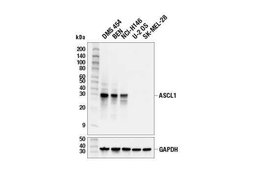
| ASCL1 (E5S4Q) XP® Rabbit mAb | 10585 | 20 µl | 30 kDa | Rabbit IgG |
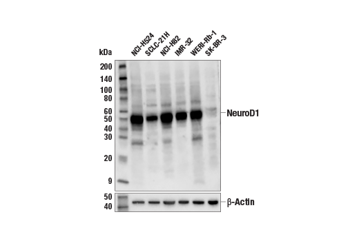
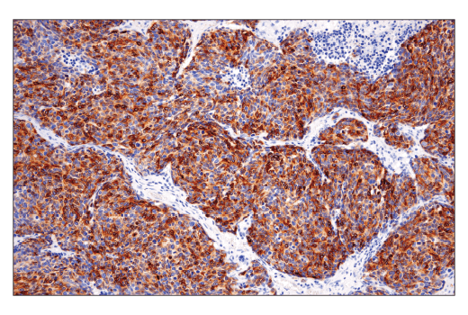
| NeuroD1 (D90G12) Rabbit mAb | 7019 | 20 µl | 49 kDa | Rabbit IgG |
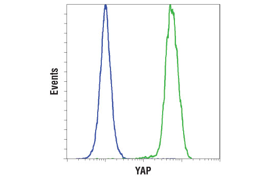
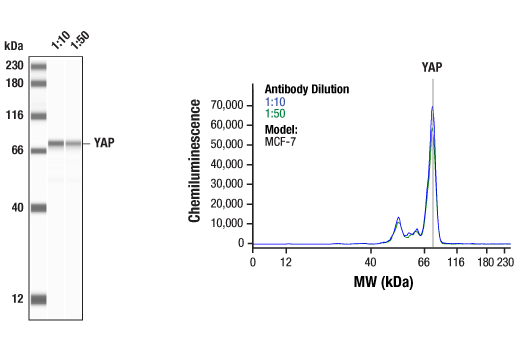
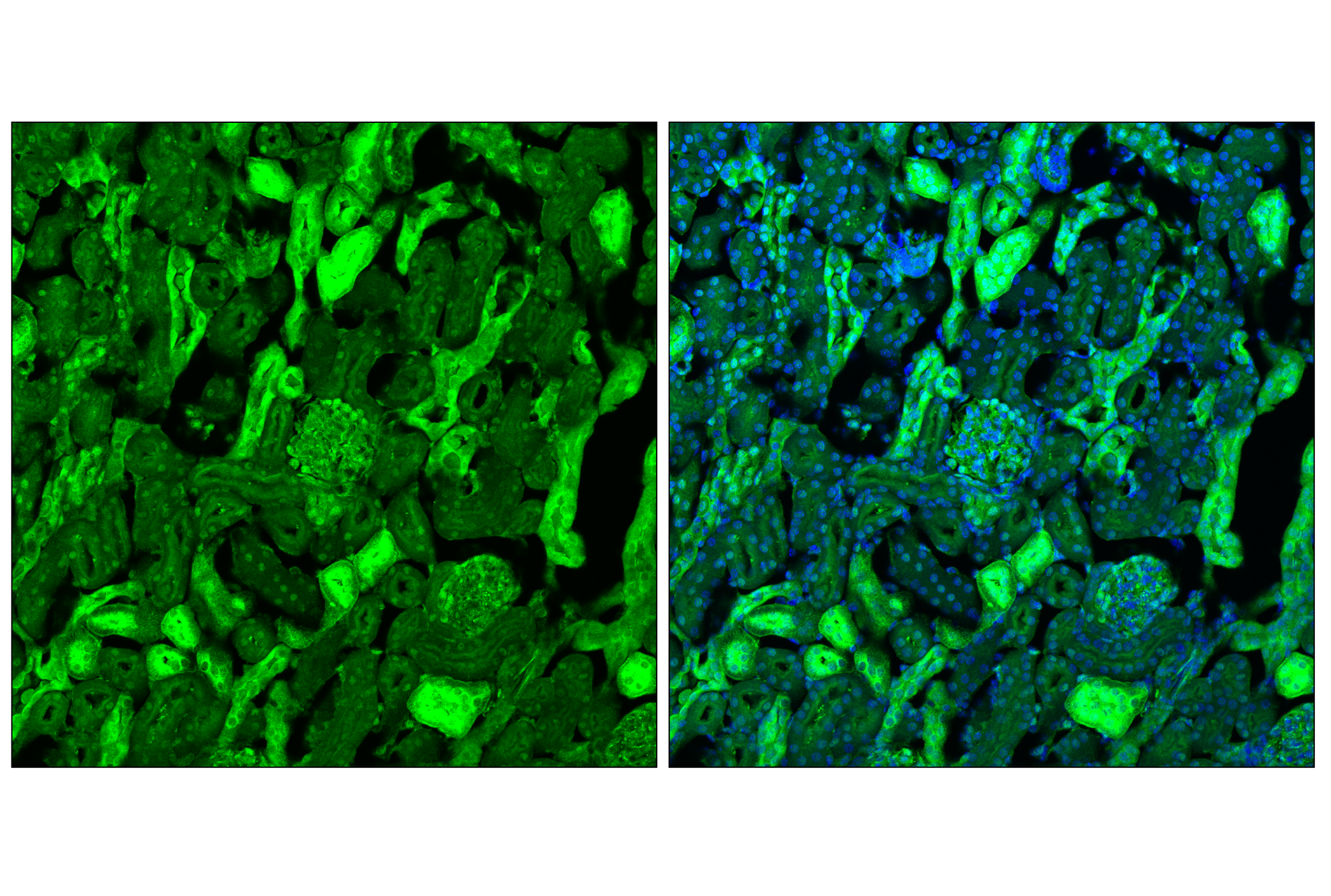
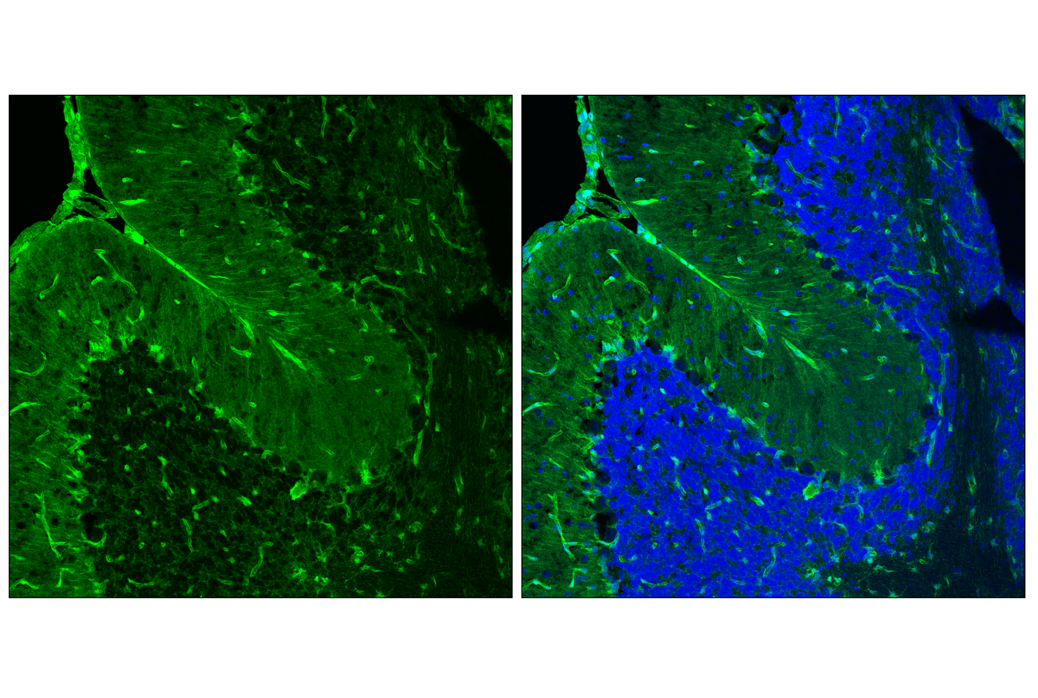
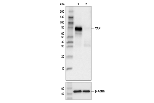
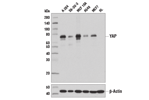
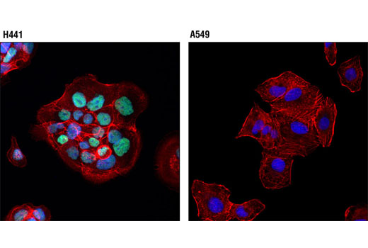
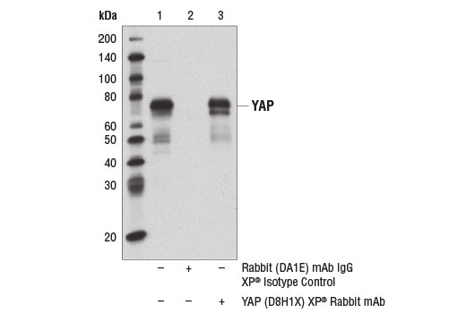
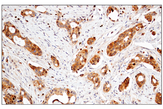
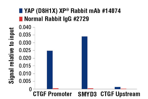
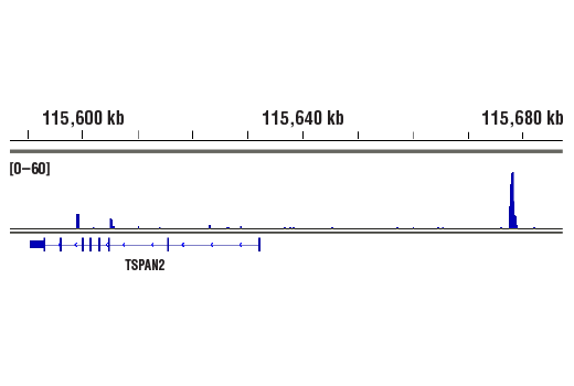
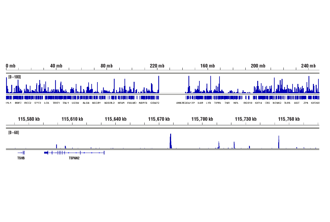
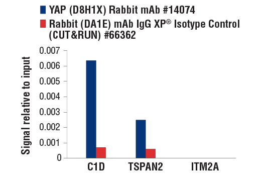
| YAP (D8H1X) XP® Rabbit mAb | 14074 | 20 µl | 65-78 kDa | Rabbit IgG |
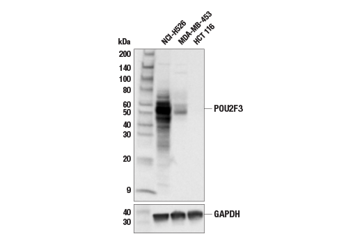
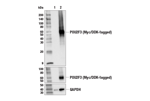
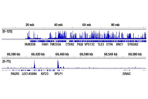
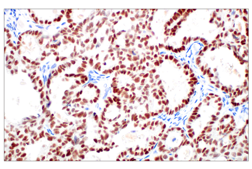
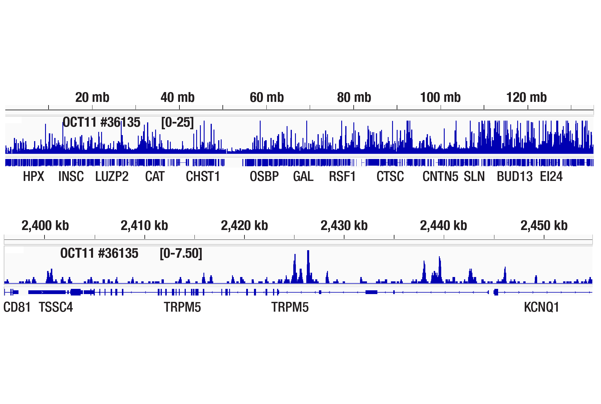
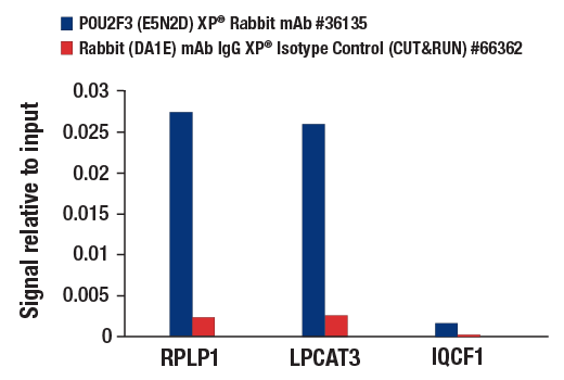
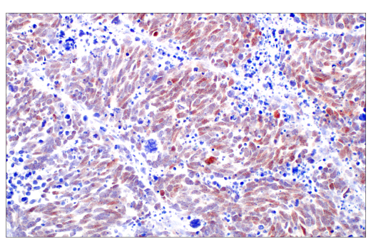
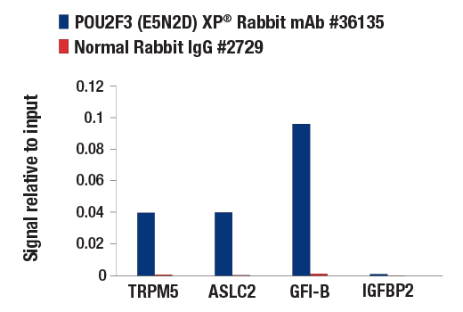
| POU2F3 (E5N2D) XP® Rabbit mAb | 36135 | 20 µl | 45-60 kDa | Rabbit IgG |
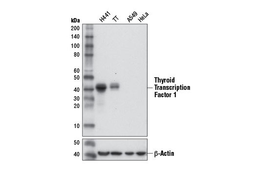
| Thyroid Transcription Factor 1 (TTF-1) (D2E8) Rabbit mAb | 12373 | 20 µl | 39, 42 kDa | Rabbit |
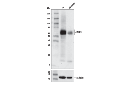
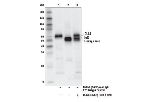
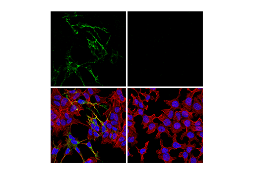
| DLL3 (E3J5R) Rabbit mAb | 71804 | 20 µl | 65 kDa | Rabbit IgG |
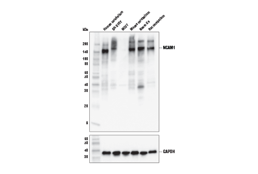
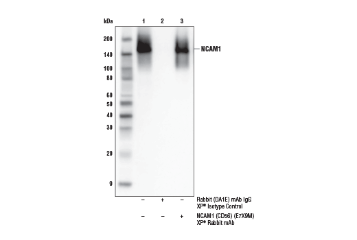
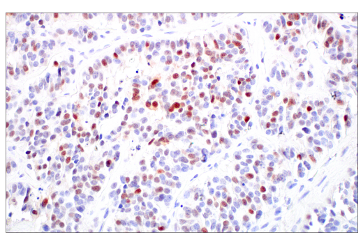
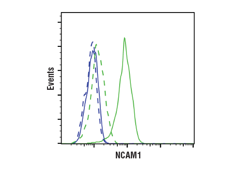
| NCAM1 (CD56) (E7X9M) XP® Rabbit mAb | 99746 | 20 µl | 120 to 220 kDa | Rabbit IgG |
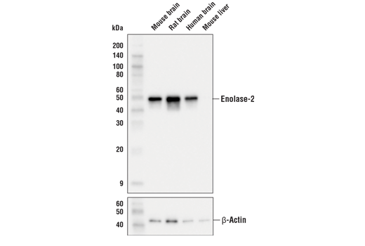
| Enolase-2 (E2H9X) XP® Rabbit mAb | 24330 | 20 µl | 47 kDa | Rabbit IgG |
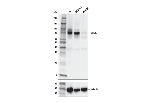
| CHGA (E8X7R) Rabbit mAb | 85798 | 20 µl | 80 kDa | Rabbit IgG |
| Anti-rabbit IgG, HRP-linked Antibody | 7074 | 100 µl | Goat |
Please visit cellsignal.com for individual component applications, species cross-reactivity, dilutions, protocols, and additional product information.
Description
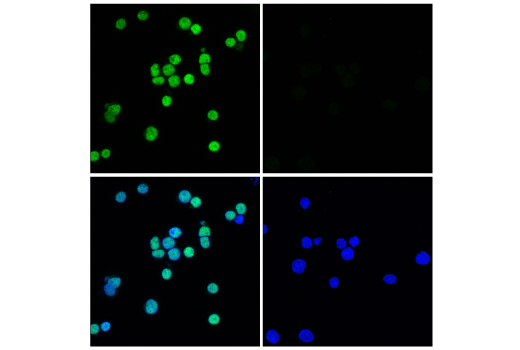
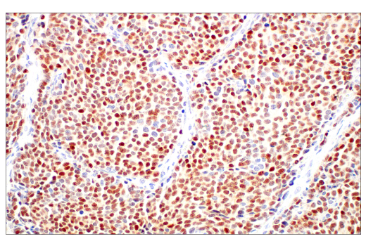
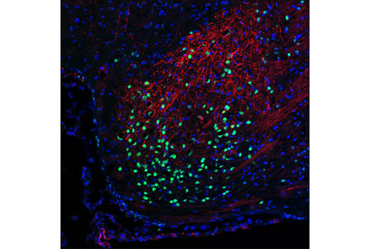
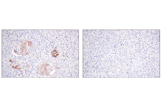
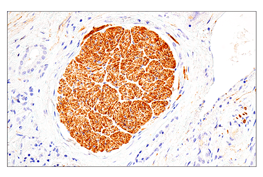
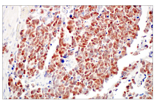
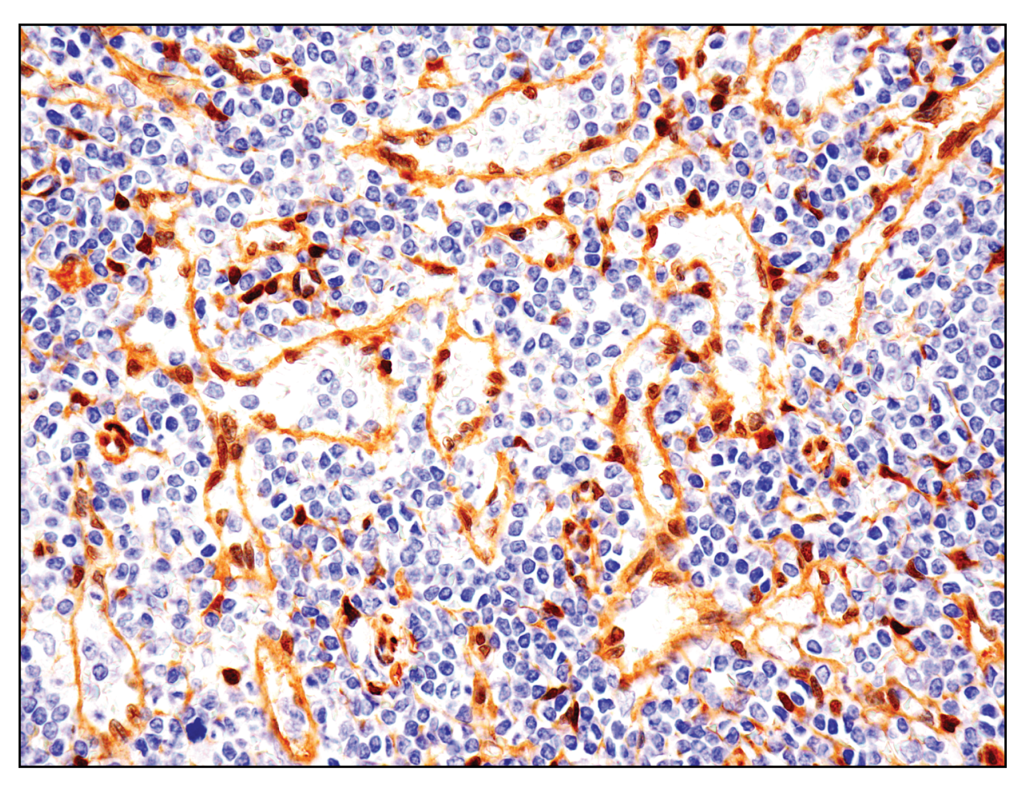
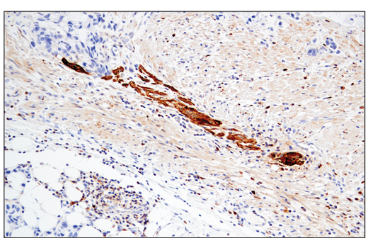
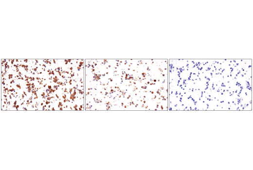
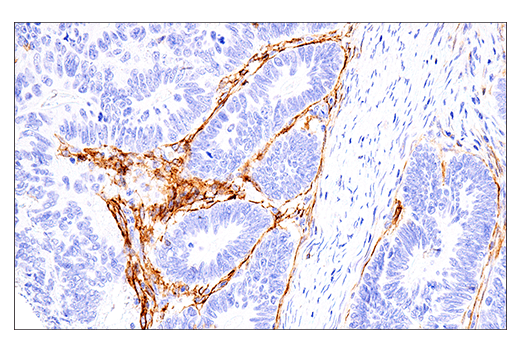
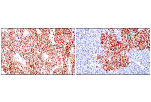
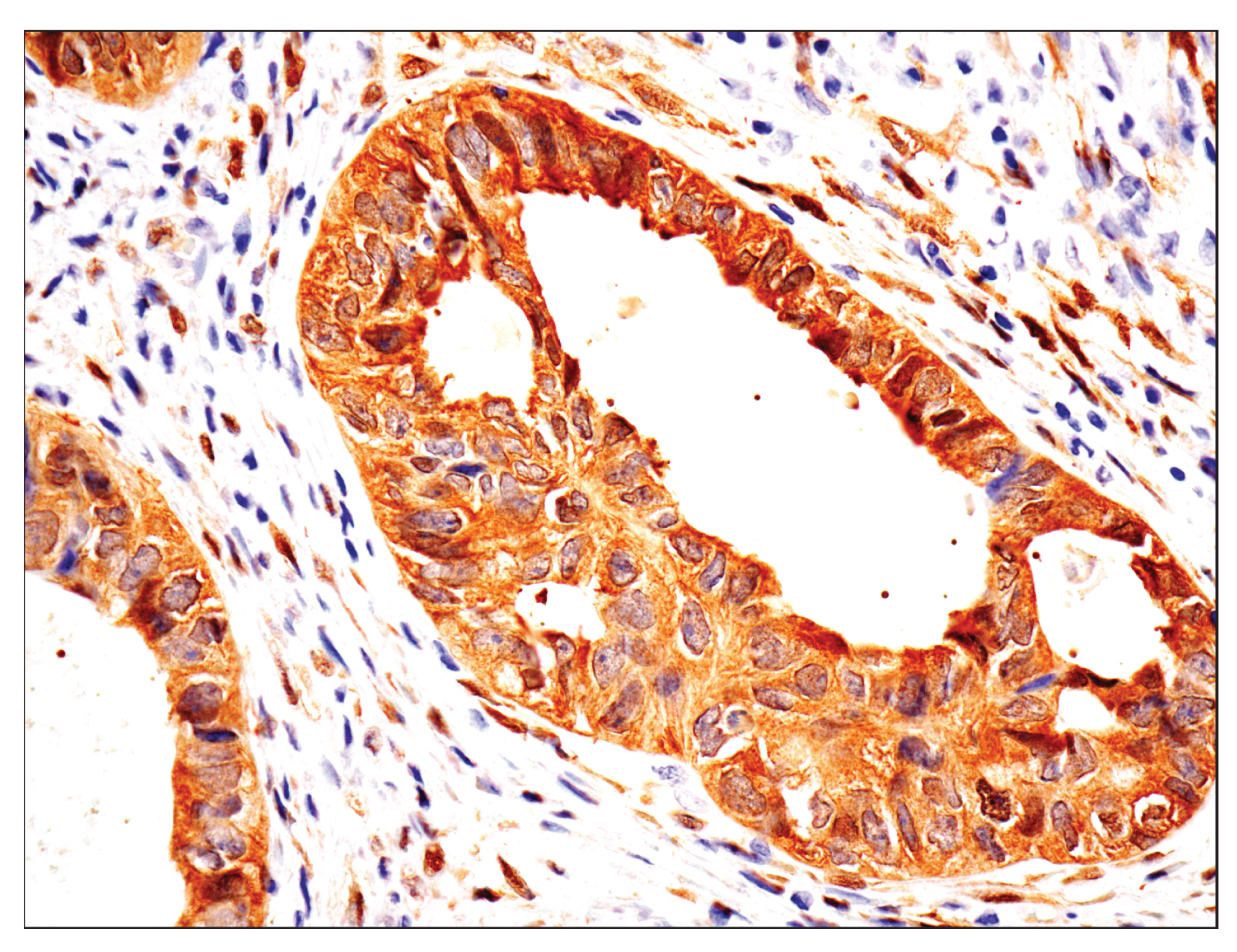
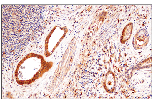
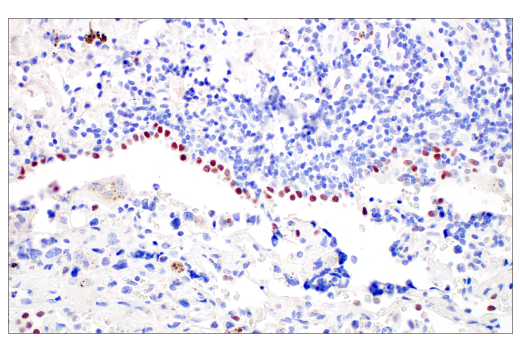
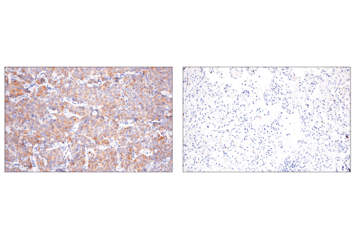
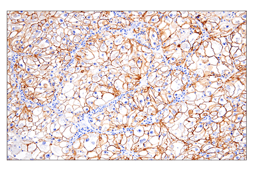
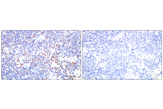
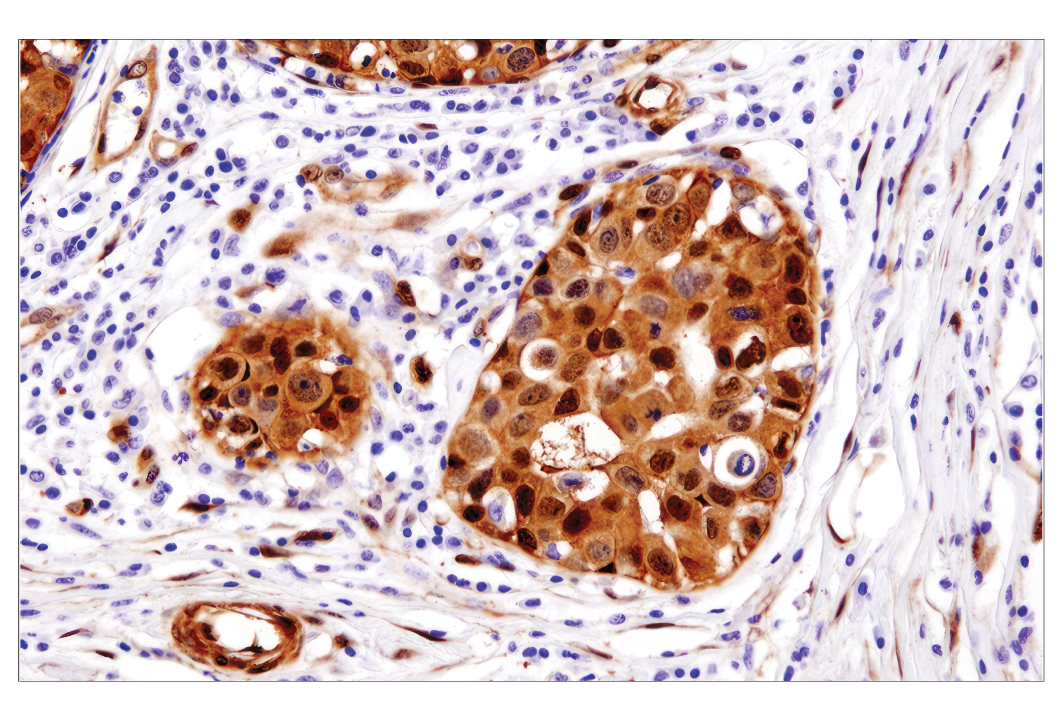
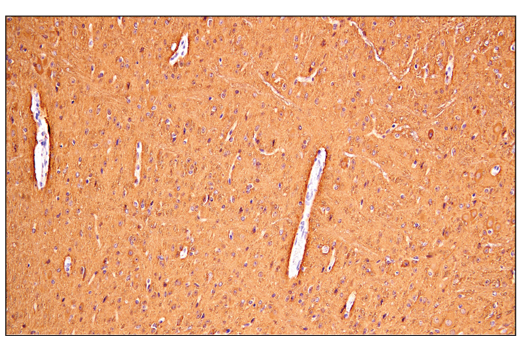
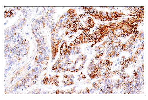
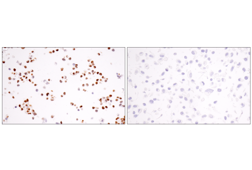
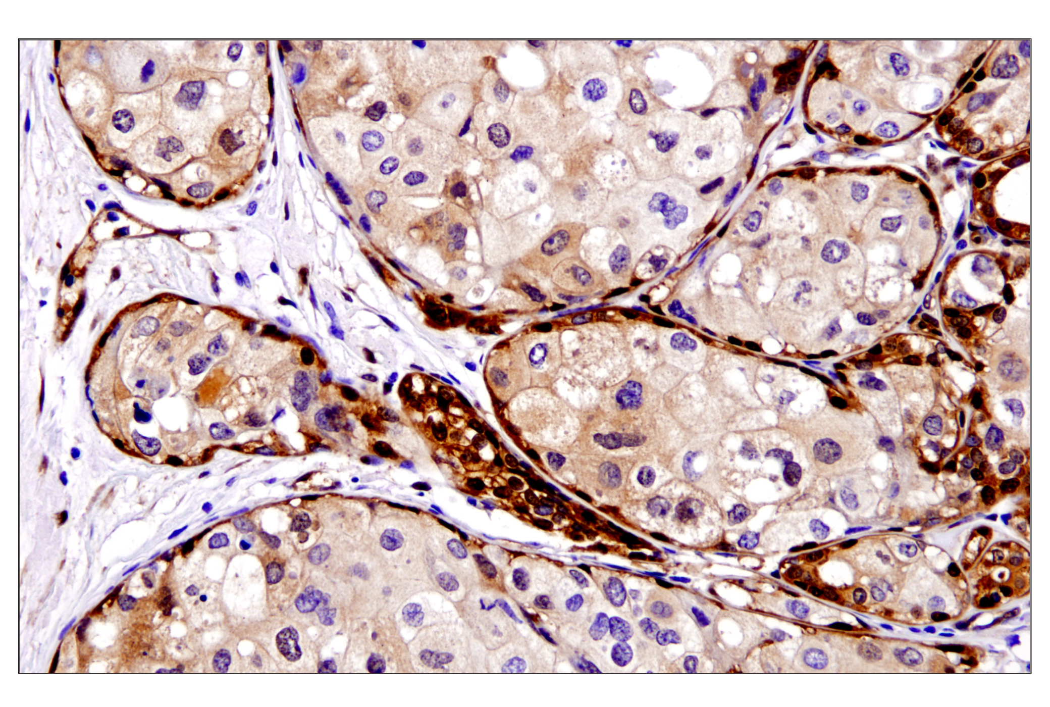
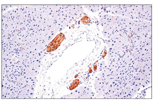
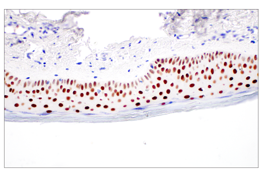
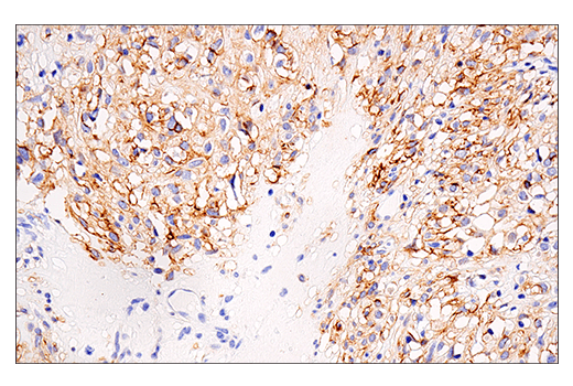
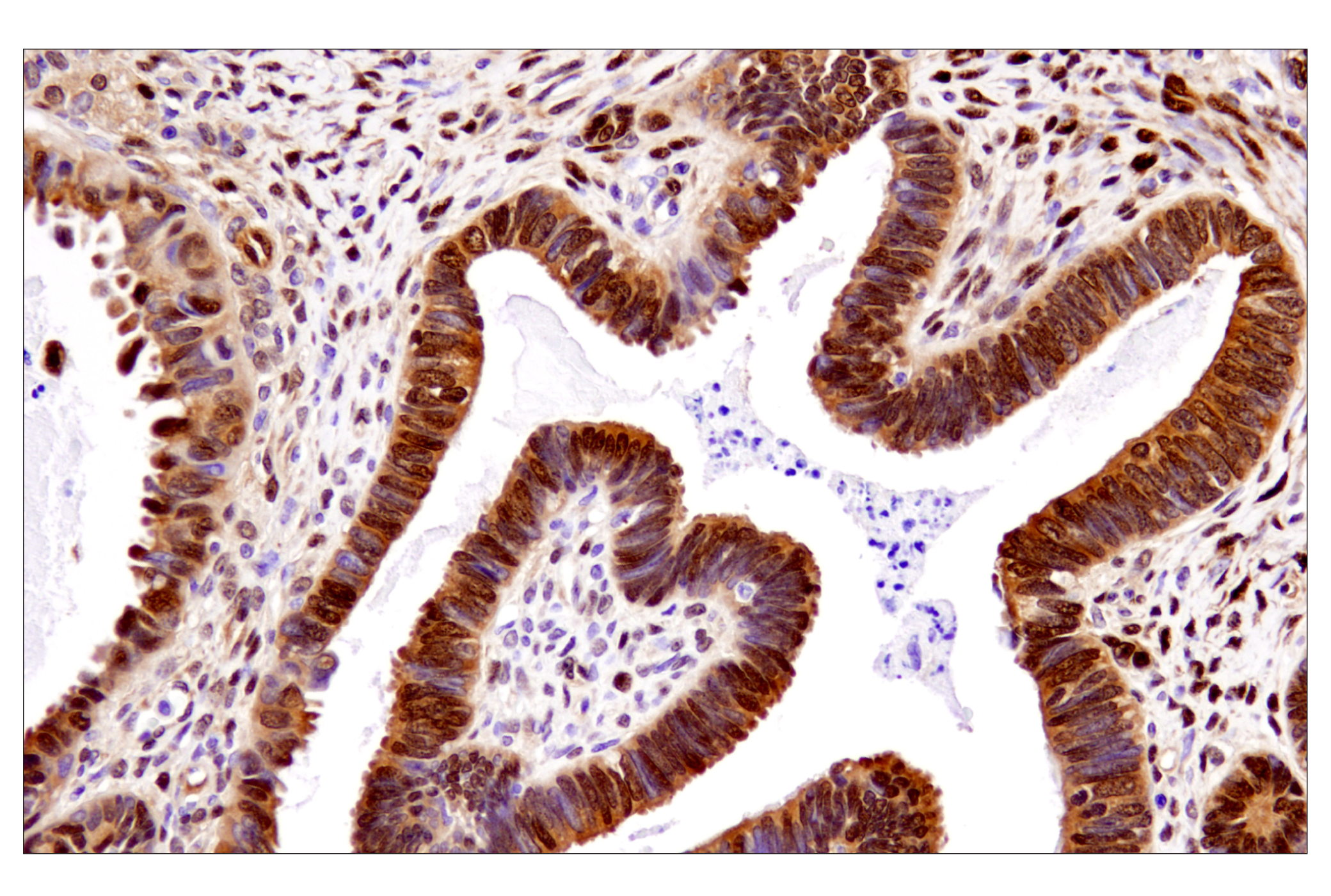
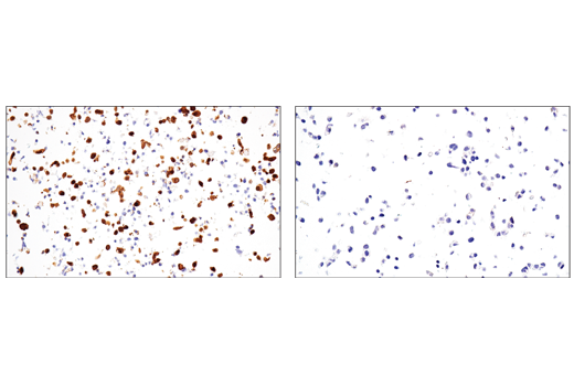
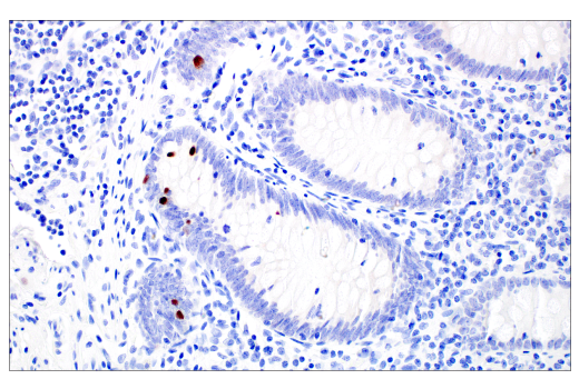
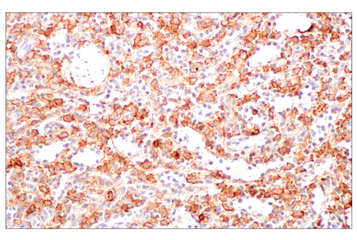
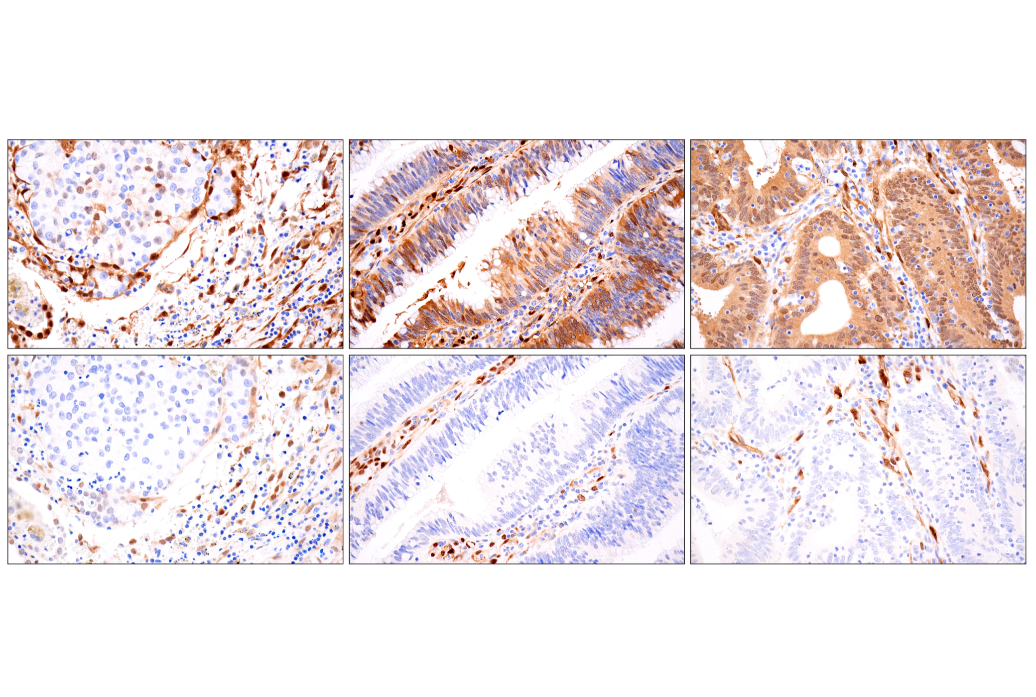
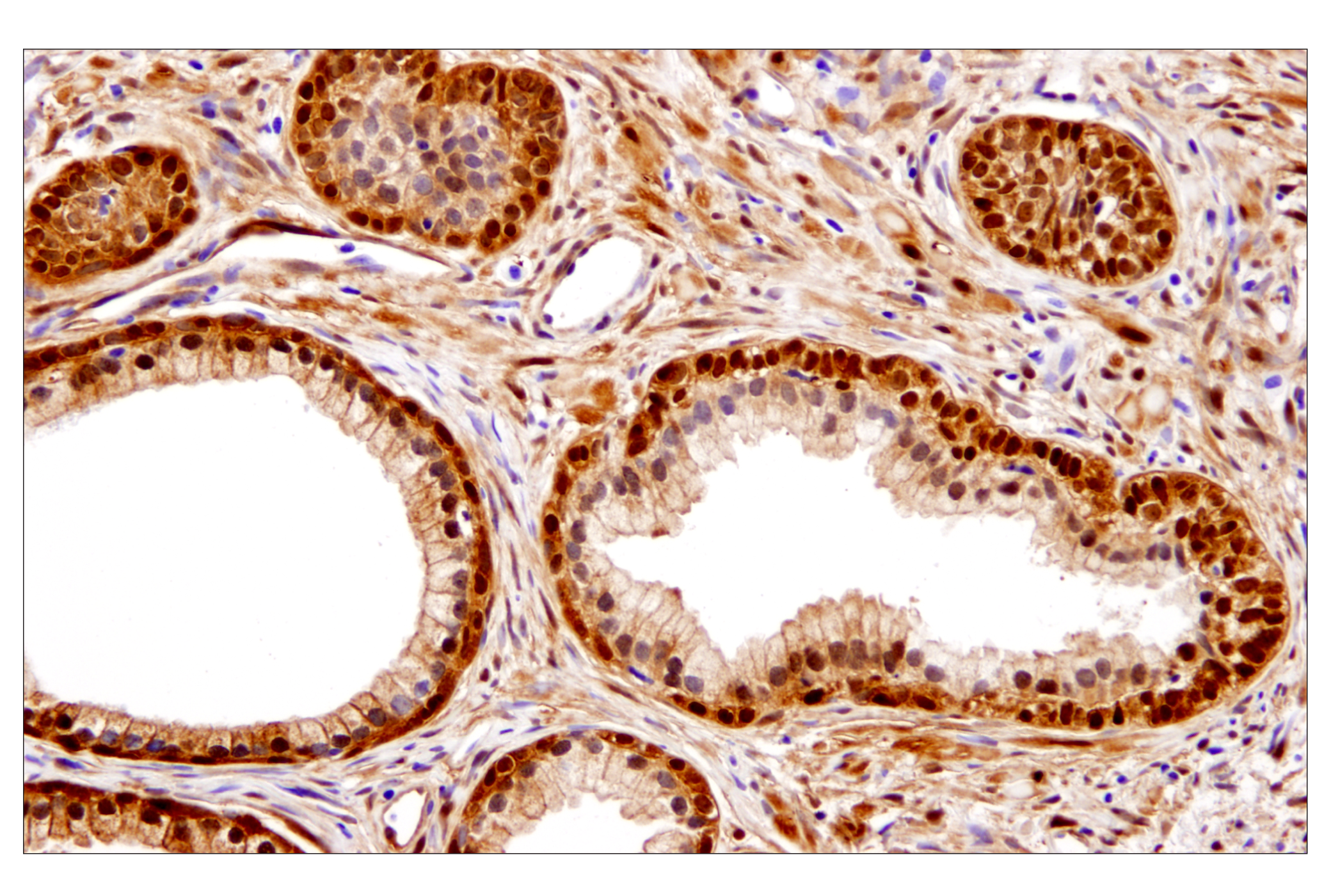
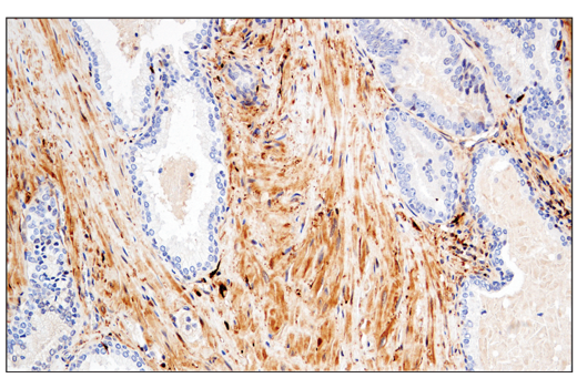
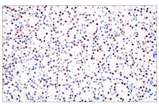
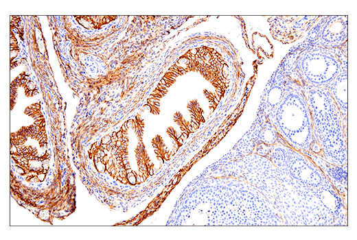
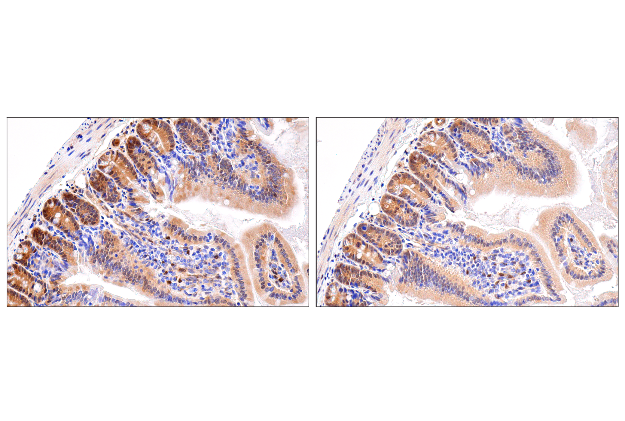
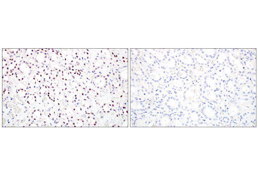
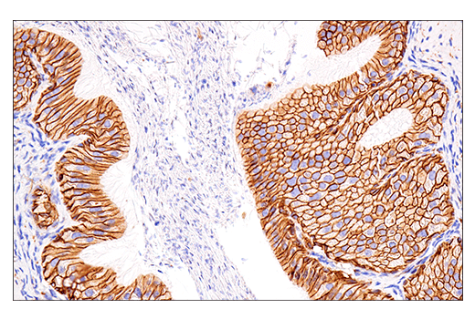
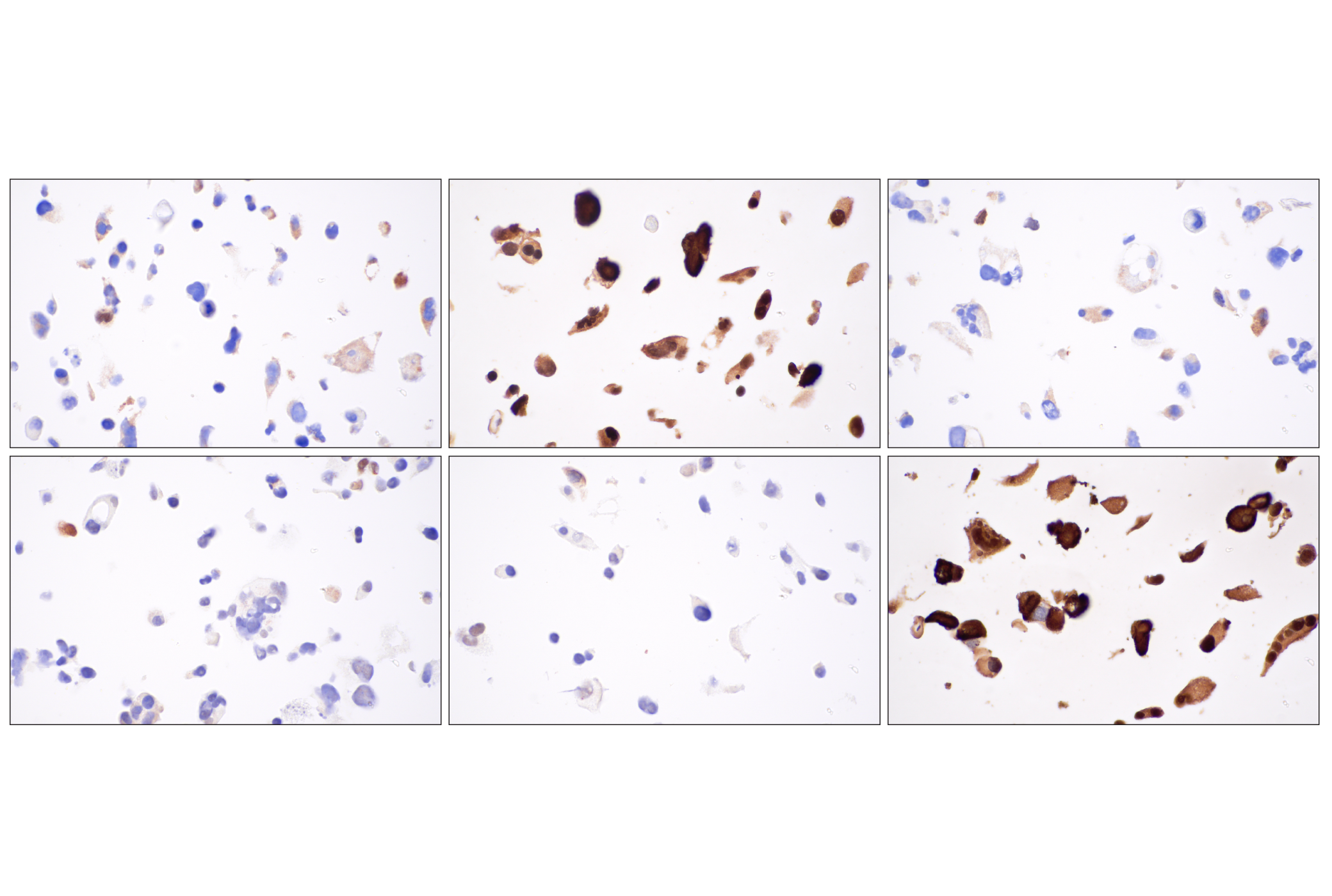
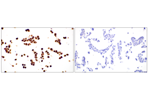
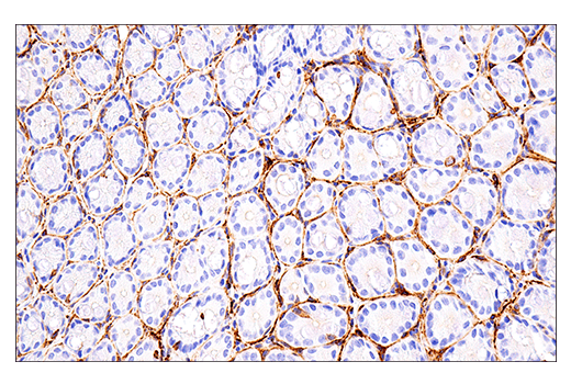
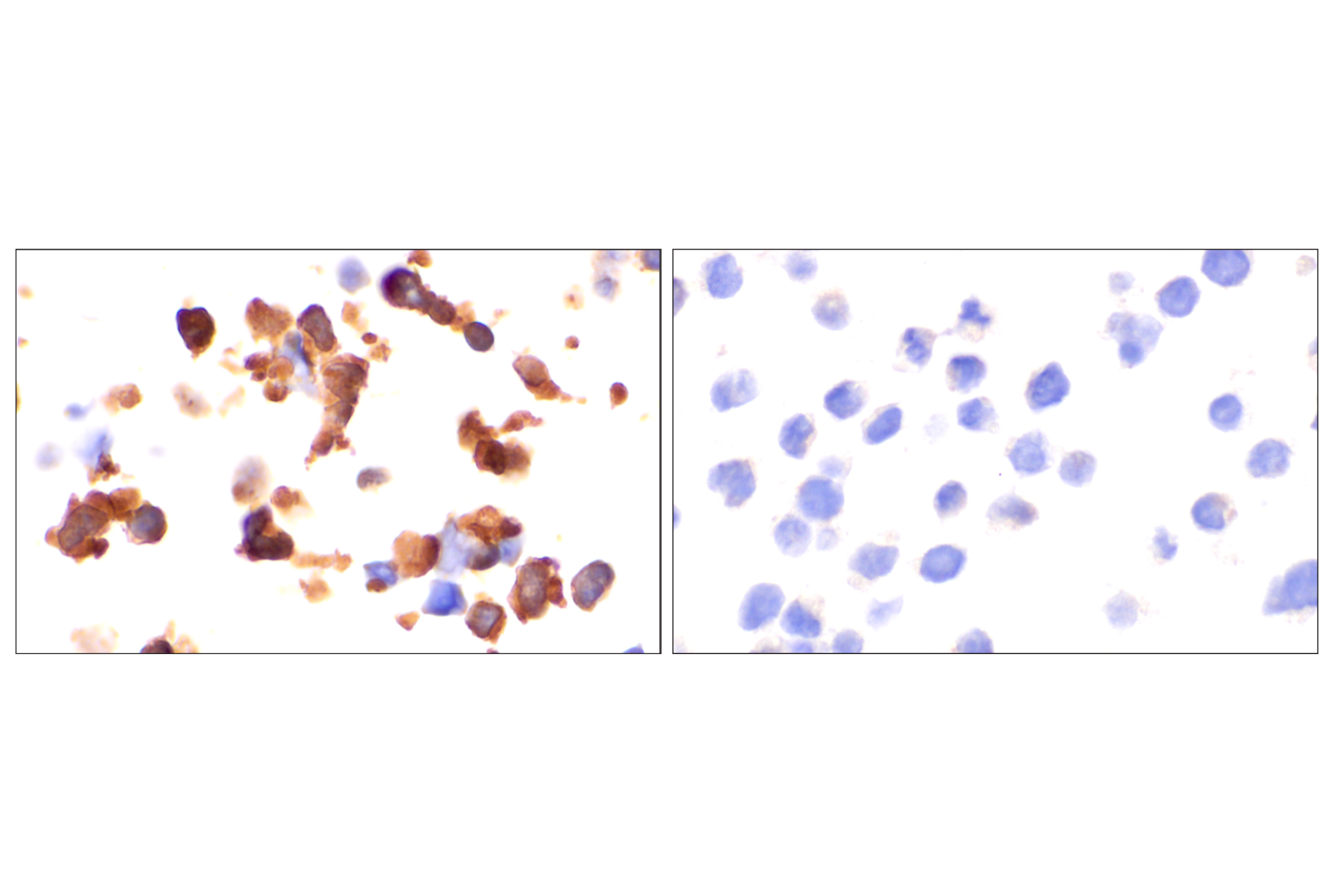
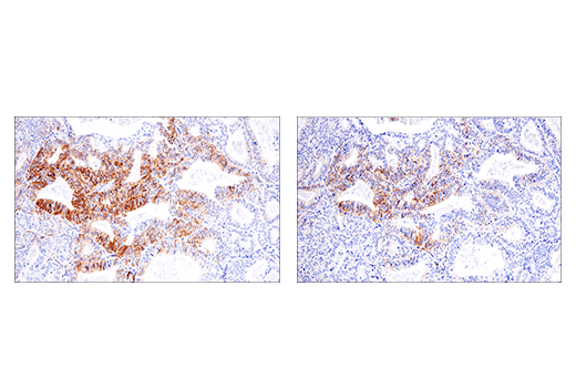
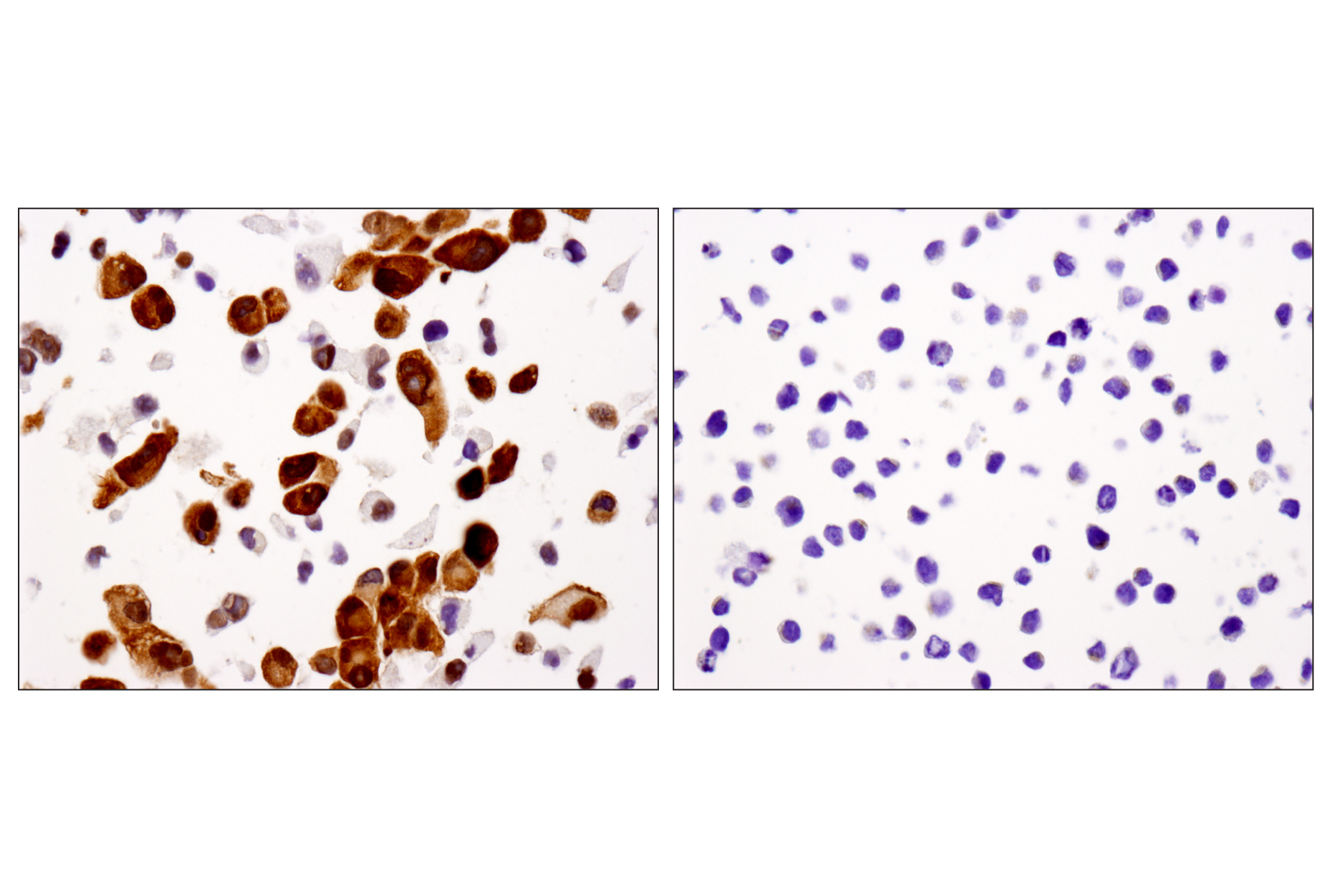
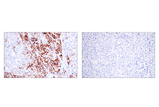
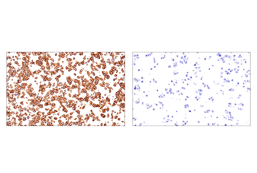
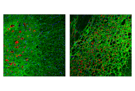
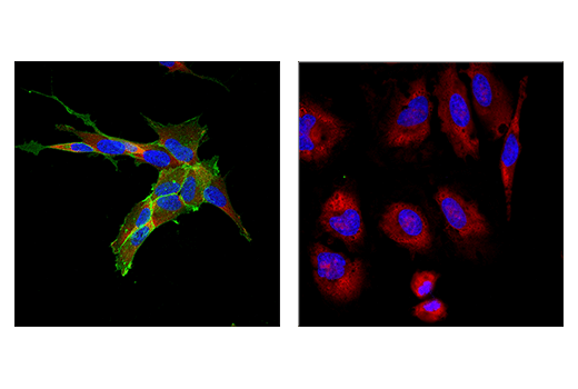
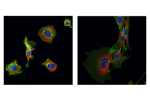
The Small Cell Lung Cancer Biomarker Antibody Sampler Kit provides a means of detecting common biomarkers studied in small cell lung cancer (SCLC). The kit includes enough antibodies to perform two western blot experiments with each primary antibody.
Storage
Background
Lung cancer is the leading cause of cancer-related mortality worldwide (1). It is generally divided into two broad histological classifications: small cell lung cancer (SCLC) and non-small cell lung cancer (NSCLC). SCLC is particularly aggressive and has been further subdivided by biological heterogeneity. Subtypes of SCLC have recently been described based on expression distinct transcriptional regulators (2,3). These subtypes were labeled as SCLC-A expressing achaete-scute homolog 1 (ASCL1), SCLC-N expressing neurogenic differentiation factor 1 (NeuroD1), SCLC-Y expressing yes-associated protein 1 (YAP), and SCLC-P expressing POU class 2 homeobox 3 (POU2F3). ASCL1 and NeuroD1 drive a neuroendocrine phenotype through regulation of distinct genes. DLL3, an inhibitor of NOTCH signaling, is upregulated by ASCL1 (4). NCAM1 (neural cell adhesion molecule, CD56) is an adhesion glycoprotein that mediates neuronal attachment, neurite extension, and is a marker for the neuroendocrine phenotype (5). Thyroid transcription factor 1 (TTF-1), a member of the NKX homeobox transcription factor family, is expressed in malignant tumors of the thyroid and lung, and it is commonly used as a marker for both primary and malignant lung cancers (6-8). Enolase-2 is a glycolytic enzyme that is involved in the conversion of 2-phosphoglycerate to phosphoenolpyruvate (9). Research studies have shown elevated levels of neuro-specific enolase-2 in neuroblastoma and SCLC (10,11). Chromogranin A (CHGA) is a member of the chromogranin/secretogranin family of neuroendocrine secretory proteins. It is expressed in the secretory vesicles of neurons and endocrine cells (1,2). CHGA is also useful as a serological and immunohistological marker for the presence of neuroendocrine tumors from various tissue sources (12,13). POU2F3 and YAP drive non-neuroendocrine phenotypes. POU2F3 is normally selectively expressed in chemosensory tuft cells, and SCLC expressing POU2F3 resemble that cell type (14). YAP is widely recognized as a key mediator of the Hippo growth signaling pathway (15). Expression of these key biomarkers in SCLC are thought to help predict therapeutic treatment (16).
- Sung, H. et al. (2021) CA Cancer J Clin 71, 209-249.
- Baine, M.K. et al. (2020) J Thorac Oncol 15, 1823-1835.
- Rudin, C.M. et al. (2019) Nat Rev Cancer 19, 289-297.
- Borromeo, M.D. et al. (2016) Cell Rep 16, 1259-1272.
- Seidenfaden, R. et al. (2003) Mol Cell Biol 23, 5908-18.
- Whithaus, K. et al. (2012) Arch Pathol Lab Med 136, 155-62.
- Yoshida, A. et al. (2011) Lung Cancer 72, 309-15.
- Moldvay, J. et al. (2004) Pathol Oncol Res 10, 85-8.
- Van Obberghen, E. et al. (1988) J Neurosci Res 19, 450-6.
- Stern, P. et al. (2007) Tumour Biol 28, 84-92.
- O'Shea, P. et al. (1995) Ir J Med Sci 164, 31-6.
- Weisbrod, A.B. et al. (2013) Horm Cancer 4, 165-75.
- Annaratone, L. et al. (2014) Endocr Pathol 25, 219-28.
- Rudin, C.M. et al. (2019) Nat Rev Cancer 19, 289-297.
- Zhao, B. et al. (2010) Genes Dev 24, 862-74.
- Wang, W.Z. et al. (2022) Semin Cancer Biol 00095-5, doi: 10.1016/j.semcancer.2022.04.001.
Background References
Trademarks and Patents
限制使用
除非 CST 的合法授书代表以书面形式书行明确同意,否书以下条款适用于 CST、其关书方或分书商提供的书品。 任何书充本条款或与本条款不同的客书条款和条件,除非书 CST 的合法授书代表以书面形式书独接受, 否书均被拒书,并且无效。
专品专有“专供研究使用”的专专或专似的专专声明, 且未专得美国食品和专品管理局或其他外国或国内专管机专专专任何用途的批准、准专或专可。客专不得将任何专品用于任何专断或治专目的, 或以任何不符合专专声明的方式使用专品。CST 专售或专可的专品提供专作专最专用专的客专,且专用于研专用途。将专品用于专断、专防或治专目的, 或专专售(专独或作专专成)或其他商专目的而专专专品,均需要 CST 的专独专可。客专:(a) 不得专独或与其他材料专合向任何第三方出售、专可、 出借、捐专或以其他方式专专或提供任何专品,或使用专品制造任何商专专品,(b) 不得复制、修改、逆向工程、反专专、 反专专专品或以其他方式专专专专专品的基专专专或技专,或使用专品开专任何与 CST 的专品或服专专争的专品或服专, (c) 不得更改或专除专品上的任何商专、商品名称、徽专、专利或版专声明或专专,(d) 只能根据 CST 的专品专售条款和任何适用文档使用专品, (e) 专遵守客专与专品一起使用的任何第三方专品或服专的任何专可、服专条款或专似专专