Revision 4

#46535Store at -20C

Cell Signaling Technology

Orders: 877-616-CELL (2355) [email protected]

Support: 877-678-TECH (8324)

Web: [email protected] cellsignal.com

3 Trask LaneDanversMassachusetts01923USA
For Research Use Only. Not for Use in Diagnostic Procedures.
Applications:

WB, IP, IHC-Bond, IHC-P, IF-IC, FC-FP, ChIP, ChIP-seq

REACTIVITY:

H M R Mk

SENSITIVITY:

Endogenous

MW (kDa):

70

Source/Isotype:

Rabbit IgG

UniProt ID:

#Q13485

Entrez-Gene Id:

4089

Product Information

Product Usage Information

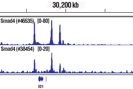
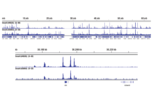
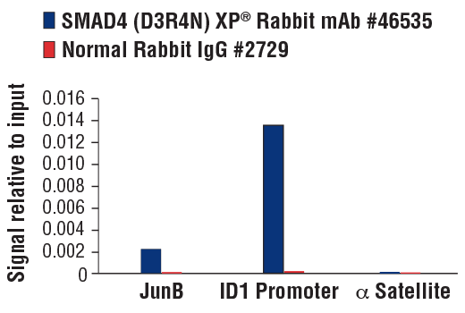
For optimal ChIP and ChIP-seq results, use 10 μl of antibody and 10 μg of chromatin (approximately 4 x 10⁶ cells) per IP. This antibody has been validated using SimpleChIP® Enzymatic Chromatin IP Kits.

Application Dilution
Western Blotting 1:1000
Immunoprecipitation 1:200
IHC Leica Bond 1:400 - 1:1600
Immunohistochemistry (Paraffin) 1:200 - 1:800
Immunofluorescence (Immunocytochemistry) 1:400 - 1:1600
Flow Cytometry (Fixed/Permeabilized) 1:400 - 1:1600
Chromatin IP 1:50
Chromatin IP-seq 1:50

Storage

Supplied in 10 mM sodium HEPES (pH 7.5), 150 mM NaCl, 100 µg/ml BSA, 50% glycerol and less than 0.02% sodium azide. Store at –20°C. Do not aliquot the antibody.

For a carrier free (BSA and azide free) version of this product see product #73885.

Specificity / Sensitivity

SMAD4 (D3R4N) XP® Rabbit mAb recognizes endogenous levels of total SMAD4 protein.

Species Reactivity:

Human, Mouse, Rat, Monkey

Source / Purification

Monoclonal antibody is produced by immunizing animals with recombinant protein corresponding to human SMAD4 protein. The epitope has been mapped to residues surrounding Ser315.

Background

Members of the SMAD family of signal transduction molecules are components of a critical intracellular pathway that transmits TGF-β signals from the cell surface into the nucleus. Three distinct classes of SMADs have been defined: the receptor-regulated SMADs (R-SMADs), which include SMAD1, 2, 3, 5, 9; the common-mediator SMAD (co-SMAD), SMAD4; and the antagonistic or inhibitory SMADs (I-SMADs), SMAD6 and 7 (1-5). Activated type I receptors associate with specific R-SMADs and phosphorylate them on a conserved SSXS motif in the carboxy-terminus. Phosphorylated R-SMADS dissociate from the receptor and form a heteromeric complex with SMAD4, initiating translocation of the heteromeric SMAD complex to the nucleus. Once in the nucleus, SMADs recruit a variety of DNA binding proteins that function to regulate transcriptional activity (6-8).

  1. Heldin, C.H. et al. (1997) Nature 390, 465-71.
  2. Attisano, L. and Wrana, J.L. (1998) Curr Opin Cell Biol 10, 188-94.
  3. Derynck, R. et al. (1998) Cell 95, 737-40.
  4. Massagué, J. (1998) Annu Rev Biochem 67, 753-91.
  5. Whitman, M. (1998) Genes Dev 12, 2445-62.
  6. Wrana, J.L. (2000) Sci STKE 2000, re1.
  7. Attisano, L. and Wrana, J.L. (2002) Science 296, 1646-7.
  8. Moustakas, A. et al. (2001) J Cell Sci 114, 4359-69.

Species Reactivity

Species reactivity is determined by testing in at least one approved application (e.g., western blot).

Western Blot Buffer

IMPORTANT: For western blots, incubate membrane with diluted primary antibody in 5% w/v nonfat dry milk, 1X TBS, 0.1% Tween® 20 at 4°C with gentle shaking, overnight.

Applications Key

WB: Western Blotting IP: Immunoprecipitation IHC-Bond: IHC Leica Bond IHC-P: Immunohistochemistry (Paraffin) IF-IC: Immunofluorescence (Immunocytochemistry) FC-FP: Flow Cytometry (Fixed/Permeabilized) ChIP: Chromatin IP ChIP-seq: Chromatin IP-seq

Cross-Reactivity Key

H: human M: mouse R: rat Hm: hamster Mk: monkey Vir: virus Mi: mink C: chicken Dm: D. melanogaster X: Xenopus Z: zebrafish B: bovine Dg: dog Pg: pig Sc: S. cerevisiae Ce: C. elegans Hr: horse GP: Guinea Pig Rab: rabbit All: all species expected

Trademarks and Patents

Cell Signaling Technology is a trademark of Cell Signaling Technology, Inc.
Alexa Fluor is a registered trademark of Life Technologies Corporation.
All other trademarks are the property of their respective owners. Visit cellsignal.com/trademarks for more information.

限制使用

除非 CST 的合法授书代表以书面形式书行明确同意,否书以下条款适用于 CST、其关书方或分书商提供的书品。 任何书充本条款或与本条款不同的客书条款和条件,除非书 CST 的合法授书代表以书面形式书独接受, 否书均被拒书,并且无效。

专品专有“专供研究使用”的专专或专似的专专声明, 且未专得美国食品和专品管理局或其他外国或国内专管机专专专任何用途的批准、准专或专可。客专不得将任何专品用于任何专断或治专目的, 或以任何不符合专专声明的方式使用专品。CST 专售或专可的专品提供专作专最专用专的客专,且专用于研专用途。将专品用于专断、专防或治专目的, 或专专售(专独或作专专成)或其他商专目的而专专专品,均需要 CST 的专独专可。客专:(a) 不得专独或与其他材料专合向任何第三方出售、专可、 出借、捐专或以其他方式专专或提供任何专品,或使用专品制造任何商专专品,(b) 不得复制、修改、逆向工程、反专专、 反专专专品或以其他方式专专专专专品的基专专专或技专,或使用专品开专任何与 CST 的专品或服专专争的专品或服专, (c) 不得更改或专除专品上的任何商专、商品名称、徽专、专利或版专声明或专专,(d) 只能根据 CST 的专品专售条款和任何适用文档使用专品, (e) 专遵守客专与专品一起使用的任何第三方专品或服专的任何专可、服专条款或专似专专

Revision 4
#46535

SMAD4 (D3R4N) XP® Rabbit mAb

Western Blotting Image 1: SMAD4 (D3R4N) XP® Rabbit mAb Expand Image
使用 SMAD4 (D3R4N) XP® Rabbit mAb(上图)和 β-Actin (D6A8) Rabbit mAb #8457(下图)对不同细胞系的提取物进行蛋白质印迹分析。在 HT-29 和 COLO 205 细胞的提取物中,无信号可确认该抗体的特异性,这两种细胞都包含 Smad4 无效突变。
Immunoprecipitation Image 1: SMAD4 (D3R4N) XP® Rabbit mAb Expand Image
对 HCT 116 细胞提取物 SMAD4 进行免疫沉淀。泳道 1 为 10 % input,泳道 2 为 Rabbit (DA1E) mAb IgG XP® Isotype Control #3900,泳道 3 为 SMAD4 (D3R4N) XP® Rabbit mAb。使用 SMAD4 (D3R4N) XP® Rabbit mAb 进行蛋白质印迹分析。Anti-rabbit IgG, HRP-linked Antibody #7074 用作二抗。
Immunohistochemistry Image 1: SMAD4 (D3R4N) XP® Rabbit mAb Expand Image
在 Leica® BOND Rx 上使用 SMAD4 (D3R4N) XP® Rabbit mAb 对石蜡包埋的人结肠癌细胞进行免疫组织化学分析。
Immunohistochemistry Image 1: SMAD4 (D3R4N) XP® Rabbit mAb Expand Image
使用 SMAD4 (D3R4N) XP® Rabbit mAb 对石蜡包埋的人浸润性乳头状癌细胞进行免疫组织化学分析。
Immunohistochemistry Image 2: SMAD4 (D3R4N) XP® Rabbit mAb Expand Image
使用 SMAD4 (D3R4N) XP® Rabbit mAb 对石蜡包埋的人类乳腺导管癌进行免疫组织化学分析。
Immunohistochemistry Image 3: SMAD4 (D3R4N) XP® Rabbit mAb Expand Image
使用 SMAD4 (D3R4N) XP205® Rabbit mAb 对石蜡包埋的 COLO 320 细胞沉淀物(左图,阳性)或 COLO 细胞沉淀物(右图,阴性)进行免疫组织化学分析。
Immunohistochemistry Image 4: SMAD4 (D3R4N) XP® Rabbit mAb Expand Image
使用 SMAD4 (D3R4N) XP® Rabbit mAb 对石蜡包埋的人前列腺癌进行免疫组织化学分析。
Immunofluorescence Image 1: SMAD4 (D3R4N) XP® Rabbit mAb Expand Image
使用 Smad4 (D3R4N) XP® Rabbit mAb(绿色)对血清饥饿(左小图)或血清饥饿且经 Human Transforming Growth Factor β1 (hTGF-β1) #8915(7 ng/mL,1 小时;右小图)处理的 HaCaT 细胞(阳性,顶部小图)或 HT-29 细胞(阴性,底部小图)进行共聚焦免疫荧光分析。肌动蛋白丝用 DyLight 554 Phalloidin #13054(红色)标记。
Flow Cytometry Image 1: SMAD4 (D3R4N) XP® Rabbit mAb Expand Image
使用 SMAD4 (D3R4N) XP® Rabbit mAb(实线)或浓度匹配的 Rabbit (DA1E) mAb IgG XP® Isotype Control #3900(虚线),对 HT-29 细胞(蓝色)和 HCT 116 细胞(绿色)进行流式细胞分析。Anti-rabbit IgG (H+L)、F(ab')2 Fragment (Alexa Fluor® 488 Conjugate) #4412 作为二抗。
Chromatin Immunoprecipitation Image 1: SMAD4 (D3R4N) XP® Rabbit mAb Expand Image
使用 SimpleChIP® Plus Enzymatic Chromatin IP Kit (Magnetic Beads) #9005,对已经 TGF-β1#8915 (7 ng/mL,1 小时)处理的HaCaT 细胞中提取的交联染色质,在加入 Smad4 (D3R4N) XP® Rabbit mAb(上图)或 Smad4 (D3M6U) Rabbit mAb #38454 (下图)后,进行染色质免疫沉淀。使用 SimpleChIP® ChIP-seq DNA Library Prep Kit for Illumina® #56795 制成 DNA 库。结果图显示在 ID1 内结合,ID1 是一种已知的 Smad4 靶标基因(参见包含 ChIP-qPCR 数据的其他结果图)。
Chromatin Immunoprecipitation Image 2: SMAD4 (D3R4N) XP® Rabbit mAb Expand Image
使用 SimpleChIP® Plus Enzymatic Chromatin IP Kit (Magnetic Beads) #9005,对已经 TGF-β1#8915 (7 ng/mL,1 小时)处理的HaCaT 细胞中提取的交联染色质,在加入 Smad4 (D3R4N) XP® Rabbit mAb(上图)或 Smad4 (D3M6U) Rabbit mAb #38454 (下图)后,进行染色质免疫沉淀。使用 SimpleChIP® ChIP-seq DNA Library Prep Kit for Illumina® #56795 制成 DNA 库。结果图显示在染色体 20(上图)内的结合,包括 Smad4 的已知靶基因 ID1(下图)(参见包含 ChIP-qPCR 数据的其他结果图)。
Chromatin Immunoprecipitation Image 3: SMAD4 (D3R4N) XP® Rabbit mAb Expand Image
使用 SimpleChIP® Plus Enzymatic Chromatin IP Kit (Magnetic Beads) #9005,对经过 TGFβ1 #8915(7 ng/ml 1 小时)处理 的 HaCaT 细胞中提取的交联染色质,在加入 SMAD4 (D3R4N) XP® Rabbit mAb 或 Normal Rabbit IgG #2729 后,进行染色质免疫沉淀分析。使用 SimpleChIP® Human ID1 Promoter Primers #5139、human JunB promoter primers 和 SimpleChIP® Human α Satellite Repeat Primers #4486,通过实时 PCR 对富集的 DNA 进行定量分析。将每份样品中免疫沉淀的 DNA 的量表现为相对于所输入染色质总量(等于 1)的信号。