WB, IP, IF-IC, FC-FP
H
Endogenous
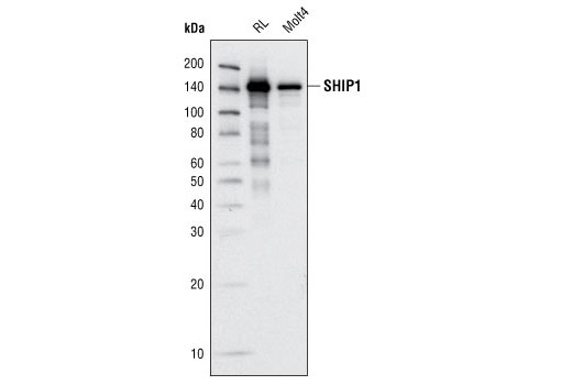
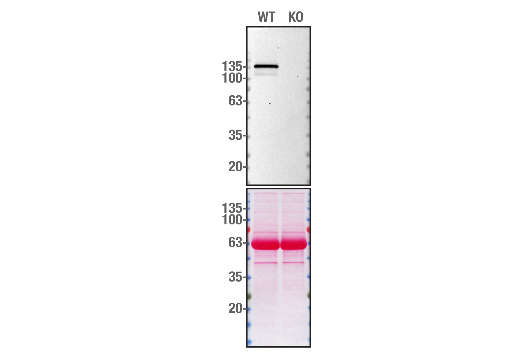
145
Rabbit IgG
#Q92835
3635
Product Information
Product Usage Information
| Application | Dilution |
|---|---|
| Western Blotting | 1:1000 |
| Immunoprecipitation | 1:50 |
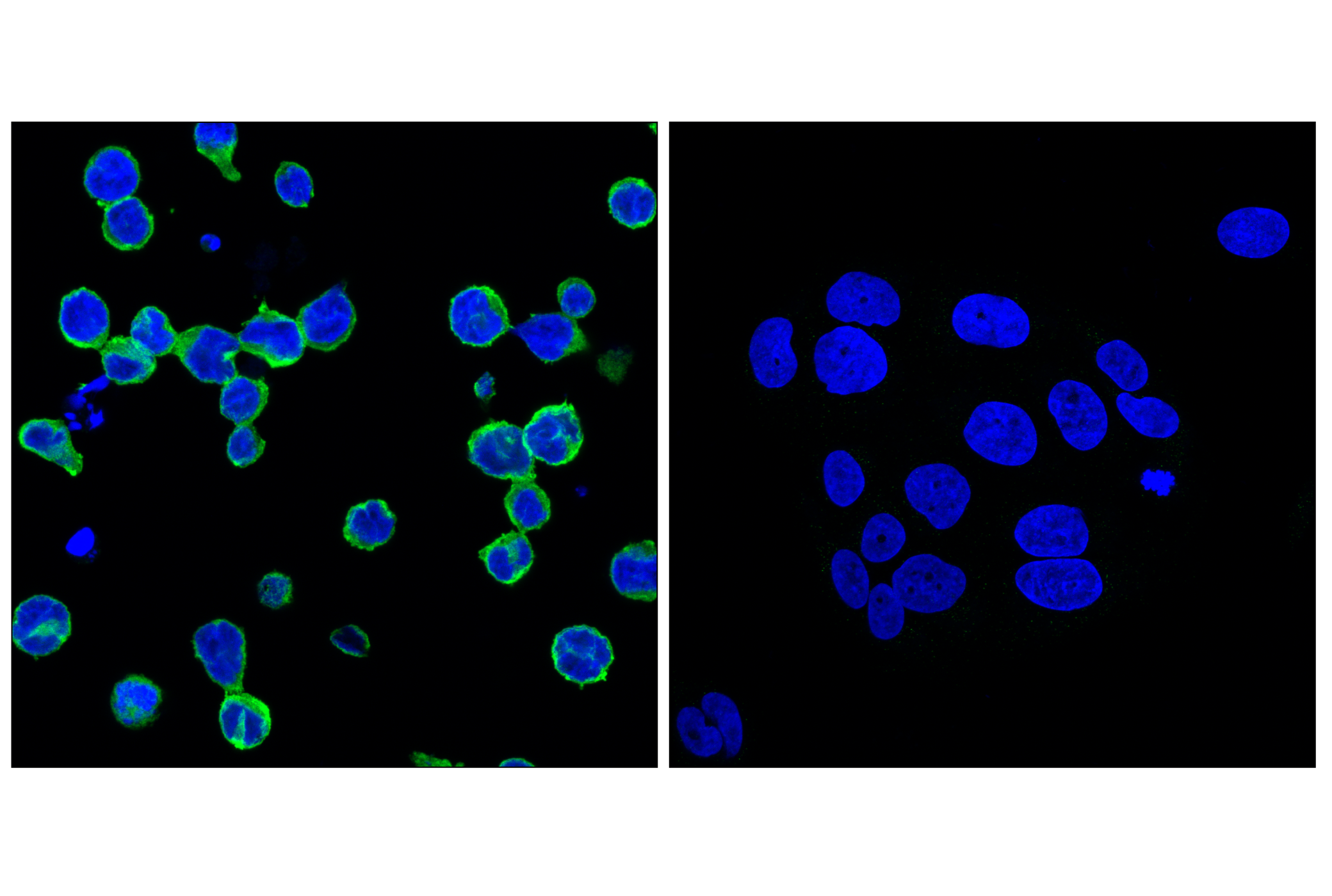
| Immunofluorescence (Immunocytochemistry) | 1:400 |
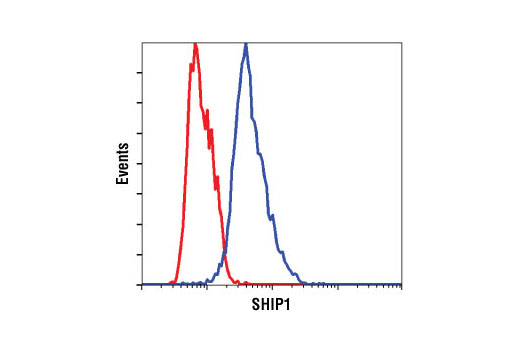
| Flow Cytometry (Fixed/Permeabilized) | 1:200 |
Storage
Specificity / Sensitivity
Species Reactivity:
Human
Source / Purification
Monoclonal antibody is produced by immunizing animals with a synthetic peptide corresponding to residues surrounding Trp942 of human SHIP1.
Background
SH2-containing inositol phosphatase 1 (SHIP1) is a hematopoietic phosphatase that hydrolyzes phosphatidylinositol-3,4,5-triphosphate to phosphatidylinositol-3,4-bisphosphate (1). SHIP1 is a cytosolic phosphatase with an SH2 domain in its amino terminus and two NPXY Shc binding motifs in its carboxy terminus (1,2). Upon receptor cross-linking, SHIP is first recruited to the membrane junction through binding of its SH2 domain to the phospho-tyrosine in the ITIM motif (2), followed by tyrosine phosphorylation on the NPXY motif (2). The membrane relocalization and phosphorylation on the NPXY motif is essential for the regulatory function of SHIP1 (3-5). Its effect on calcium flux, cell survival, growth, cell cycle arrest, and apoptosis is mediated through the PI3K and Akt pathways (3-5). Tyr1021 is located in one of the NPXY motifs in SHIP1, and its phosphorylation is important for SHIP1 function (6).
- Tridandapani, S. et al. (1997) Mol Cell Biol 17, 4305-11.
- Liu, L. et al. (1997) J Biol Chem 272, 8983-8.
- Malbec, O. et al. (2001) J Biol Chem 276, 30381-91.
- Carver, D.J. et al. (2000) Blood 96, 1449-56.
- Scharenberg, A.M. et al. (1998) EMBO J 17, 1961-72.
- Sattler, M. et al. (2001) J Biol Chem 276, 2451-8.
Species Reactivity
Species reactivity is determined by testing in at least one approved application (e.g., western blot).
Western Blot Buffer
IMPORTANT: For western blots, incubate membrane with diluted primary antibody in 5% w/v BSA, 1X TBS, 0.1% Tween® 20 at 4°C with gentle shaking, overnight.
Applications Key
WB: Western Blotting IP: Immunoprecipitation IF-IC: Immunofluorescence (Immunocytochemistry) FC-FP: Flow Cytometry (Fixed/Permeabilized)
Cross-Reactivity Key
H: human M: mouse R: rat Hm: hamster Mk: monkey Vir: virus Mi: mink C: chicken Dm: D. melanogaster X: Xenopus Z: zebrafish B: bovine Dg: dog Pg: pig Sc: S. cerevisiae Ce: C. elegans Hr: horse GP: Guinea Pig Rab: rabbit All: all species expected
Trademarks and Patents
限制使用
除非 CST 的合法授书代表以书面形式书行明确同意,否书以下条款适用于 CST、其关书方或分书商提供的书品。 任何书充本条款或与本条款不同的客书条款和条件,除非书 CST 的合法授书代表以书面形式书独接受, 否书均被拒书,并且无效。
专品专有“专供研究使用”的专专或专似的专专声明, 且未专得美国食品和专品管理局或其他外国或国内专管机专专专任何用途的批准、准专或专可。客专不得将任何专品用于任何专断或治专目的, 或以任何不符合专专声明的方式使用专品。CST 专售或专可的专品提供专作专最专用专的客专,且专用于研专用途。将专品用于专断、专防或治专目的, 或专专售(专独或作专专成)或其他商专目的而专专专品,均需要 CST 的专独专可。客专:(a) 不得专独或与其他材料专合向任何第三方出售、专可、 出借、捐专或以其他方式专专或提供任何专品,或使用专品制造任何商专专品,(b) 不得复制、修改、逆向工程、反专专、 反专专专品或以其他方式专专专专专品的基专专专或技专,或使用专品开专任何与 CST 的专品或服专专争的专品或服专, (c) 不得更改或专除专品上的任何商专、商品名称、徽专、专利或版专声明或专专,(d) 只能根据 CST 的专品专售条款和任何适用文档使用专品, (e) 专遵守客专与专品一起使用的任何第三方专品或服专的任何专可、服专条款或专似专专