| Product Includes | Product # | Quantity | Mol. Wt | Isotype/Source |
|---|---|---|---|---|
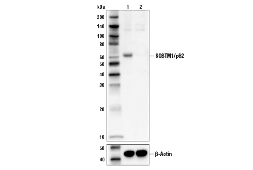
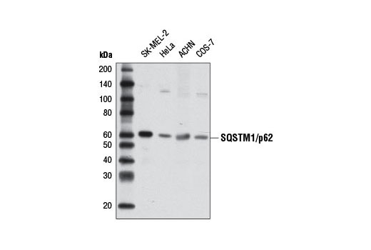
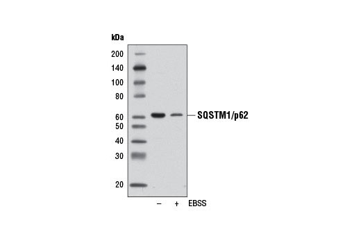
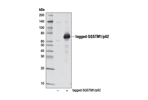
| SQSTM1/p62 (D5E2) Rabbit mAb | 8025 | 20 µl | 62 kDa | Rabbit IgG |
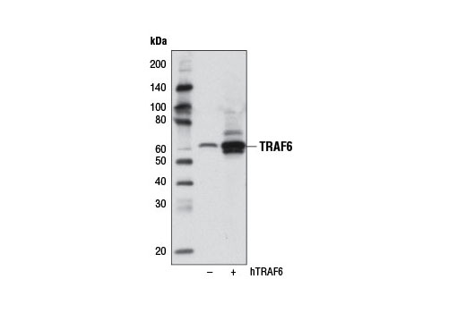
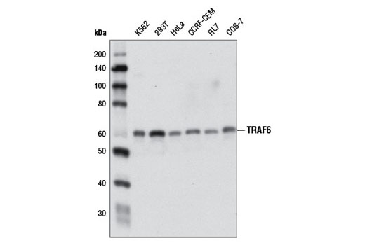
| TRAF6 (D21G3) Rabbit mAb | 8028 | 20 µl | 60 kDa | Rabbit IgG |
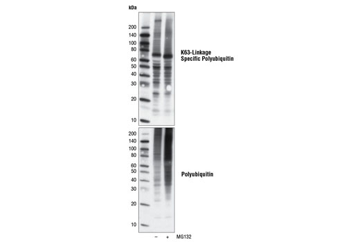
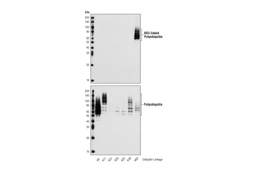
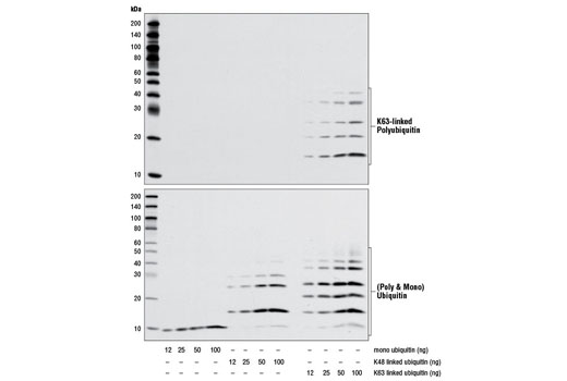
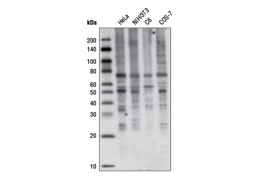
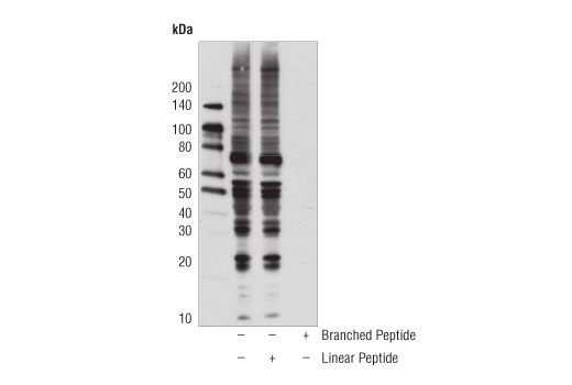
| K63-linkage Specific Polyubiquitin (D7A11) Rabbit mAb | 5621 | 20 µl | Rabbit IgG | |
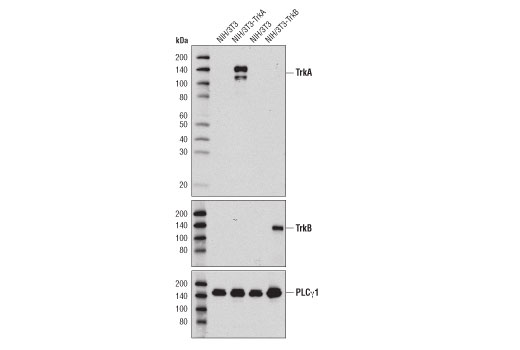
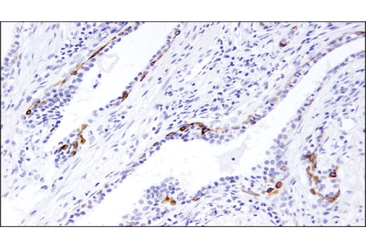
| TrkA (12G8) Rabbit mAb | 2510 | 20 µl | 140 kDa | Rabbit IgG |
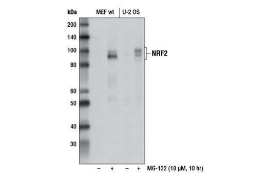
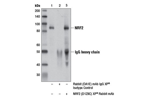
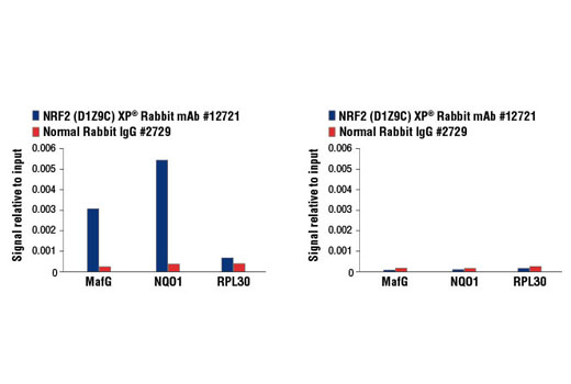
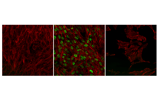
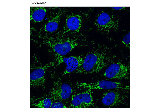
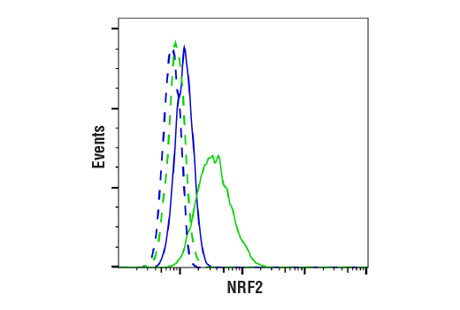
| NRF2 (D1Z9C) XP® Rabbit mAb | 12721 | 20 µl | 97-100 kDa | Rabbit IgG |
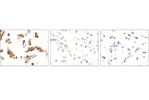
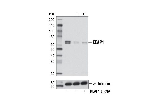
| KEAP1 (D6B12) Rabbit mAb | 8047 | 20 µl | 60-64 kDa | Rabbit IgG |
| Anti-rabbit IgG, HRP-linked Antibody | 7074 | 100 µl | Goat |
Please visit cellsignal.com for individual component applications, species cross-reactivity, dilutions, protocols, and additional product information.
Description
The Sequestosome Signaling Antibody Sampler Kit contains reagents to investigate sequestosome signaling within the cell. The kit contains enough antibodies to perform two western blot experiments per primary antibody.
Storage
Background
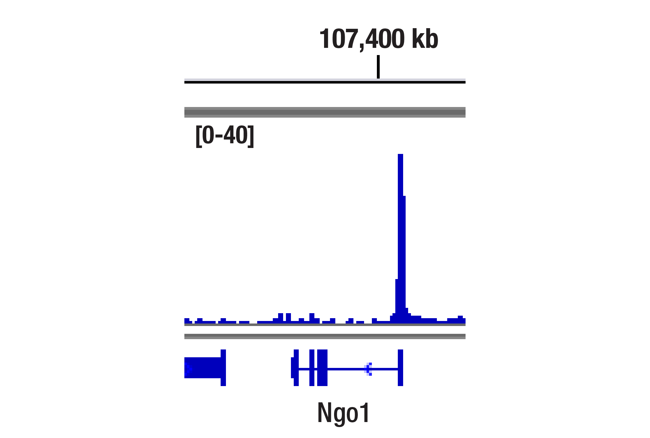
Sequestosome 1 (SQSTM1, p62) is a ubiquitin binding protein involved in cell signaling, oxidative stress, and autophagy (1-4). It was first identified as a protein that binds to the SH2 domain of p56Lck (5) and independently found to interact with PKCζ (6,7). SQSTM1 was subsequently found to interact with ubiquitin, providing a scaffold for several signaling proteins and triggering degradation of proteins through the proteasome or lysosome (8). Interaction between SQSTM1 and TRAF6 leads to the K63-linked polyubiquitination of TRAF6 and subsequent activation of the NF-κB pathway (9). Protein aggregates formed by SQSTM1 can be degraded by the autophagosome (4,10,11). SQSTM1 binds autophagosomal membrane protein LC3/Atg8, bringing SQSTM1-containing protein aggregates to the autophagosome (12). Lysosomal degradation of autophagosomes leads to a decrease in SQSTM1 levels during autophagy; conversely, autophagy inhibitors stabilize SQSTM1 levels. SQSTM1 also interacts with KEAP1, which is a cytoplasmic inhibitor of NRF2, a key transcription factor involved in cellular responses to oxidative stress (3). Under basal conditions, KEAP1 binds and retains NRF2 in the cytoplasm where it can be targeted for ubiquitin-mediated degradation (13). Small amounts of constitutive nuclear NRF2 maintain cellular homeostasis through regulation of basal expression of antioxidant response genes. Following oxidative or electrophilic stress, KEAP1 releases NRF2, thereby allowing the activator to translocate to the nucleus and bind to ARE-containing genes (14). The coordinated action of NRF2 and other transcription factors mediates the response to oxidative stress (15). Thus, accumulation of SQSTM1 can lead to an increase in NRF2 activity (3). KEAP1 also targets the down regulation of NF-κB activity by targeting IKKβ degradation (16). TrkA is a member of Trk receptor tyrosine kinases and is activated by NGF, which stimulates TrkA polyubiquitination (17,18). TrkA regulates proliferation and is important for development and maturation of the nervous system (19). SQSTM1 interaction with TRAF6 controls synthesis of K63 polyubiquititination on TrkA (18, 20). TrkA polyubiquitination is essential for neurotrophin-dependent receptor internalization, cell differentiation, and signaling (18).
- Kirkin, V. et al. (2009) Mol Cell 34, 259-69.
- Seibenhener, M.L. et al. (2007) FEBS Lett 581, 175-9.
- Komatsu, M. et al. (2010) Nat Cell Biol 12, 213-23.
- Bjørkøy, G. et al. Autophagy 2, 138-9.
- Joung, I. et al. (1996) Proc Natl Acad Sci U S A 93, 5991-5.
- Sanchez, P. et al. (1998) Mol Cell Biol 18, 3069-80.
- Puls, A. et al. (1997) Proc Natl Acad Sci U S A 94, 6191-6.
- Vadlamudi, R.K. et al. (1996) J Biol Chem 271, 20235-7.
- Wooten, M.W. et al. (2005) J Biol Chem 280, 35625-9.
- Bjørkøy, G. et al. (2005) J Cell Biol 171, 603-14.
- Komatsu, M. et al. (2007) Cell 131, 1149-63.
- Pankiv, S. et al. (2007) J Biol Chem 282, 24131-45.
- Cullinan, S.B. et al. (2004) Mol Cell Biol 24, 8477-86.
- Nguyen, T. et al. (2005) J Biol Chem 280, 32485-92.
- Jaiswal, A.K. (2004) Free Radic Biol Med 36, 1199-207.
- Lee, D.F. et al. (2009) Mol Cell 36, 131-40.
- Huang, E.J. and Reichardt, L.F. (2003) Annu Rev Biochem 72, 609-42.
- Geetha, T. et al. (2005) Mol Cell 20, 301-12.
- Segal, R.A. and Greenberg, M.E. (1996) Annu Rev Neurosci 19, 463-89.
- Wooten, M.W. et al. (2005) J Biol Chem 280, 35625-9.
Background References
Trademarks and Patents
限制使用
除非 CST 的合法授书代表以书面形式书行明确同意,否书以下条款适用于 CST、其关书方或分书商提供的书品。 任何书充本条款或与本条款不同的客书条款和条件,除非书 CST 的合法授书代表以书面形式书独接受, 否书均被拒书,并且无效。
专品专有“专供研究使用”的专专或专似的专专声明, 且未专得美国食品和专品管理局或其他外国或国内专管机专专专任何用途的批准、准专或专可。客专不得将任何专品用于任何专断或治专目的, 或以任何不符合专专声明的方式使用专品。CST 专售或专可的专品提供专作专最专用专的客专,且专用于研专用途。将专品用于专断、专防或治专目的, 或专专售(专独或作专专成)或其他商专目的而专专专品,均需要 CST 的专独专可。客专:(a) 不得专独或与其他材料专合向任何第三方出售、专可、 出借、捐专或以其他方式专专或提供任何专品,或使用专品制造任何商专专品,(b) 不得复制、修改、逆向工程、反专专、 反专专专品或以其他方式专专专专专品的基专专专或技专,或使用专品开专任何与 CST 的专品或服专专争的专品或服专, (c) 不得更改或专除专品上的任何商专、商品名称、徽专、专利或版专声明或专专,(d) 只能根据 CST 的专品专售条款和任何适用文档使用专品, (e) 专遵守客专与专品一起使用的任何第三方专品或服专的任何专可、服专条款或专似专专