Revision 1
#42397
Store at -20C
877-616-CELL (2355)
877-678-TECH (8324)
3 Trask Lane | Danvers | Massachusetts | 01923 | USA
For Research Use Only. Not for Use in Diagnostic Procedures.
Applications:
W, IHC-P, IF-F, IF-IC
Reactivity:
H M R
Sensitivity:
Endogenous
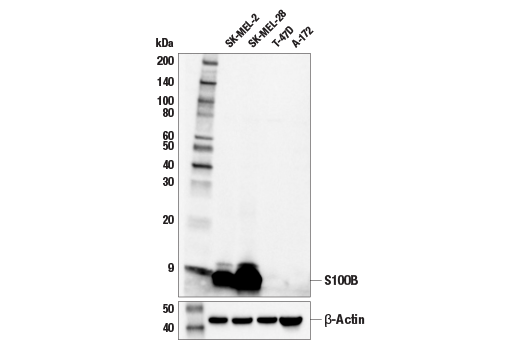
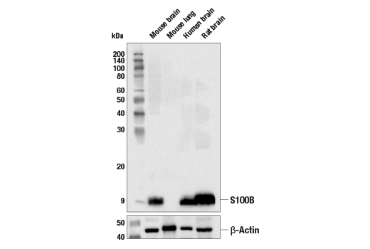
MW (kDa):
10
Source/Isotype:
Rabbit IgG
UniProt ID:
#P04271
Entrez-Gene Id:
6285
Product Usage Information
This product is the carrier-free version of product #90393. All data were generated using the same antibody clone in the standard formulation which contains BSA and glycerol.
This formulation is ideal for use with technologies requiring specialized or custom antibody labeling, including fluorophores, metals, lanthanides, and oligonucleotides. It is not recommended for ChIP, ChIP-seq, CUT&RUN, or CUT&Tag assays. If you require a carrier-free formulation for chromatin profiling, please contact us. Optimal dilutions/concentrations should be determined by the end user.
BSA and Azide Free antibodies are quality control tested by size exclusion chromatography (SEC) to determine antibody integrity.Formulation
Supplied in 1X PBS (10 mM Na2HPO4, 3 mM KCl, 2 mM KH2PO4, and 140 mM NaCl (pH 7.8)). BSA and Azide Free.
For standard formulation of this product see product #90393.
Storage
Specificity/Sensitivity
Source / Purification
Background
Each S100 monomer bears two EF-hand motifs and can bind up to two molecules of calcium (or other divalent cations in some instances). Structural evidence shows that S100 proteins form antiparallel homo- or heterodimers that coordinate binding partner proximity in a calcium-dependent (and sometimes calcium-independent) manner. Although structurally and functionally similar, individual members show restricted tissue distribution, are localized in specific cellular compartments, and display unique protein binding partners, which suggests that each plays a specific role in various signaling pathways. In addition to an intracellular role, some S100 proteins have been shown to act as receptors for extracellular ligands or are secreted and exhibit cytokine-like activities (1-4).
S100B is abundantly expressed in astrocytes and is commonly used as an astrocytic marker in studies of the mammalian CNS. S100B is also expressed in immature and mature myelinating oligodendrocytes that are chondroitin sulfate proteoglycan (NG2)-positive (5).
Species Reactivity
Species reactivity is determined by testing in at least one approved application (e.g., western blot).
Applications Key
W: Western Blotting IHC-P: Immunohistochemistry (Paraffin) IF-F: Immunofluorescence (Frozen) IF-IC: Immunofluorescence (Immunocytochemistry)
Cross-Reactivity Key
H: Human M: Mouse R: Rat
Trademarks and Patents
Cell Signaling Technology is a trademark of Cell Signaling Technology, Inc.
Alexa Fluor is a registered trademark of Life Technologies Corporation.
All other trademarks are the property of their respective owners. Visit cellsignal.com/trademarks for more information.
Limited Uses
Except as otherwise expressly agreed in a writing signed by a legally authorized representative of CST, the following terms apply to Products provided by CST, its affiliates or its distributors. Any Customer's terms and conditions that are in addition to, or different from, those contained herein, unless separately accepted in writing by a legally authorized representative of CST, are rejected and are of no force or effect.
Products are labeled with For Research Use Only or a similar labeling statement and have not been approved, cleared, or licensed by the FDA or other regulatory foreign or domestic entity, for any purpose. Customer shall not use any Product for any diagnostic or therapeutic purpose, or otherwise in any manner that conflicts with its labeling statement. Products sold or licensed by CST are provided for Customer as the end-user and solely for research and development uses. Any use of Product for diagnostic, prophylactic or therapeutic purposes, or any purchase of Product for resale (alone or as a component) or other commercial purpose, requires a separate license from CST. Customer shall (a) not sell, license, loan, donate or otherwise transfer or make available any Product to any third party, whether alone or in combination with other materials, or use the Products to manufacture any commercial products, (b) not copy, modify, reverse engineer, decompile, disassemble or otherwise attempt to discover the underlying structure or technology of the Products, or use the Products for the purpose of developing any products or services that would compete with CST products or services, (c) not alter or remove from the Products any trademarks, trade names, logos, patent or copyright notices or markings, (d) use the Products solely in accordance with CST Product Terms of Sale and any applicable documentation, and (e) comply with any license, terms of service or similar agreement with respect to any third party products or services used by Customer in connection with the Products.
Revision 1
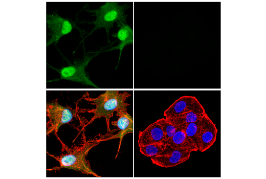
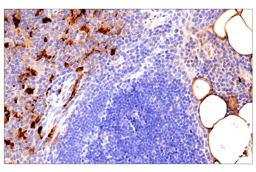
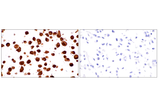
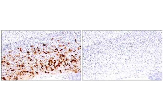
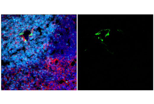
Immunohistochemical analysis of paraffin-embedded normal human spleen using S100B (E7C3A) Rabbit mAb. Data were generated using the standard formulation of this product.
Revision 1
Revision 1
Revision 1
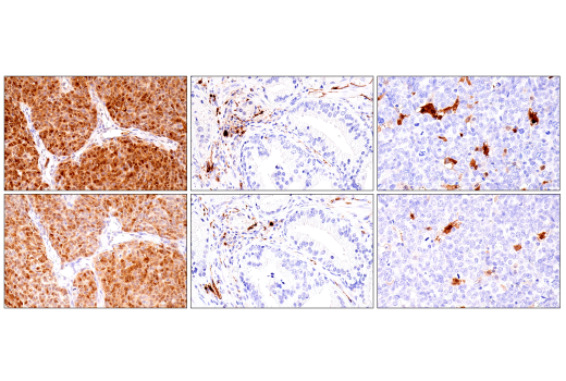
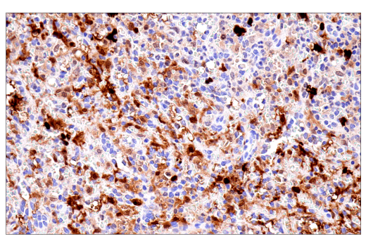
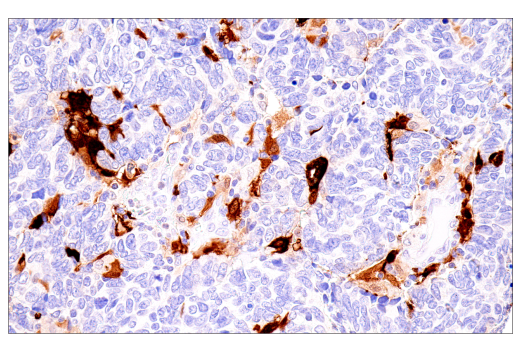
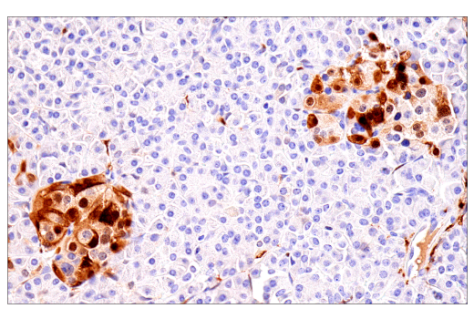
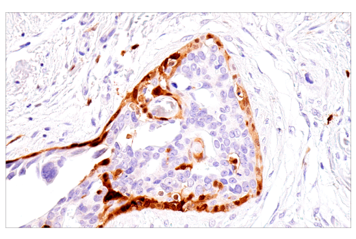
Immunohistochemical analysis of paraffin-embedded human ductal breast carcinoma using S100B (E7C3A) Rabbit mAb. Data were generated using the standard formulation of this product.
Revision 1
Revision 1