Revision 1

#8348Store at -20C

1 个试剂盒

(9 x 20 microliters)

Cell Signaling Technology

Orders: 877-616-CELL (2355) [email protected]

Support: 877-678-TECH (8324)

Web: [email protected] cellsignal.com

3 Trask LaneDanversMassachusetts01923USA
For Research Use Only. Not for Use in Diagnostic Procedures.
Product Includes Product # Quantity Mol. Wt Isotype/Source
MDA-5 (D74E4) Rabbit mAb 5321 20 µl 135 kDa Rabbit IgG
Rig-I (D14G6) Rabbit mAb 3743 20 µl 102 kDa Rabbit IgG
MAVS Antibody 3993 20 µl 75, 52 kDa Rabbit 
IRF-3 (D6I4C) XP® Rabbit mAb 11904 20 µl 50-55 kDa Rabbit IgG
TBK1/NAK (D1B4) Rabbit mAb 3504 20 µl 84 kDa Rabbit IgG
Phospho-TBK1/NAK (Ser172) (D52C2) XP® Rabbit mAb 5483 20 µl 84 kDa Rabbit IgG
Phospho-IRF-3 (Ser396) (4D4G) Rabbit mAb 4947 20 µl 45-55 kDa Rabbit IgG
Phospho-IKKε (Ser172) (D1B7) Rabbit mAb 8766 20 µl 80 kDa Rabbit IgG
IKKε (D20G4) Rabbit mAb 2905 20 µl 80 kDa Rabbit IgG
Anti-rabbit IgG, HRP-linked Antibody 7074 100 µl Goat 

Please visit cellsignal.com for individual component applications, species cross-reactivity, dilutions, protocols, and additional product information.

Description

The Rig-I Pathway Antibody Sampler Kit provides an economical means to evaluate the activation state and total protein levels of multiple members of the Rig-I pathway including Rig-I, MDA-5, MAVS, IRF-3, TBK1/NAK, and IKKε. The kit includes enough primary antibody to perform two western blot experiments per antibody.

Storage

Supplied in 10 mM sodium HEPES (pH 7.5), 150 mM NaCl, 100 µg/ml BSA, 50% glycerol and less than 0.02% sodium azide. Store at –20°C. Do not aliquot the antibody.

Background

Antiviral innate immunity depends on the combination of parallel pathways triggered by virus detecting proteins in the Toll-like receptor (TLR) family and RNA helicases, such as Rig-I (retinoic acid-inducible gene I) and MDA-5 (melanoma differentiation-associated antigen 5), which promote the transcription of type I interferons (IFN) and antiviral enzymes (1-3). TLRs and helicase proteins contain sites that recognize the molecular patterns of different virus types, including DNA, single-stranded RNA (ssRNA), double-stranded RNA (dsRNA), and glycoproteins. These antiviral proteins are found in different cell compartments; TLRs (i.e. TLR3, TLR7, TLR8, and TLR9) are expressed on endosomal membranes and helicases are localized to the cytoplasm. Rig-I expression is induced by retinoic acid, LPS, IFN, and viral infection (4,5). Both Rig-I and MDA-5 share a DExD/H-box helicase domain that detects viral dsRNA and two amino-terminal caspase recruitment domains (CARD) that are required for triggering downstream signaling (4-7). Rig-I binds both dsRNA and viral ssRNA that contains a 5'-triphosphate end not seen in host RNA (8,9). Though structurally related, Rig-I and MDA-5 detect a distinct set of viruses (10,11). The CARD domain of the helicases, which is sufficient to generate signaling and IFN production, is recruited to the CARD domain of the MAVS/VISA/Cardif/IPS-1 mitochondrial protein, which triggers activation of NF-κB, TBK1/IKKε, and IRF-3/IRF-7 (12-15).

  1. Yoneyama, M. and Fujita, T. (2007) J Biol Chem 282, 15315-8.
  2. Meylan, E. and Tschopp, J. (2006) Mol Cell 22, 561-9.
  3. Thompson, A.J. and Locarnini, S.A. (2007) Immunol Cell Biol 85, 435-45.
  4. Imaizumi, T. et al. (2002) Biochem Biophys Res Commun 292, 274-9.
  5. Zhang, X. et al. (2000) Microb Pathog 28, 267-78.
  6. Yoneyama, M. et al. (2005) J Immunol 175, 2851-8.
  7. Yoneyama, M. et al. (2004) Nat Immunol 5, 730-7.
  8. Hornung, V. et al. (2006) Science 314, 994-7.
  9. Pichlmair, A. et al. (2006) Science 314, 997-1001.
  10. Kato, H. et al. (2006) Nature 441, 101-5.
  11. Childs, K. et al. (2007) Virology 359, 190-200.
  12. Meylan, E. et al. (2005) Nature 437, 1167-72.
  13. Xu, L.G. et al. (2005) Mol Cell 19, 727-40.
  14. Kawai, T. et al. (2005) Nat Immunol 6, 981-8.
  15. Seth, R.B. et al. (2005) Cell 122, 669-82.

Background References

    Trademarks and Patents

    Cell Signaling Technology is a trademark of Cell Signaling Technology, Inc.
    All other trademarks are the property of their respective owners. Visit cellsignal.com/trademarks for more information.

    限制使用

    除非 CST 的合法授书代表以书面形式书行明确同意,否书以下条款适用于 CST、其关书方或分书商提供的书品。 任何书充本条款或与本条款不同的客书条款和条件,除非书 CST 的合法授书代表以书面形式书独接受, 否书均被拒书,并且无效。

    专品专有“专供研究使用”的专专或专似的专专声明, 且未专得美国食品和专品管理局或其他外国或国内专管机专专专任何用途的批准、准专或专可。客专不得将任何专品用于任何专断或治专目的, 或以任何不符合专专声明的方式使用专品。CST 专售或专可的专品提供专作专最专用专的客专,且专用于研专用途。将专品用于专断、专防或治专目的, 或专专售(专独或作专专成)或其他商专目的而专专专品,均需要 CST 的专独专可。客专:(a) 不得专独或与其他材料专合向任何第三方出售、专可、 出借、捐专或以其他方式专专或提供任何专品,或使用专品制造任何商专专品,(b) 不得复制、修改、逆向工程、反专专、 反专专专品或以其他方式专专专专专品的基专专专或技专,或使用专品开专任何与 CST 的专品或服专专争的专品或服专, (c) 不得更改或专除专品上的任何商专、商品名称、徽专、专利或版专声明或专专,(d) 只能根据 CST 的专品专售条款和任何适用文档使用专品, (e) 专遵守客专与专品一起使用的任何第三方专品或服专的任何专可、服专条款或专似专专
    Revision 1
    #8348

    Rig-I Pathway Antibody Sampler Kit

    Rig-I Pathway Antibody Sampler Kit: Image 1 Expand Image
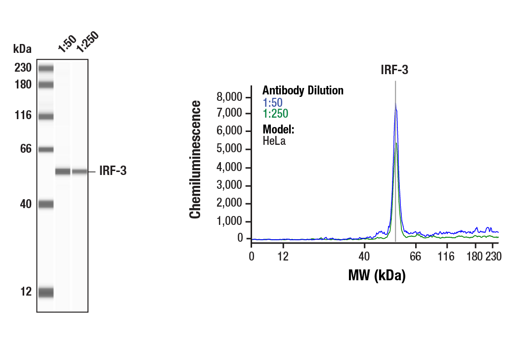
    使用 IRF-3 (D6I4C) XP ® Rabbit mAb #11904 对 HeLa 细胞的裂解物 (1 mg/mL) 进行 Simple Western™ 分析。虚拟泳道式图像(左图)显示一抗稀释比例为 1:50 和 1:250 时的靶标条带(如图所示)。对应的电泳图(右图)为一抗稀释比例在 1:50(蓝线)和 1:250(绿线)时沿毛细血管内分子量的化学发光结果。在还原条件下,使用 12-230 kDa 分离模块在 ProteinSimple(BioTechne 品牌)的 Jess™ Simple Western 仪器上进行该实验。
    Rig-I Pathway Antibody Sampler Kit: Image 2 Expand Image
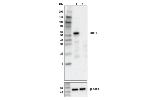
    使用 IRF-3 (D6I4C) XP® Rabbit mAb, #11904(上图)或 β-actin (13E5) Rabbit mAb, #4970(下图)对对照型 HeLa 细胞(泳道 1) 或 IRF-3 敲除型 HeLa 细胞(泳道 2)的提取物进行蛋白质印迹分析。IRF-3 敲除型 HeLa 细胞中没有信号,这证实了该抗体对 IRF-3 的特异性。
    Rig-I Pathway Antibody Sampler Kit: Image 3 Expand Image
    使用 IKKε (D20G4) Rabbit mAb 对重组 GST-IKKε #7553 和 Ramos 细胞和 RL 细胞的裂解物进行蛋白质印迹分析。
    Rig-I Pathway Antibody Sampler Kit: Image 4 Expand Image
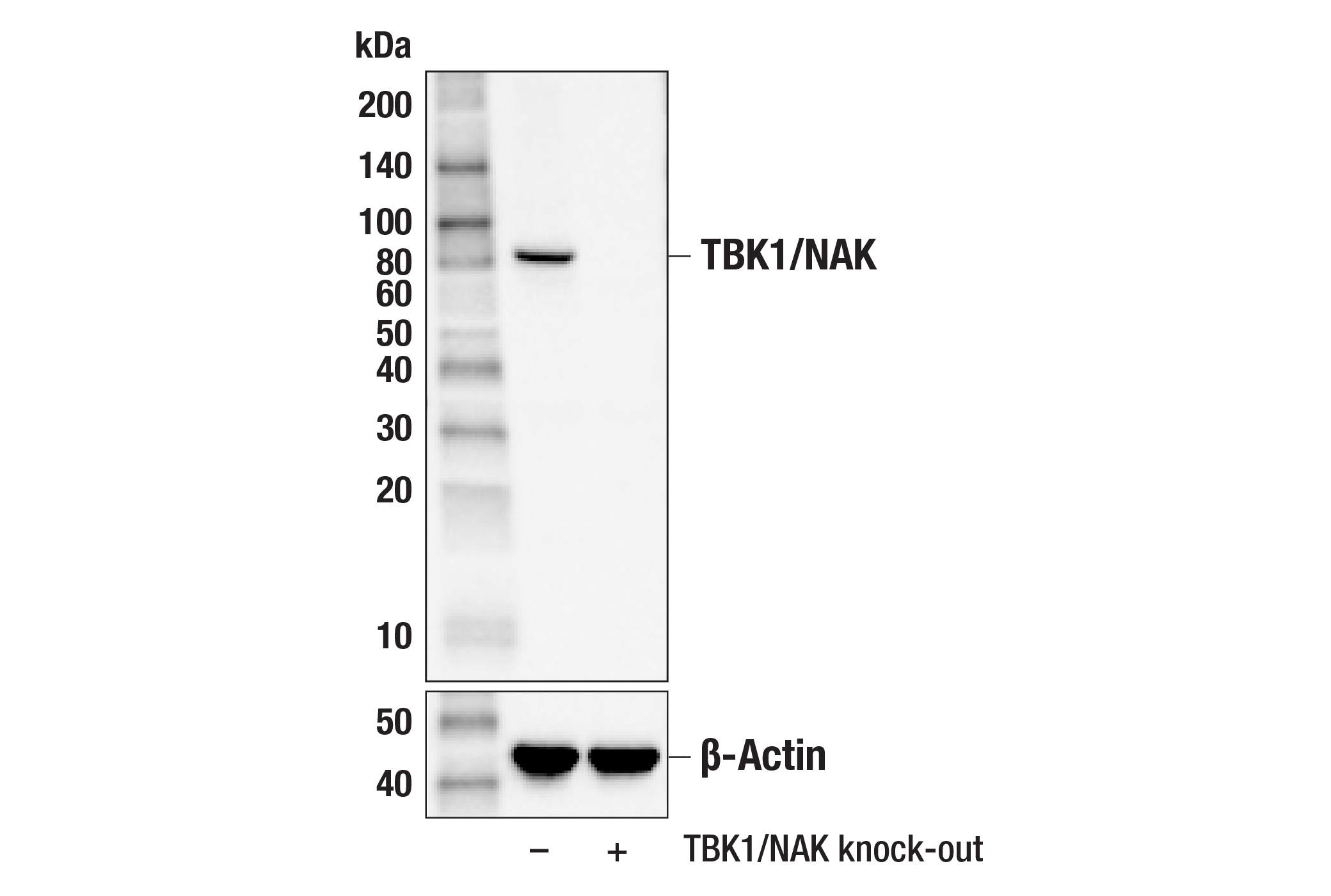
    使用 TBK/NAK (D1B4) 兔单克隆抗体 #3504(上图)或 β-肌动蛋白 (D6A8) 兔单克隆抗体 #8457(下图)对未经处理 (-) 的或 TBK1/NAK 敲除型 (+) HCT116 细胞提取物进行蛋白印迹分析。
    Rig-I Pathway Antibody Sampler Kit: Image 5 Expand Image
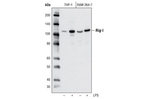
    使用 Rig-I (D14G6) Rabbit mAb 对未经或已经 LPS(1 ug/ml,过夜)处理的分化 THP-1 和 RAW 264.7 细胞进行蛋白质印迹分析。
    Rig-I Pathway Antibody Sampler Kit: Image 6 Expand Image
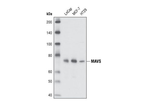
    使用 MAVS Antibody 对 LNCaP 细胞系、MCF-7 细胞系和 HT29 细胞系提取物进行蛋白质印迹分析。
    Rig-I Pathway Antibody Sampler Kit: Image 7 Expand Image
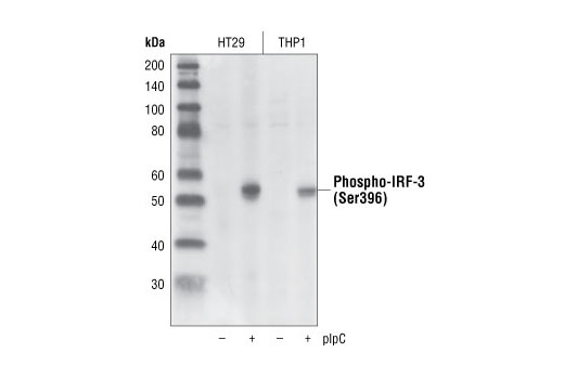
    使用 Phospho-IRF-3 (Ser396) (4D4G) Rabbit mAb,对对照品经 plpC 转染 (1 小时) 的 HT29 和 THP1 细胞提取物进行蛋白质印迹分析。
    Rig-I Pathway Antibody Sampler Kit: Image 8 Expand Image
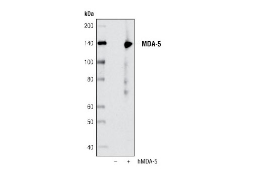
    使用 MDA-5 (D74E4) Rabbit mAb,对 293T 细胞提取物进行蛋白质印迹分析,而 293T 细胞经转染空载 (-) 或使用人类 MDA-5 (+) 进行转染。
    Rig-I Pathway Antibody Sampler Kit: Image 9 Expand Image
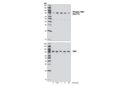
    使用 Phospho-TBK1/NAK (Ser172) (D52C2) XP® Rabbit mAb(上图)或总 TBK1/NAK (D1B4) Rabbit mAb #3504(下图)对用 TPA #4174(80 nM,过夜)分化后用 LPS (1 μg/ml) 处理长达 24 小时的 THP-1 细胞的提取物进行蛋白质印迹分析。
    Rig-I Pathway Antibody Sampler Kit: Image 10 Expand Image
    一抗与靶标蛋白结合之后,与偶联 HRP 的二抗形成复合体。添加 LumiGLO®,在酶催化分解期间发光。
    Rig-I Pathway Antibody Sampler Kit: Image 11 Expand Image
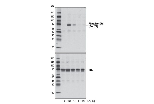
    使用 Phospho-IKKε (Ser172) (D1B7) Rabbit mAb (上图) 或 IKKε (D20G4) Rabbit mAb #2905(下图),对经 TPA #4174(80nM, 过夜) 分化之后以 LPS (1 μg/ml, 指定时间) 处理的 THP-1 细胞提取物进行蛋白质印迹分析。
    Rig-I Pathway Antibody Sampler Kit: Image 12 Expand Image
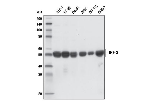
    使用 IRF-3 (D6I4C) XP® Rabbit mAb 对不同细胞系提取物进行蛋白质印迹分析。
    Rig-I Pathway Antibody Sampler Kit: Image 13 Expand Image
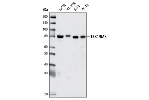
    使用 TBK1/NAK (D1B4) Rabbit mAb 对不同细胞系的提取物进行蛋白质印迹分析。
    Rig-I Pathway Antibody Sampler Kit: Image 14 Expand Image
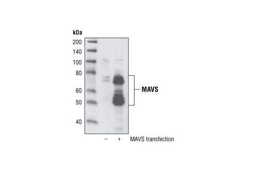
    使用 MAVS Antibody 对转染空载或转染人MAVS 的 HeLa 细胞提取物进行蛋白质印迹分析。
    Rig-I Pathway Antibody Sampler Kit: Image 15 Expand Image
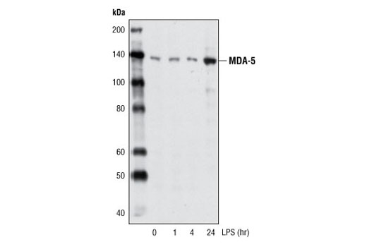
    使用 MDA-5 (D74E4) Rabbit mAb,对未处理或用脂多糖(1 μg/ml,按所示时间处理)处理的已分化 THP-1 细胞提取物,进行蛋白质印迹分析。
    Rig-I Pathway Antibody Sampler Kit: Image 16 Expand Image
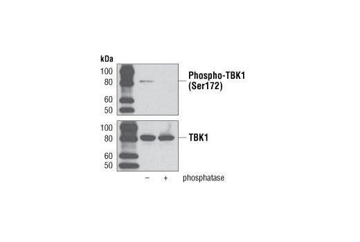
    使用 Phospho-TBK1/NAK (Ser172) (D52C2) XP® Rabbit mAb(上图)或总 TBK1/NAK (D1B4) Rabbit mAb #3504(下图)对用 TPA #4174(80 nM,过夜)分化后用 LPS(1 μg/ml,1 小时)处理、未经处理或已经磷酸酶处理的 THP-1 细胞的提取物进行蛋白质印迹分析。
    Rig-I Pathway Antibody Sampler Kit: Image 17 Expand Image
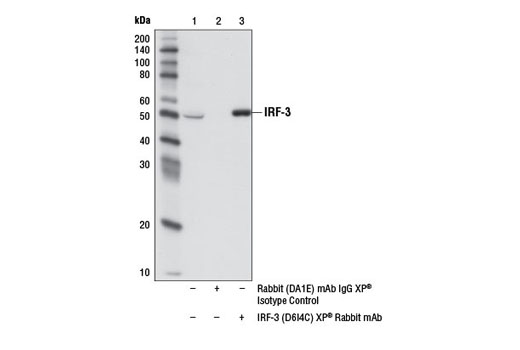
    使用 Rabbit (DA1E) mAb IgG XP® Isotype Control #3900(泳道 2)或 IRF-3 (D6I4C) XP® Rabbit mAb(泳道 3),对 THP-1 细胞提取物的 IRF-3进行免疫沉淀。泳道 1 是 10% 输入对照。使用 IRF-3 (D6I4C) XP® Rabbit mAb 进行蛋白质印迹分析。使用 Mouse Anti-rabbit IgG (Conformation Specific) (L27A9) mAb #3678 和 Anti-mouse IgG, HRP-linked Antibody #7076 作为二抗。
    Rig-I Pathway Antibody Sampler Kit: Image 18 Expand Image
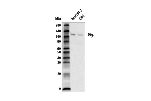
    使用 Rig-I (D14G6) Rabbit mAb 对 Raw264.7 和 CHO 细胞的提取物进行蛋白质印迹分析。
    Rig-I Pathway Antibody Sampler Kit: Image 19 Expand Image
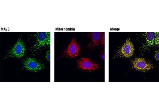
    使用 MAVS Antibody(绿色)对 MCF-7 细胞进行共聚焦免疫荧光分析,从而证实与 MitoTracker® Red CMXRos(红色)标记的线粒体存在共定位。蓝色伪彩 = DRAQ5 (DNA 荧光染料)。
    Rig-I Pathway Antibody Sampler Kit: Image 20 Expand Image
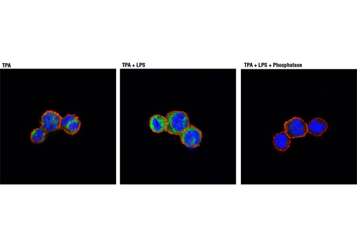
    使用 Phospho-TBK1/NAK (Ser172) (D52C2) XP® Rabbit mAb(绿色)对用 TPA #4174(80nM,过夜)(左图)处理后用 LPS(1μg/ml,1 小时)(中图)处理或用 LPS 和 λ 磷酸酶处理(右图)的 THP-1 细胞进行共聚焦免疫荧光分析。肌动蛋白丝用 DY-554 Phalloidin(红色)标记。蓝色伪彩 = DRAQ5® #4084(DNA 荧光染料)。
    Rig-I Pathway Antibody Sampler Kit: Image 21 Expand Image
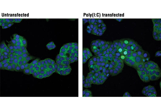
    使用 IRF-3 (D6I4C) XP® Rabbit mAb(绿色)对未经(左)或经 Poly (I:C)(2.5 μg/ml,6 小时;右)转染的 HT-29 细胞进行共聚焦免疫荧光分析。蓝色伪彩 = DRAQ5® #4084(DNA 荧光染料)。
    Rig-I Pathway Antibody Sampler Kit: Image 22 Expand Image
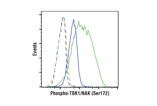
    使用 Phospho-TBK1/NAK (Ser172) (D52C2) XP® Rabbit mAb(实线)或浓度匹配的 Rabbit (DA1E) mAb IgG XP® Isotype Control #3900(虚线)对用 TPA(80 nM,4 天)#9905 分化、未经处理(蓝色)或已经过 LPS(1 ug/ml,1 小时;绿色)#14011 处理的 THP-1 细胞进行流式细胞分析。Anti-rabbit IgG (H+L), F(ab')2 Fragment (Alexa Fluor® 488 Conjugate) #4412 用作二抗。