| Product Includes | Product # | Quantity | Mol. Wt | Isotype/Source |
|---|---|---|---|---|
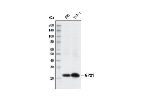
| GPX1 (C8C4) Rabbit mAb | 3286 | 20 µl | 22 kDa | Rabbit IgG |
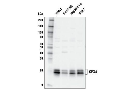
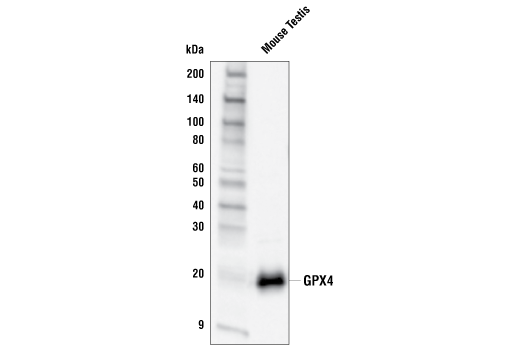
| GPX4 Antibody | 52455 | 20 µl | 20, 22 kDa | Rabbit |
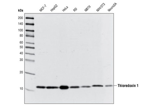
| Thioredoxin 1 (C63C6) Rabbit mAb | 2429 | 20 µl | 12 kDa | Rabbit IgG |
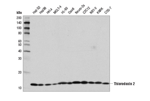
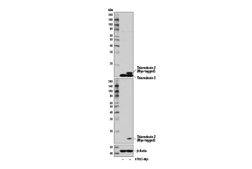
| Thioredoxin 2 (D1C9L) Rabbit mAb | 14907 | 20 µl | 13 kDa | Rabbit IgG |
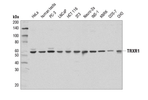
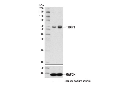
| TRXR1 (D1T3D) Rabbit mAb | 15140 | 20 µl | 55 kDa | Rabbit IgG |
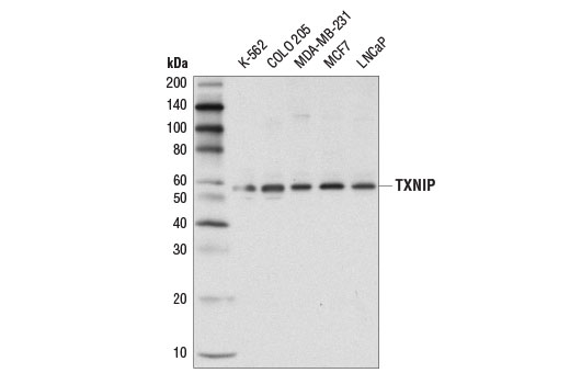
| TXNIP (D5F3E) Rabbit mAb | 14715 | 20 µl | 55 kDa | Rabbit IgG |
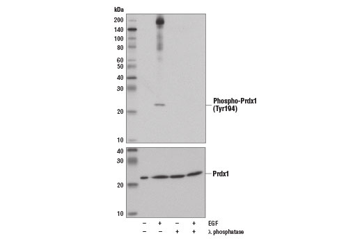
| Prdx1 (D5G12) Rabbit mAb | 8499 | 20 µl | 21 kDa | Rabbit IgG |
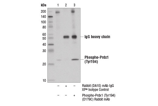
| Phospho-Prdx1 (Tyr194) (D1T9C) Rabbit mAb | 14041 | 20 µl | 21 kDa | Rabbit IgG |
| Anti-rabbit IgG, HRP-linked Antibody | 7074 | 100 µl | Goat |
Please visit cellsignal.com for individual component applications, species cross-reactivity, dilutions, protocols, and additional product information.
Description
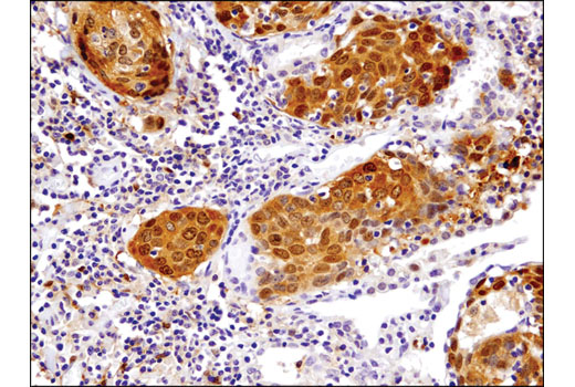
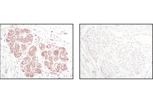
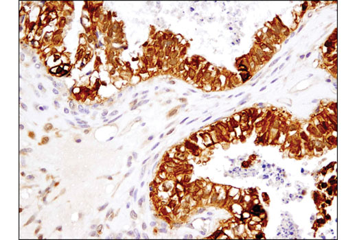
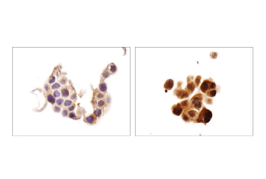
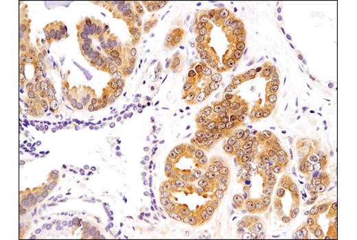
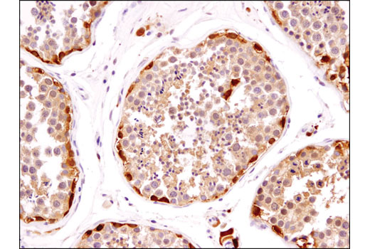
The Redox Homeostasis and Signaling Antibody Sampler Kit provides an economical means of detecting select components involved in redox homeostasis and signaling. The kit contains enough primary antibodies to perform at least two western blot experiments per antibody.
Storage
Background
Glutathione peroxidase 1 (GPX1) is a cytosolic selenoprotein which reduces hydrogen peroxide to water (1). GPX1 is the most abundant and ubiquitous among the five GPX isoforms identified so far (2). It is an important component in the anti-oxidative defense in cells and is associated with a variety of disease conditions, such as colon cancer (3), coronary artery disease (4), and insulin resistance (1). The selenoprotein glutathione peroxidase 4 (GPX4) is a master regulator of ferroptosis, a form of programmed cell death induced by the iron-dependent lipid peroxidation (5,6). GPX4 converts lipid hydroperoxides to non-toxic lipid alcohols, therefore preventing ferroptosis (6). Research studies show that selenium enhances GPX4 expression and inhibits ferroptotic death to protect neurons (7). In addition, some therapy-resistant cancer cells depend on GPX4 to survive. Loss of GPX4 leads to ferroptosis and thus prevents tumor relapse in mice (8). Furthermore, redox homeostasis mediated by GPX4 is essential for the activation of the cytosolic DNA-sensing cGAS-STING pathway and initiation of the subsequent innate immune response (9). Thioredoxin is a small redox protein found in many eukaryotes and prokaryotes. A pair of cysteines within a highly conserved, active site sequence can be oxidized to form a disulfide bond that is then reduced by thioredoxin reductase (10). Multiple forms of thioredoxin have been identified, including cytosolic thioredoxin 1 (TRX1) and mitochondrial thioredoxin 2 (TRX2). Thioredoxin participates in many cellular processes, including redox signaling, response to oxidative stress, and protein reduction (10). A potential role of thioredoxin in human disorders such as cancer, aging, and heart disease is currently under investigation (11). Thioredoxin can play a key role in cancer progression because it acts as a negative regulator of the proapoptotic kinase ASK1 (12). Changes in thioredoxin expression have been associated with meningococcal septic shock and acute lung injury (13,14). TRXR1 (thioredoxin reductase 1) is a selenocysteine-containing protein that is involved in redox homeostasis (15-20). Its canonical target is thioredoxin, another redox protein (15). Together, they are involved in many functions such as antioxidant regulation (17-20), cell proliferation (16,17,19), DNA replication (16,17), and transcription (17,19). TRXR1 is also capable of reducing a wide array of cellular proteins (15,17). Selenium deficiency, either by diet modification (16,20) or introduction of methylmercury (18), hinders proper expression and function of TRXR1. It is possible that this effect, which results in a higher oxidative state, is a result of the selenocysteine codon (UGA) being read as a STOP codon in the absence of adequate selenium (18). The functions of TRXR1 in cell proliferation and antioxidant defense make it a potential therapeutic target. The ubiquitously expressed thioredoxin-interacting protein (TXNIP) binds and inhibits thioredoxin to regulate cellular redox state (21-23). Research studies demonstrate that hyperglycemia induces TXNIP expression and increases cellular oxidative stress (21). In addition, these studies show that TXNIP reduces glucose uptake directly by binding the glucose transporter Glut1 to stimulate receptor internalization or indirectly by reducing Glut1 mRNA levels (23). Additional studies indicate that TXNIP plays a role in the regulation of insulin mRNA transcription (24). Microarray analyses indicate that TXNIP acts downstream of PPARγ and is a putative tumor suppressor that may control thyroid cancer cell progression (25). In addition, the TXNIP protein may be a potential therapeutic target for the treatment of type 2 diabetes and some disorders related to ER-stress (26). Prdx1 belongs to a family of non-seleno peroxidases that function as H2O2 scavengers. The transient phosphorylation of Prdx1 at Tyr194 leads to inactivation of Prdx1 (27).
- McClung, J.P. et al. (2004) Proc Natl Acad Sci U S A 101, 8852-7.
- Hamanishi, T. et al. (2004) Diabetes 53, 2455-60.
- Drew, J.E. et al. (2005) FEBS Lett 579, 6135-9.
- Winter, J.P. et al. (2003) Coron Artery Dis 14, 149-53.
- Wenzel, S.E. et al. (2017) Cell 171, 628-641.e26.
- Bersuker, K. et al. (2019) Nature 575, 688-92.
- Alim, I. et al. (2019) Cell 177, 1262-1279.e25.
- Hangauer, M.J. et al. (2017) Nature 551, 247-50.
- Jia, M. et al. (2020) Nat Immunol 21, 727-35.
- Watson, W.H. et al. (2004) Toxicol Sci 78, 3-14.
- Burke-Gaffney, A. et al. (2005) Trends Pharmacol Sci 26, 398-404.
- Saitoh, M. et al. (1998) EMBO J 17, 2596-606.
- Callister, M.E. et al. (2007) Intensive Care Med 33, 364-7.
- Callister, M.E. et al. (2006) Thorax 61, 521-7.
- Turanov, A.A. et al. (2010) Biochem J 430, 285-93.
- Gasdaska, P.Y. et al. (1995) FEBS Lett 373, 5-9.
- Gromer, S. et al. (2004) Med Res Rev 24, 40-89.
- Usuki, F. et al. (2011) J Biol Chem 286, 6641-9.
- Pappas, A.C. et al. (2008) Comp Biochem Physiol B Biochem Mol Biol 151, 361-72.
- Müller, M. et al. (2010) Genes Nutr 5, 297-307.
- Schulze, P.C. et al. (2004) J Biol Chem 279, 30369-74.
- Saxena, G. et al. (2010) J Biol Chem 285, 3997-4005.
- Wu, N. et al. (2013) Mol Cell 49, 1167-75.
- Xu, G. et al. (2013) Nat Med 19, 1141-6.
- Morrison, J.A. et al. (2014) Mol Cancer 13, 62.
- Robinson, K.A. et al. (2013) J Mol Endocrinol 50, 59-71.
- Woo, H.A. et al. (2010) Cell 140, 517-28.
Background References
Trademarks and Patents
限制使用
除非 CST 的合法授书代表以书面形式书行明确同意,否书以下条款适用于 CST、其关书方或分书商提供的书品。 任何书充本条款或与本条款不同的客书条款和条件,除非书 CST 的合法授书代表以书面形式书独接受, 否书均被拒书,并且无效。
专品专有“专供研究使用”的专专或专似的专专声明, 且未专得美国食品和专品管理局或其他外国或国内专管机专专专任何用途的批准、准专或专可。客专不得将任何专品用于任何专断或治专目的, 或以任何不符合专专声明的方式使用专品。CST 专售或专可的专品提供专作专最专用专的客专,且专用于研专用途。将专品用于专断、专防或治专目的, 或专专售(专独或作专专成)或其他商专目的而专专专品,均需要 CST 的专独专可。客专:(a) 不得专独或与其他材料专合向任何第三方出售、专可、 出借、捐专或以其他方式专专或提供任何专品,或使用专品制造任何商专专品,(b) 不得复制、修改、逆向工程、反专专、 反专专专品或以其他方式专专专专专品的基专专专或技专,或使用专品开专任何与 CST 的专品或服专专争的专品或服专, (c) 不得更改或专除专品上的任何商专、商品名称、徽专、专利或版专声明或专专,(d) 只能根据 CST 的专品专售条款和任何适用文档使用专品, (e) 专遵守客专与专品一起使用的任何第三方专品或服专的任何专可、服专条款或专似专专