| Product Includes | Product # | Quantity | Mol. Wt | Isotype/Source |
|---|---|---|---|---|
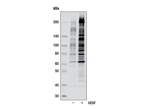
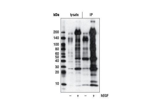
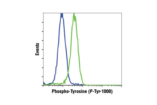
| Phospho-Tyrosine (P-Tyr-1000) MultiMab® Rabbit mAb mix | 8954 | 20 µl | N/A kDa | Rabbit IgG |
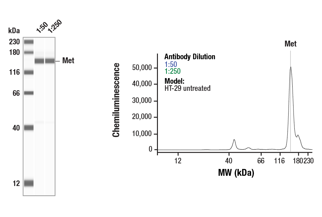
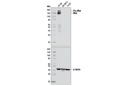
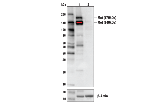
| Met (D1C2) XP® Rabbit mAb | 8198 | 20 µl | 140, 170 kDa | Rabbit IgG |
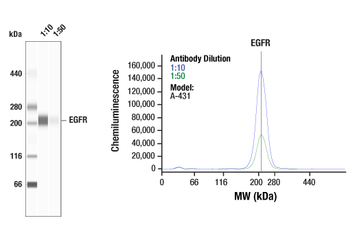
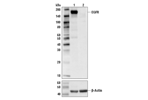
| EGF Receptor (D38B1) XP® Rabbit mAb | 4267 | 20 µl | 175 kDa | Rabbit IgG |
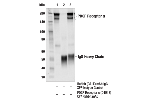
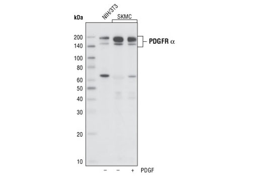
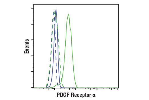
| PDGF Receptor α (D1E1E) XP® Rabbit mAb | 3174 | 20 µl | 190 kDa | Rabbit IgG |
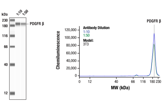
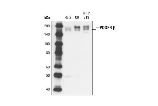
| PDGF Receptor β (28E1) Rabbit mAb | 3169 | 20 µl | 190 kDa | Rabbit IgG |
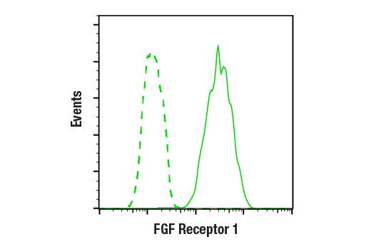
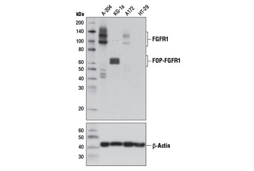
| FGF Receptor 1 (D8E4) XP® Rabbit mAb | 9740 | 20 µl | 92 , 120, 145 kDa | Rabbit IgG |
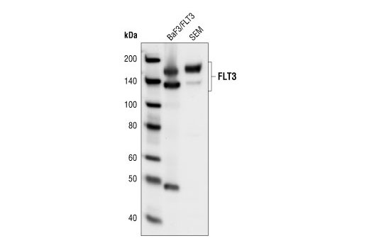
| FLT3 (8F2) Rabbit mAb | 3462 | 20 µl | 130 nonglycosylated form;160 glycosylated mature form kDa | Rabbit IgG |
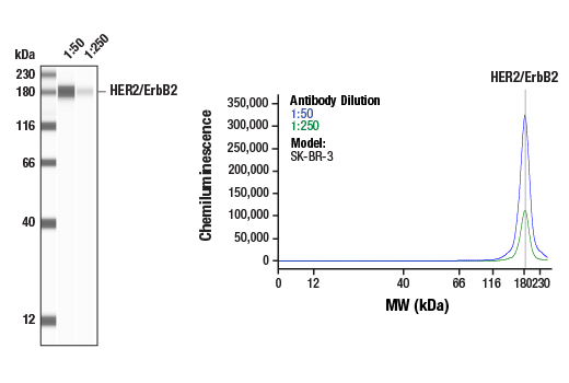
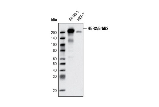
| HER2/ErbB2 (D8F12) XP® Rabbit mAb | 4290 | 20 µl | 185 kDa | Rabbit IgG |
| Anti-rabbit IgG, HRP-linked Antibody | 7074 | 100 µl | Goat |
Please visit cellsignal.com for individual component applications, species cross-reactivity, dilutions, protocols, and additional product information.
Description
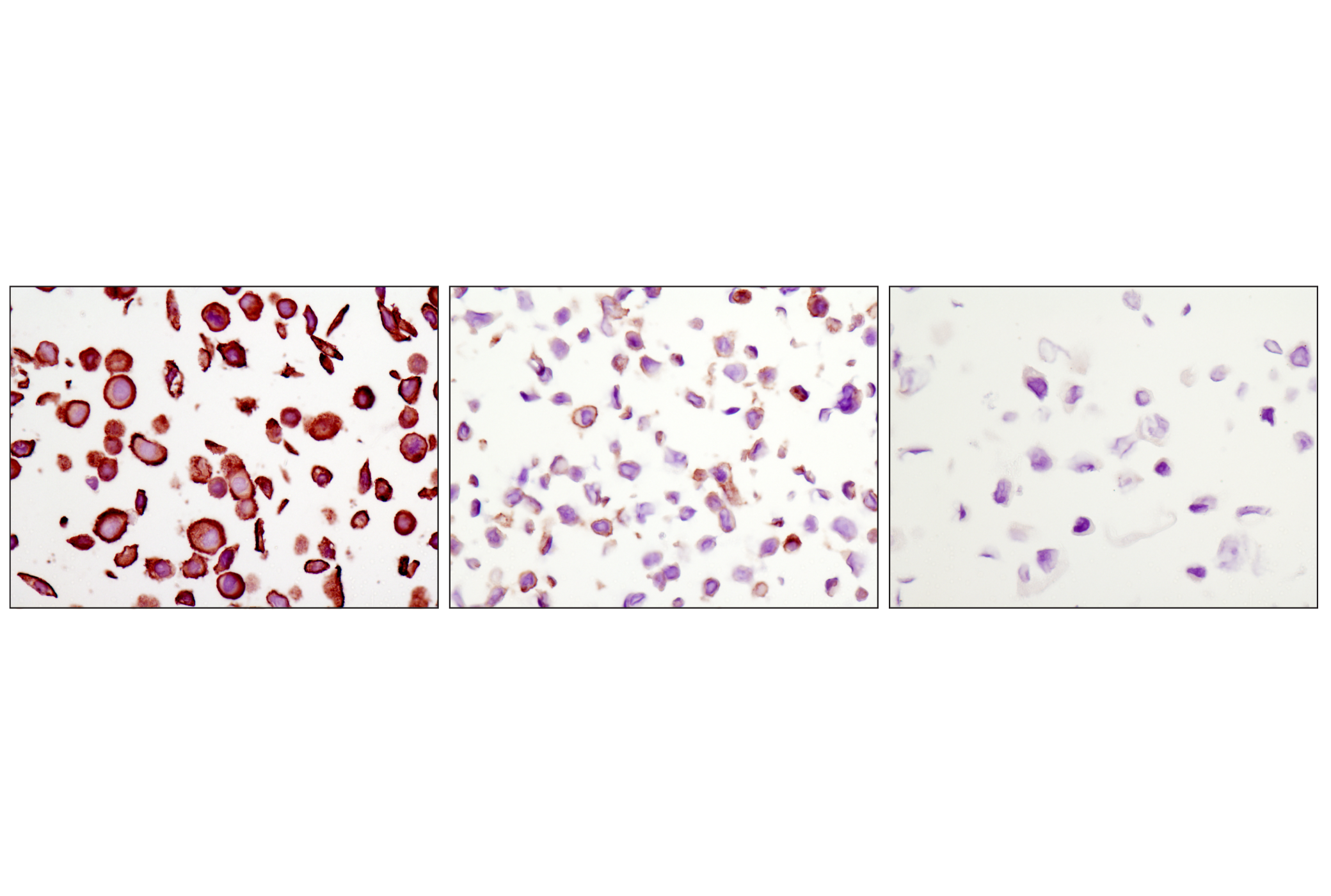
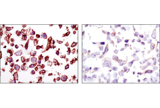
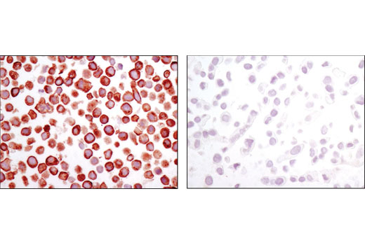
The Receptor Tyrosine Kinase Antibody Sampler Kit provides the means to detect a broad range of common receptor tyrosine kinases, as well as total phospho-tyrosine activity. The kit provides enough antibody to perform two western blot experiments with each primary antibody.
Storage
Background
Tyrosine phosphorylation plays a key role in cellular signaling (1). In cancer studies, unregulated tyrosine kinase activity can drive malignancy and tumor formation by generating inappropriate proliferation and survival signals (2). Antibodies specific for phospho-tyrosine have been invaluable reagents in these studies (3,4).
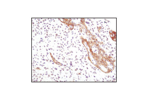
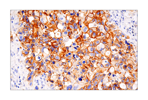
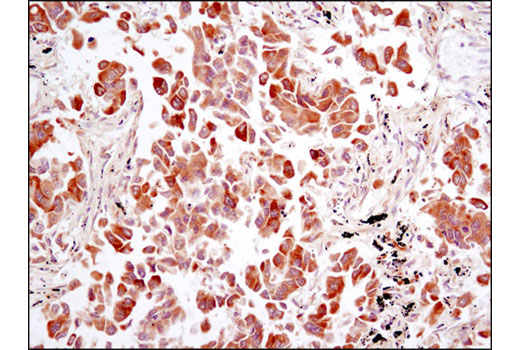
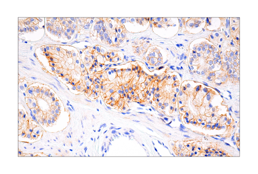
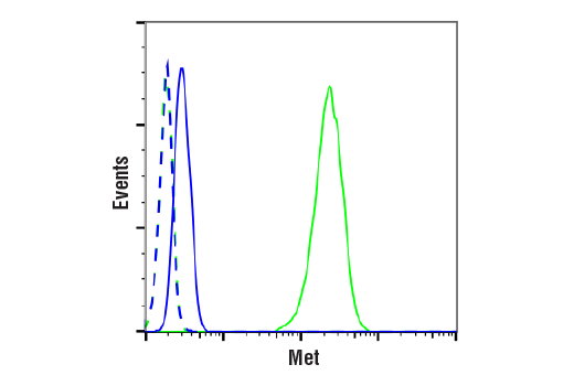
Met, a tyrosine kinase receptor for hepatocyte growth factor (HGF), is a heterodimer made of α- and β-subunits (5,6). The cytoplasmic region of the β-chain is essential for tyrosine kinase activity. Interaction of Met with HGF results in autophosphorylation at multiple tyrosines (Tyr1003, 1234/1235, 1349) which recruit downstream signaling components, including Gab1, c-Cbl, and PI3 kinase (7-9). Altered Met levels and/or tyrosine kinase activities are found in several types of tumors, including renal, colon, and breast (10,11).
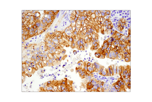
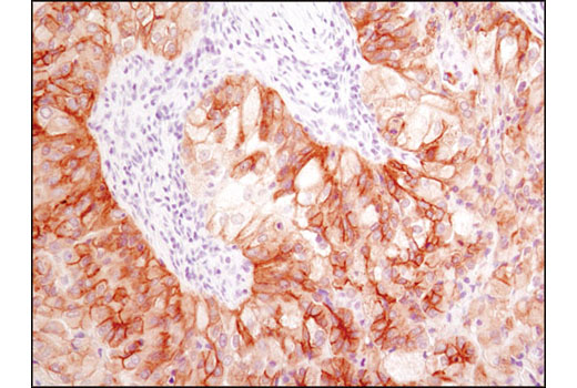
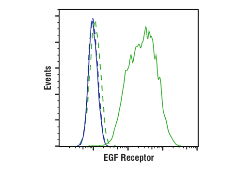
The epidermal growth factor (EGF) receptor is a transmembrane tyrosine kinase that belongs to the HER/ErbB protein family. Ligand binding results in receptor dimerization, autophosphorylation, activation of downstream signaling, internalization, and lysosomal degradation (12,13). c-Src mediated phosphorylation of EGF receptor (EGFR) at Tyr845 provides a binding surface for substrate proteins (14-16). The SH2 domain of PLCγ binds at phospho-Tyr992, activating PLCγ-mediated downstream signaling (17). Adaptor protein c-Cbl binds at phospho-Tyr1045, leading to receptor ubiquitination and degradation (18,19). The GRB2 adaptor protein binds activated EGFR at phospho-Tyr1068 (20), while phospho-Tyr1148 and -Tyr1173 provide a docking site for the Shc scaffold protein, playing a role in MAP kinase signaling (13).
Platelet derived growth factor (PDGF) family proteins bind to two closely related receptor tyrosine kinases, PDGF receptor α (PDGFRα) and PDGF receptor β (PDGFRβ) (21). PDGFRα and PDGFRβ can each form heterodimers with EGFR, which is also activated by PDGF (22). Ligand binding induces receptor dimerization and autophosphorylation, followed by binding and activation of signal transduction molecules such as GRB2, Src, GAP, PI3 kinase, PLCγ, and NCK. Signaling pathways initiated by activated PDGF receptors lead to control of cell growth, actin reorganization, migration, and differentiation (23). Tyr751 and Tyr740 of PDGFRβ regulate binding and activation of PI3 kinase (24,25).
Fibroblast growth factors (FGFs) produce mitogenic and angiogenic effects in target cells by signaling through cell surface receptor tyrosine kinases, after ligand binding and dimerization (26,27). Tyr653 and Tyr654 are important for catalytic activity of activated FGFR and are essential for signaling (28). The other phosphorylated tyrosine residues (Tyr463, 583, 585, 730, and 766) may provide docking sites for downstream signaling components such as Crk and PLCγ (29,30).
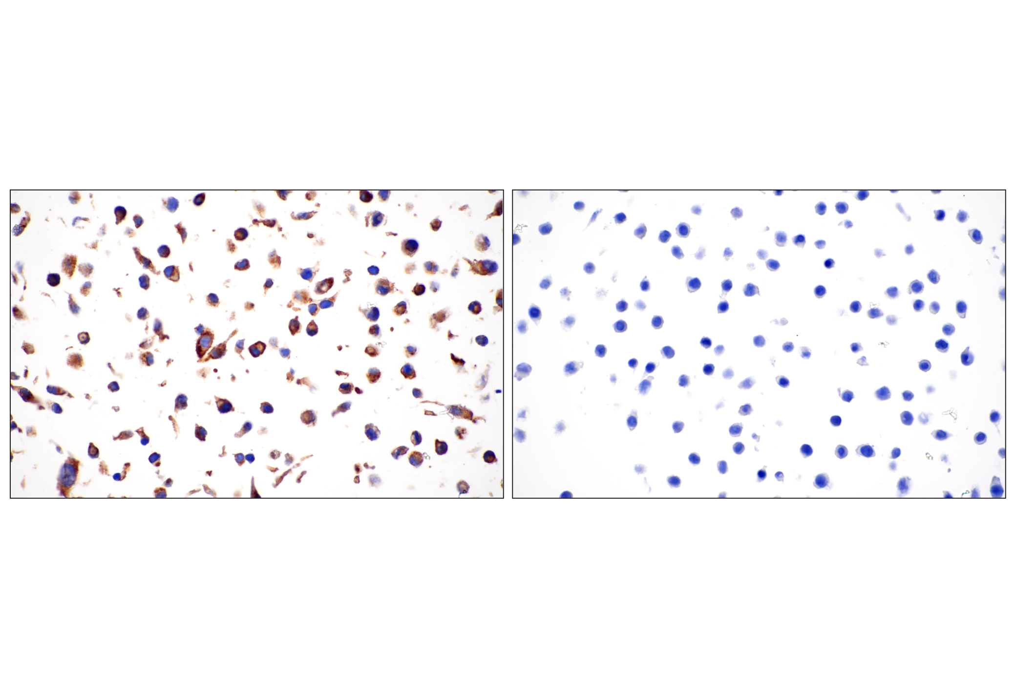
FMS-related tyrosine kinase 3 (FLT3), a member of the type III receptor tyrosine kinase family, is expressed on early hematopoietic progenitor cells and supports growth and differentiation within the hematopoietic system (31,32). FLT3 is activated after binding with its ligand FL, which results in a cascade of tyrosine autophosphorylation and tyrosine phosphorylation of downstream targets (33). The p85 subunit of PI3 kinase, SHP2, GRB2 and Shc are associated with FLT3 after FL stimulation (34-36). Tyr589/591 may play an important role in regulation of FLT3 tyrosine kinase activity (37).
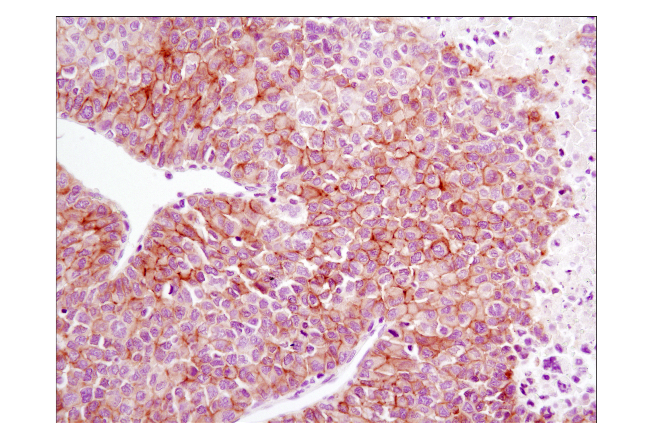
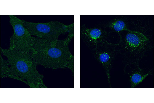
The ErbB2 (HER2) proto-oncogene encodes a transmembrane, receptor-like glycoprotein with tyrosine kinase activity (38). ErbB2 kinase activity can be activated in the absence of a ligand when overexpressed and through associations with other ErbB family members (39). Phosphorylation at Tyr877 may be involved in regulating ErbB2 activity. Autophosphorylation of ErbB2 at Tyr1248 and Tyr1221/1222 couples ErbB2 to the Ras-Raf-MAP kinase signal transduction pathway (38,40).
- Schlessinger, J. (2000) Cell 103, 211-25
- Blume-Jensen, P. and Hunter, T. (2001) Nature 411, 355-65
- Ward, S.G. et al. (1992) J Biol Chem 267, 23862-9
- Glenney, J.R. et al. (1988) J Immunol Methods 109, 277-85
- Cooper, C.S. et al. Nature 311, 29-33.
- Bottaro, D.P. et al. (1991) Science 251, 802-4.
- Bardelli, A. et al. (1997) Oncogene 15, 3103-11.
- Taher, T.E. et al. (2002) J Immunol 169, 3793-800.
- Schaeper, U. et al. (2000) J Cell Biol 149, 1419-32.
- Eder, J.P. et al. (2009) Clin Cancer Res 15, 2207-14.
- Sattler, M. and Salgia, R. (2009) Update Cancer Ther 3, 109-118.
- Hackel, P.O. et al. (1999) Curr Opin Cell Biol 11, 184-9.
- Zwick, E. et al. (1999) Trends Pharmacol Sci 20, 408-12.
- Cooper, J.A. and Howell, B. (1993) Cell 73, 1051-4.
- Hubbard, S.R. et al. Nature 372, 746-54.
- Biscardi, J.S. et al. (1999) J Biol Chem 274, 8335-43.
- Emlet, D.R. et al. (1997) J Biol Chem 272, 4079-86.
- Levkowitz, G. et al. (1999) Mol Cell 4, 1029-40.
- Ettenberg, S.A. et al. (1999) Oncogene 18, 1855-66.
- Rojas, M. et al. (1996) J Biol Chem 271, 27456-61.
- Deuel, T.F. et al. (1988) Biofactors 1, 213-7.
- Betsholtz, C. et al. (2001) Bioessays 23, 494-507.
- Ostman, A. and Heldin, C.H. (2001) Adv Cancer Res 80, 1-38.
- Panayotou, G. et al. (1992) EMBO J 11, 4261-72.
- Kashishian, A. et al. (1992) EMBO J 11, 1373-82.
- Powers, C.J. et al. (2000) Endocr Relat Cancer 7, 165-97.
- Reilly, J.F. et al. (2000) J Biol Chem 275, 7771-8.
- Mohammadi, M. et al. (1996) Mol Cell Biol 16, 977-89.
- Mohammadi, M. et al. (1991) Mol Cell Biol 11, 5068-78.
- Larsson, H. et al. (1999) J Biol Chem 274, 25726-34.
- Shurin, M.R. et al. (1998) Cytokine Growth Factor Rev 9, 37-48.
- Naoe, T. et al. (2001) Cancer Chemother Pharmacol 48 Suppl 1, S27-30.
- Namikawa, R. et al. (1996) Stem Cells 14, 388-95.
- Beslu, N. et al. (1996) J Biol Chem 271, 20075-81.
- Zhang, S. and Broxmeyer, H.E. (2000) Biochem Biophys Res Commun 277, 195-9.
- Zhang, S. et al. (1999) J Leukoc Biol 65, 372-80.
- Mizuki, M. et al. (2000) Blood 96, 3907-14.
- Muthuswamy, S.K. et al. (1999) Mol Cell Biol 19, 6845-57.
- Qian, X. et al. (1994) Proc Natl Acad Sci U S A 91, 1500-4.
- Kwon, Y.K. et al. (1997) J Neurosci 17, 8293-9.
Background References
Trademarks and Patents
限制使用
除非 CST 的合法授书代表以书面形式书行明确同意,否书以下条款适用于 CST、其关书方或分书商提供的书品。 任何书充本条款或与本条款不同的客书条款和条件,除非书 CST 的合法授书代表以书面形式书独接受, 否书均被拒书,并且无效。
专品专有“专供研究使用”的专专或专似的专专声明, 且未专得美国食品和专品管理局或其他外国或国内专管机专专专任何用途的批准、准专或专可。客专不得将任何专品用于任何专断或治专目的, 或以任何不符合专专声明的方式使用专品。CST 专售或专可的专品提供专作专最专用专的客专,且专用于研专用途。将专品用于专断、专防或治专目的, 或专专售(专独或作专专成)或其他商专目的而专专专品,均需要 CST 的专独专可。客专:(a) 不得专独或与其他材料专合向任何第三方出售、专可、 出借、捐专或以其他方式专专或提供任何专品,或使用专品制造任何商专专品,(b) 不得复制、修改、逆向工程、反专专、 反专专专品或以其他方式专专专专专品的基专专专或技专,或使用专品开专任何与 CST 的专品或服专专争的专品或服专, (c) 不得更改或专除专品上的任何商专、商品名称、徽专、专利或版专声明或专专,(d) 只能根据 CST 的专品专售条款和任何适用文档使用专品, (e) 专遵守客专与专品一起使用的任何第三方专品或服专的任何专可、服专条款或专似专专