Revision 1
Cell Signaling Technology

Orders: 877-616-CELL (2355) [email protected]

Support: 877-678-TECH (8324)

Web: [email protected] cellsignal.com

3 Trask LaneDanversMassachusetts01923USA
For Research Use Only. Not for Use in Diagnostic Procedures.
:

:

:

:

UniProt ID:

#P06400

Entrez-Gene Id:

5925

Product Information

Storage

Supplied in 10 mM sodium HEPES (pH 7.5), 150 mM NaCl, 100 µg/ml BSA, 50% glycerol and less than 0.02% sodium azide. Store at –20°C. Do not aliquot the antibody.

Specificity / Sensitivity

Phospho-Rb (Ser780) (D59B7) Rabbit mAb and Phospho-Rb (Ser807/811) (D20B12) XP® Rabbit mAb detect endogenous levels of Rb only when phosphorylated at Ser780 and Ser807/811, respectively. Rb (4H1) Mouse mAb detects endogenous levels of total Rb protein. The antibody does not cross-react with the Rb homologues p107 or p130, or with other proteins.

Source / Purification

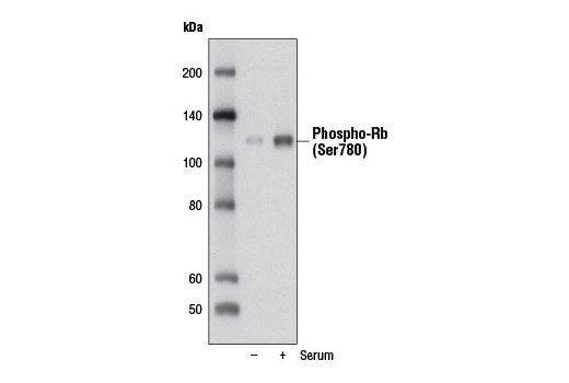
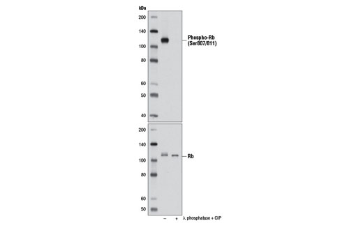
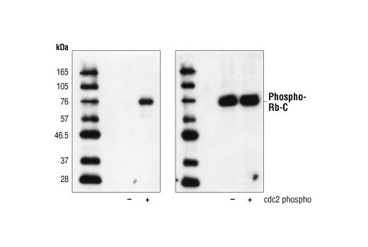
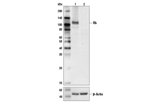
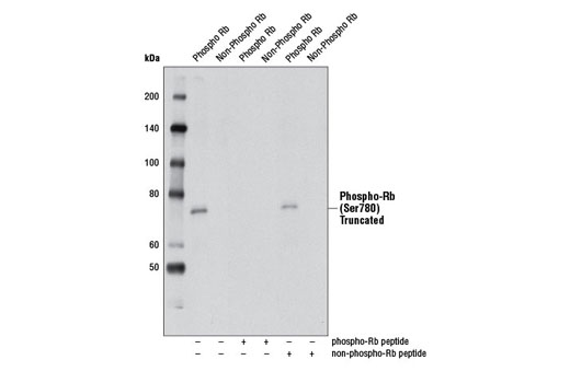
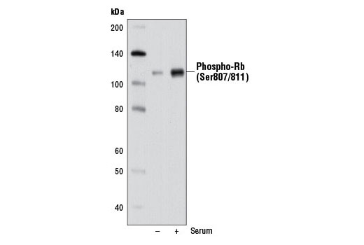
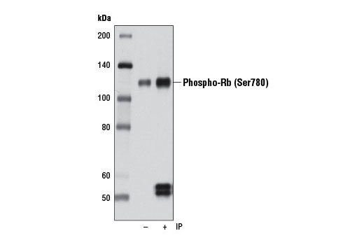
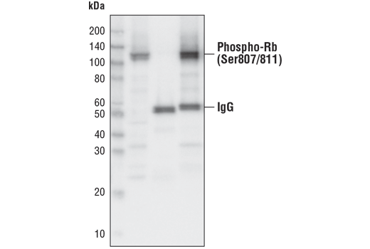
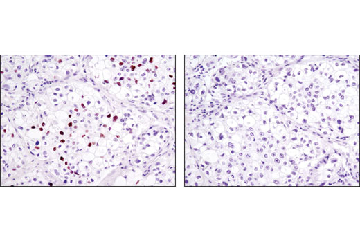
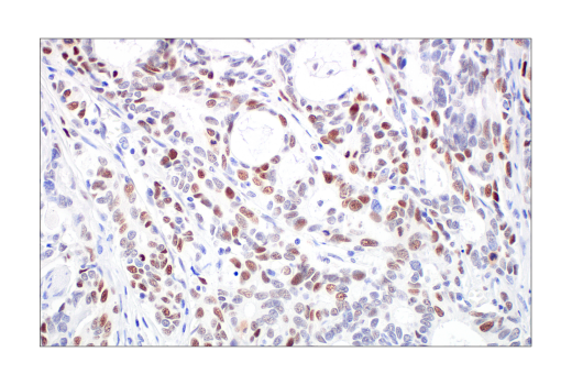
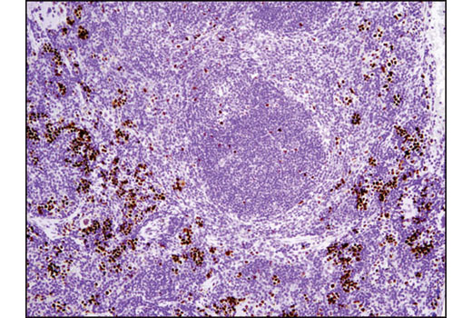
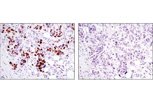
Phospho-Rb (Ser780) (D59B7) Rabbit mAb is produced by immunizing animals with a synthetic peptide corresponding to residues surrounding Ser780 of human Rb protein. Phospho-Rb (Ser807/811) (D20B12) XP® Rabbit mAb is produced by immunizing animals with a synthetic peptide corresponding to residues surrounding Ser807/811 of human Rb protein. Rb (4H1) monoclonal antibody is produced by immunizing animals with Rb-C Fusion Protein #6022, which contains residues 701-928 of human Rb .

Product Description

The PhosphoPlus® Rb (Ser780, Ser807/811) Antibody Kit provides reagents and protocols to investigate cell cycle progression within cells. The kit contains a total Rb antibody, two distinct phospho-Rb antibodies, and Rb control proteins along with secondary antibodies and reagents to perform up to 10 western blots.

Background

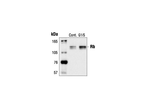
The retinoblastoma tumor suppressor protein Rb regulates cell proliferation by controlling progression through the restriction point within the G1-phase of the cell cycle (1). Rb has three functionally distinct binding domains and interacts with critical regulatory proteins including the E2F family of transcription factors, c-Abl tyrosine kinase, and proteins with a conserved LXCXE motif (2-4). Cell cycle-dependent phosphorylation by a CDK inhibits Rb target binding and allows cell cycle progression (5). Rb inactivation and subsequent cell cycle progression likely requires an initial phosphorylation by cyclin D-CDK4/6 followed by cyclin E-CDK2 phosphorylation (6). Specificity of different CDK/cyclin complexes has been observed in vitro (6-8) and cyclin D1 is required for Ser780 phosphorylation in vivo (9).

  1. Sherr, C.J. (1996) Science 274, 1672-7.
  2. Nevins, J.R. (1992) Science 258, 424-9.
  3. Welch, P.J. and Wang, J.Y. (1993) Cell 75, 779-90.
  4. Hu, Q.J. et al. (1990) EMBO J 9, 1147-55.
  5. Knudsen, E.S. and Wang, J.Y. (1997) Mol Cell Biol 17, 5771-83.
  6. Lundberg, A.S. and Weinberg, R.A. (1998) Mol Cell Biol 18, 753-61.
  7. Connell-Crowley, L. et al. (1997) Mol Biol Cell 8, 287-301.
  8. Kitagawa, M. et al. (1996) EMBO J 15, 7060-9.
  9. Geng, Y. et al. (2001) Proc Natl Acad Sci USA 98, 194-9.

Species Reactivity

Species reactivity is determined by testing in at least one approved application (e.g., western blot).

Cross-Reactivity Key

H: human M: mouse R: rat Hm: hamster Mk: monkey Vir: virus Mi: mink C: chicken Dm: D. melanogaster X: Xenopus Z: zebrafish B: bovine Dg: dog Pg: pig Sc: S. cerevisiae Ce: C. elegans Hr: horse GP: Guinea Pig Rab: rabbit All: all species expected

Trademarks and Patents

Cell Signaling Technology is a trademark of Cell Signaling Technology, Inc.
PhosphoPlus is a registered trademark of Cell Signaling Technology, Inc.
All other trademarks are the property of their respective owners. Visit cellsignal.com/trademarks for more information.

限制使用

除非 CST 的合法授书代表以书面形式书行明确同意,否书以下条款适用于 CST、其关书方或分书商提供的书品。 任何书充本条款或与本条款不同的客书条款和条件,除非书 CST 的合法授书代表以书面形式书独接受, 否书均被拒书,并且无效。

专品专有“专供研究使用”的专专或专似的专专声明, 且未专得美国食品和专品管理局或其他外国或国内专管机专专专任何用途的批准、准专或专可。客专不得将任何专品用于任何专断或治专目的, 或以任何不符合专专声明的方式使用专品。CST 专售或专可的专品提供专作专最专用专的客专,且专用于研专用途。将专品用于专断、专防或治专目的, 或专专售(专独或作专专成)或其他商专目的而专专专品,均需要 CST 的专独专可。客专:(a) 不得专独或与其他材料专合向任何第三方出售、专可、 出借、捐专或以其他方式专专或提供任何专品,或使用专品制造任何商专专品,(b) 不得复制、修改、逆向工程、反专专、 反专专专品或以其他方式专专专专专品的基专专专或技专,或使用专品开专任何与 CST 的专品或服专专争的专品或服专, (c) 不得更改或专除专品上的任何商专、商品名称、徽专、专利或版专声明或专专,(d) 只能根据 CST 的专品专售条款和任何适用文档使用专品, (e) 专遵守客专与专品一起使用的任何第三方专品或服专的任何专可、服专条款或专似专专

Revision 1
#9306

PhosphoPlus® Rb (Ser780, Ser807/811) Antibody Kit

PhosphoPlus® Rb (Ser780, Ser807/811) Antibody Kit: Image 1 Expand Image
Confocal immunofluorescent analysis of fixed frozen mouse small intestine labeled with Phospho-Rb (Ser807/811) (D20B12) XP® Rabbit mAb (left, green) and co-labeled with DyLight 554 Phalloidin #13054 (right, red) and ProLong® Gold Antifade Reagent with DAPI #8961 (right, blue).
PhosphoPlus® Rb (Ser780, Ser807/811) Antibody Kit: Image 2 Expand Image
Confocal immunofluorescent analysis of fixed frozen mouse testis labeled with Phospho-Rb (Ser807/811) (D20B12) XP® Rabbit mAb (left, green) and co-labeled with DyLight 554 Phalloidin #13054 (right, red) and ProLong® Gold Antifade Reagent with DAPI #8961 (right, blue).
PhosphoPlus® Rb (Ser780, Ser807/811) Antibody Kit: Image 3 Expand Image
Confocal immunofluorescent analysis of fixed frozen mouse thymus labeled with Phospho-Rb (Ser807/811) (D20B12) XP® Rabbit mAb (left, green) and co-labeled with DyLight 554 Phalloidin #13054 (right, red) and ProLong® Gold Antifade Reagent with DAPI #8961 (right, blue).
PhosphoPlus® Rb (Ser780, Ser807/811) Antibody Kit: Image 4 Expand Image
Simple Western™ analysis of lysates (0.1 mg/mL) from MCF-7 cells using Phospho-Rb (Ser807/811) (D20B12) XP® Rabbit mAb #8516. 虚拟泳道视图(左图)显示一抗稀释比例为 1:10 时的靶标条带(如图所示)。The corresponding electropherogram view (right) plots chemiluminescence by molecular weight along the capillary at 1:10 dilution of primary antibody. 在还原条件下,使用 12-230 kDa 分离模块在 ProteinSimple(BioTechne 品牌)的 Jess™ Simple Western 仪器上进行该实验。
PhosphoPlus® Rb (Ser780, Ser807/811) Antibody Kit: Image 5 Expand Image
一抗与靶标蛋白结合之后,与偶联 HRP 的二抗形成复合体。添加 LumiGLO®,在酶催化分解期间发光。
PhosphoPlus® Rb (Ser780, Ser807/811) Antibody Kit: Image 6 Expand Image
一抗与靶标蛋白结合之后,与偶联 HRP 的二抗形成复合体。添加 LumiGLO®,在酶催化分解期间发光。
PhosphoPlus® Rb (Ser780, Ser807/811) Antibody Kit: Image 7 Expand Image
一抗与靶标蛋白结合之后,与偶联 HRP 的二抗形成复合体。添加 LumiGLO*,它可在酶催化的分解期间发光。
PhosphoPlus® Rb (Ser780, Ser807/811) Antibody Kit: Image 8 Expand Image
使用 Phospho-Rb (Ser780) (D59B7) Rabbit mAb 对血清饥饿 3 天 (-) 或血清饥饿 3 天后用 10% 血清处理 2 天 (+) 的 WI-38 细胞的提取物进行蛋白质印迹分析。
PhosphoPlus® Rb (Ser780, Ser807/811) Antibody Kit: Image 9 Expand Image
使用 Phospho-Rb (Ser807/811) (D20B12) XP® Rabbit mAb(上)或 Rb (4H1) Mouse mAb #9309(下),对未经处理 (-) 或经小牛小肠磷酸酶 (CIP) 和 λ 磷酸酶 (+) 处理的 MCF7 细胞提取物进行蛋白质印迹分析。
PhosphoPlus® Rb (Ser780, Ser807/811) Antibody Kit: Image 10 Expand Image
使用 Phospho-Rb (Ser795) Antibody #9301(左图)或对照抗体(右图),对经 cdc2/周期素 B 蛋白激酶 (New England Biolabs #P6020) 体外磷酸化之前 (-) 和之后 (+) 的 Rb-C Fusion Protein #6022(Rb 的氨基酸 701-928 融合到 MBP)进行蛋白质印迹分析。
PhosphoPlus® Rb (Ser780, Ser807/811) Antibody Kit: Image 11 Expand Image
使用 Rb (4H1) Mouse mAb #9309(上图)和 β-actin (D6A8) Rabbit mAb #8457(下图)对 HeLa 细胞(泳道 1)或 Rb 敲除型细胞(泳道 2)的提取物进行蛋白印迹分析。Rb 敲除型 HeLa 细胞中没有信号,这证实了抗体对 Rb 的特异性。
PhosphoPlus® Rb (Ser780, Ser807/811) Antibody Kit: Image 12 Expand Image
使用 Phospho-Rb (Ser780) (D59B7) Rabbit mAb 对有或没有 Rb 封闭肽的磷酸化或非磷酸化重组截短型 Rb 进行蛋白质印迹分析。
PhosphoPlus® Rb (Ser780, Ser807/811) Antibody Kit: Image 13 Expand Image
使用 Phospho-Rb (Ser807/811) (D20B12) XP® Rabbit mAb,对 WI-38 细胞提取物进行蛋白质印迹分析,而 WI-38 细胞经血清饥饿 3 天 (-) 或血清饥饿 3 天后用 10% 的血清处理 2 天 (+)。
PhosphoPlus® Rb (Ser780, Ser807/811) Antibody Kit: Image 14 Expand Image
使用 Rb (4H1) Mouse mAb #9309 和 α-Tubulin (11H10) Rabbit mAb #2125 ,对转染 100 nM SignalSilence® Control siRNA (Fluorescein Conjugate) #6201 (-)、SignalSilence® Rb siRNA I #6451 (+) 或 SignalSilence® Rb siRNA II (+) 的 Hela 细胞提取物进行蛋白质印迹分析。Rb (4H1) Mouse mAb 确认 Rb 表达沉默,而 α-Tubulin (11H10) Rabbit mAb 则用于对照上样量和 Rb siRNA 的特异性。
PhosphoPlus® Rb (Ser780, Ser807/811) Antibody Kit: Image 15 Expand Image
使用 Phospho-Rb (Ser780) (D59B7) Rabbit mAb(泳道 2)对 WI-38 细胞提取物磷酸-Rb (Ser780) 进行免疫沉淀。使用同一抗体进行蛋白质进行印迹分析。泳道 1 是 10% 输入对照。
PhosphoPlus® Rb (Ser780, Ser807/811) Antibody Kit: Image 16 Expand Image
使用 Rabbit (DA1E) mAb IgG XP® Isotype Control #3900(泳道 2)或 Phospho-Rb (Ser807/811) (D20B12) XP® Rabbit mAb(泳道 3),对 Cos 细胞提取物磷酸 Rb (Ser807/811) 进行免疫沉淀。泳道 1 是 10% 输入对照。使用 Phospho-Rb (Ser807/811) (D20B12) XP® Rabbit mAb 进行蛋白质印迹分析。
PhosphoPlus® Rb (Ser780, Ser807/811) Antibody Kit: Image 17 Expand Image
使用 Rb (4H1) Mouse mAb 对未经或已经羟基脲处理 (G1/S) 的 COS-7 细胞提取物进行蛋白质印迹分析。
PhosphoPlus® Rb (Ser780, Ser807/811) Antibody Kit: Image 18 Expand Image
使用 Rb (4H1) Mouse mAb 对石蜡包埋的且显示胞核定位的人乳腺癌细胞进行免疫组织化学分析。
PhosphoPlus® Rb (Ser780, Ser807/811) Antibody Kit: Image 19 Expand Image
使用 Phospho-Rb (Ser807/811) (D20B12) XP® Rabbit mAb 对石蜡包埋的人结肠癌进行免疫组织化学分析。
PhosphoPlus® Rb (Ser780, Ser807/811) Antibody Kit: Image 20 Expand Image
使用 Rb (4H1) Mouse mAb 对石蜡包埋的人肺癌细胞进行免疫组织化学分析。
PhosphoPlus® Rb (Ser780, Ser807/811) Antibody Kit: Image 21 Expand Image
使用 Phospho-Rb (Ser807/811) (D20B12) XP® Rabbit mAb,对对照性(左)或经 λ 磷酸酶处理(右)的石蜡包埋的肺癌进行免疫组织化学分析。
PhosphoPlus® Rb (Ser780, Ser807/811) Antibody Kit: Image 22 Expand Image
使用 Rb (4H1) Mouse mAb 对石蜡包埋的人食道癌细胞进行免疫组织化学分析。
PhosphoPlus® Rb (Ser780, Ser807/811) Antibody Kit: Image 23 Expand Image
使用 Phospho-Rb (Ser807/811) (D20B12) XP® Rabbit mAb 对石蜡包埋的小鼠脾脏细胞进行免疫组织化学分析。
PhosphoPlus® Rb (Ser780, Ser807/811) Antibody Kit: Image 24 Expand Image
使用 Rb (4H1) Mouse mAb 对石蜡包埋的人 T 细胞淋巴瘤细胞进行免疫组织化学分析。
PhosphoPlus® Rb (Ser780, Ser807/811) Antibody Kit: Image 25 Expand Image
在对照肽(左)或抗原特异性肽(右)存在的情况下,使用 Phospho-Rb (Ser807/811) (D20B12) XP® Rabbit mAb,对石蜡包埋的人卵巢浆液性腺癌进行免疫组织化学分析。
PhosphoPlus® Rb (Ser780, Ser807/811) Antibody Kit: Image 26 Expand Image
使用 Rb (4H1) Mouse mAb 对石蜡包埋的人胃腺癌细胞进行免疫组织化学分析。
PhosphoPlus® Rb (Ser780, Ser807/811) Antibody Kit: Image 27 Expand Image
使用 Phospho-Rb (Ser807/Ser811) (D20B12) XP® Rabbit mAb(绿色),对未经处理(上)或经 λ 磷酸酶处理(下)的 MCF7(左)和 BT-549(右)细胞进行共聚焦免疫荧光分析。使用 DY-554 phalloidin(红色)标记肌动蛋白纤丝。蓝色伪彩 = DRAQ5® #4084(DNA 荧光染料)。
PhosphoPlus® Rb (Ser780, Ser807/811) Antibody Kit: Image 28 Expand Image
使用 Rb (4H1) Mouse mAb(绿色)对 SH-SY5Y 细胞进行共聚焦免疫荧光图像分析。肌动蛋白纤丝用 Alexa Fluor®555 phalloidin(红色)进行标记。
PhosphoPlus® Rb (Ser780, Ser807/811) Antibody Kit: Image 29 Expand Image
使用 Phospho-Rb (Ser807/811) (D20B12) XP® Rabbit mAb 和 Propidium Iodide (PI/RNase Staining Solution #4087对 Jurkat 细胞进行流式细胞分析,以测量 DNA 含量。Anti-rabbit IgG (H+L), F(ab')2 Fragment (Alexa Fluor® 488 Conjugate) #4412 用作二抗。
PhosphoPlus® Rb (Ser780, Ser807/811) Antibody Kit: Image 30 Expand Image
以碘化丙啶(DNA 含量)作为对照,使用 Rb (4H1) Mouse mAb 对 Jurkat 细胞进行流式细胞分析。该盒表示 Rb 阳性细胞。
PhosphoPlus® Rb (Ser780, Ser807/811) Antibody Kit: Image 31 Expand Image
使用 SimpleChIP® Enzymatic Chromatin IP Kit (Magnetic Beads) #9003,对 Raji 细胞中提取的交联染色质,在加入 Rb (4H1) Mouse mAb 或 Normal Rabbit IgG #2729 后,进行染色质免疫沉淀。使用 SimpleChIP® Human Timeless Intron 1 Primers #7001、人 DHFR 启动子引物和 SimpleChIP® Human α Satellite Repeat Primers #4486,通过实时 PCR 对富集 DNA 进行量化。将每份样品中免疫沉淀的 DNA 的量表现为相对于所输入染色质总量(等于 1)的信号。