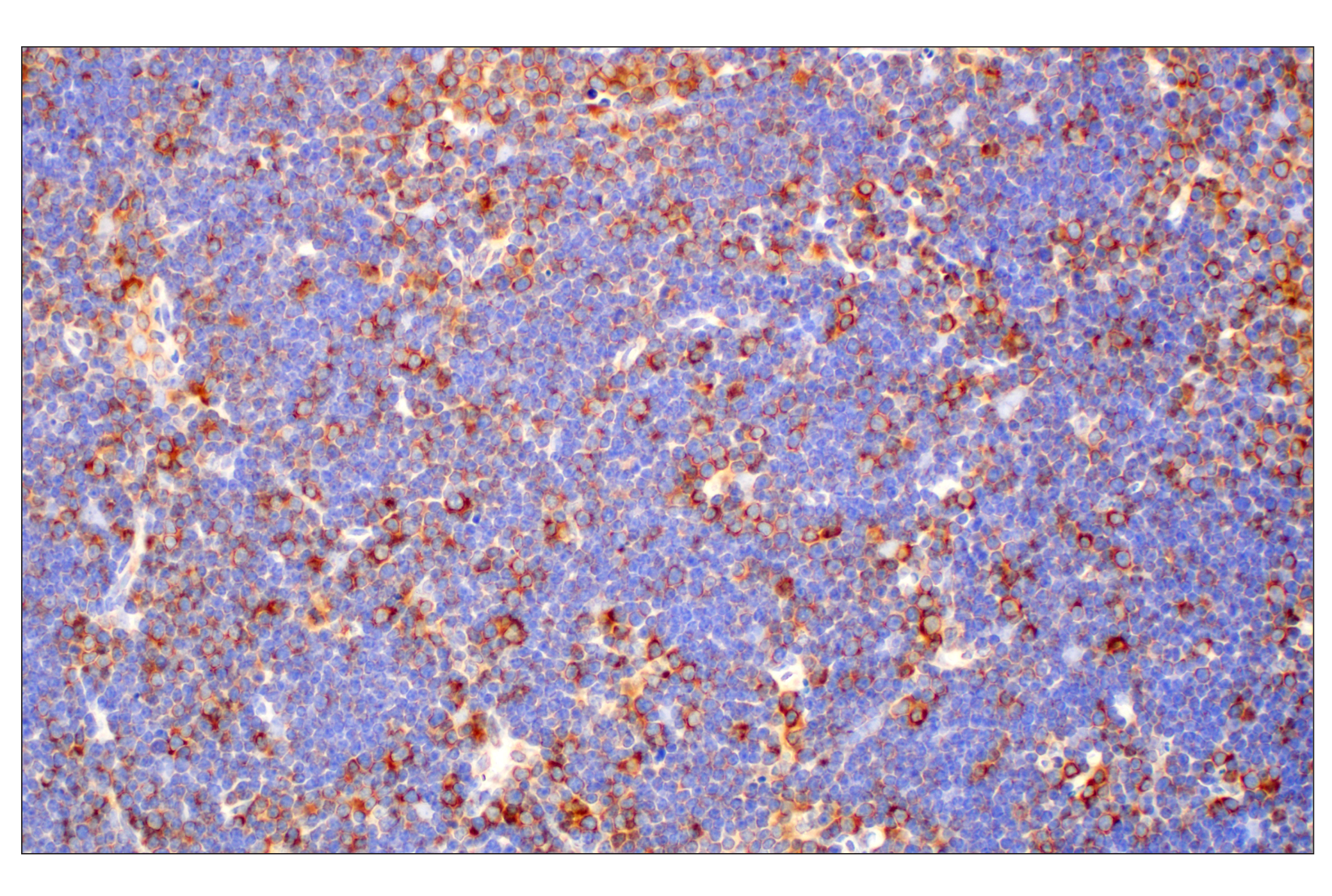
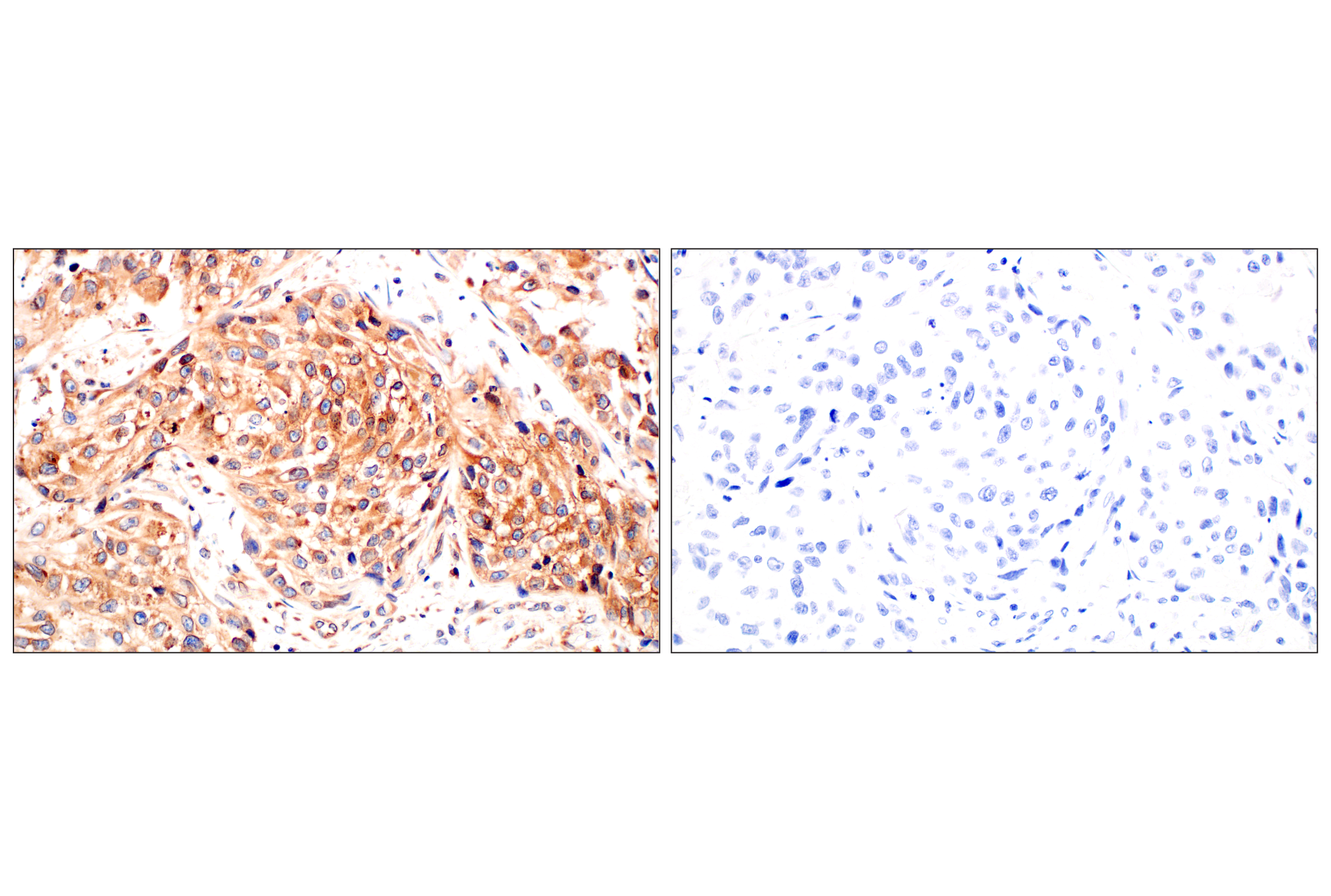
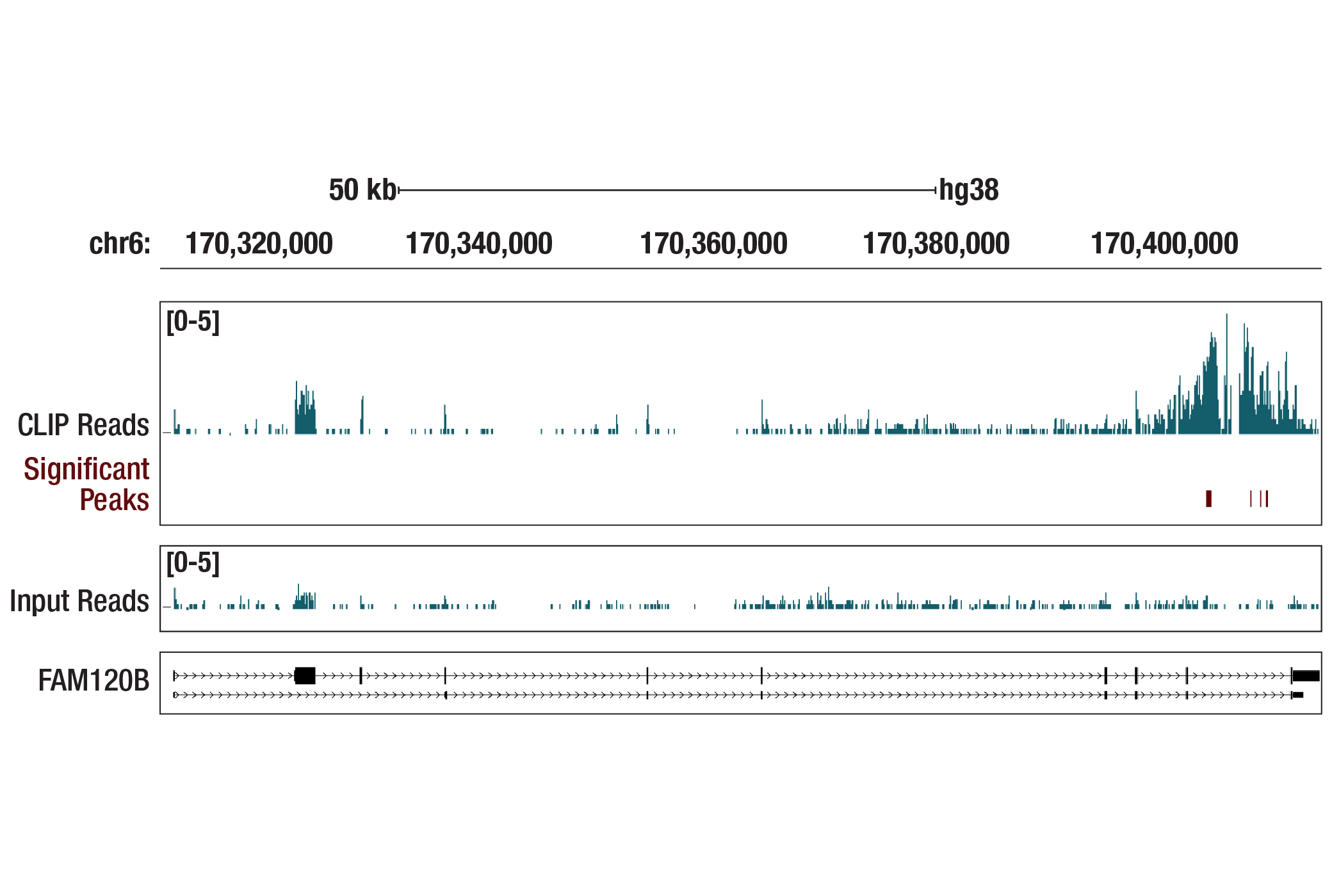
WB, IP, IHC-P, eCLIP
H M R Mk Dm
Endogenous
32
Rabbit IgG
#P63244
10399
Product Information
Product Usage Information
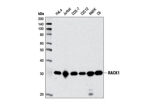
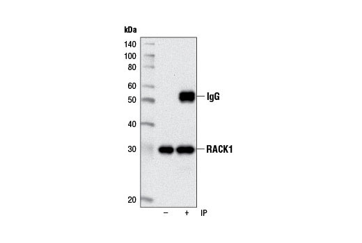
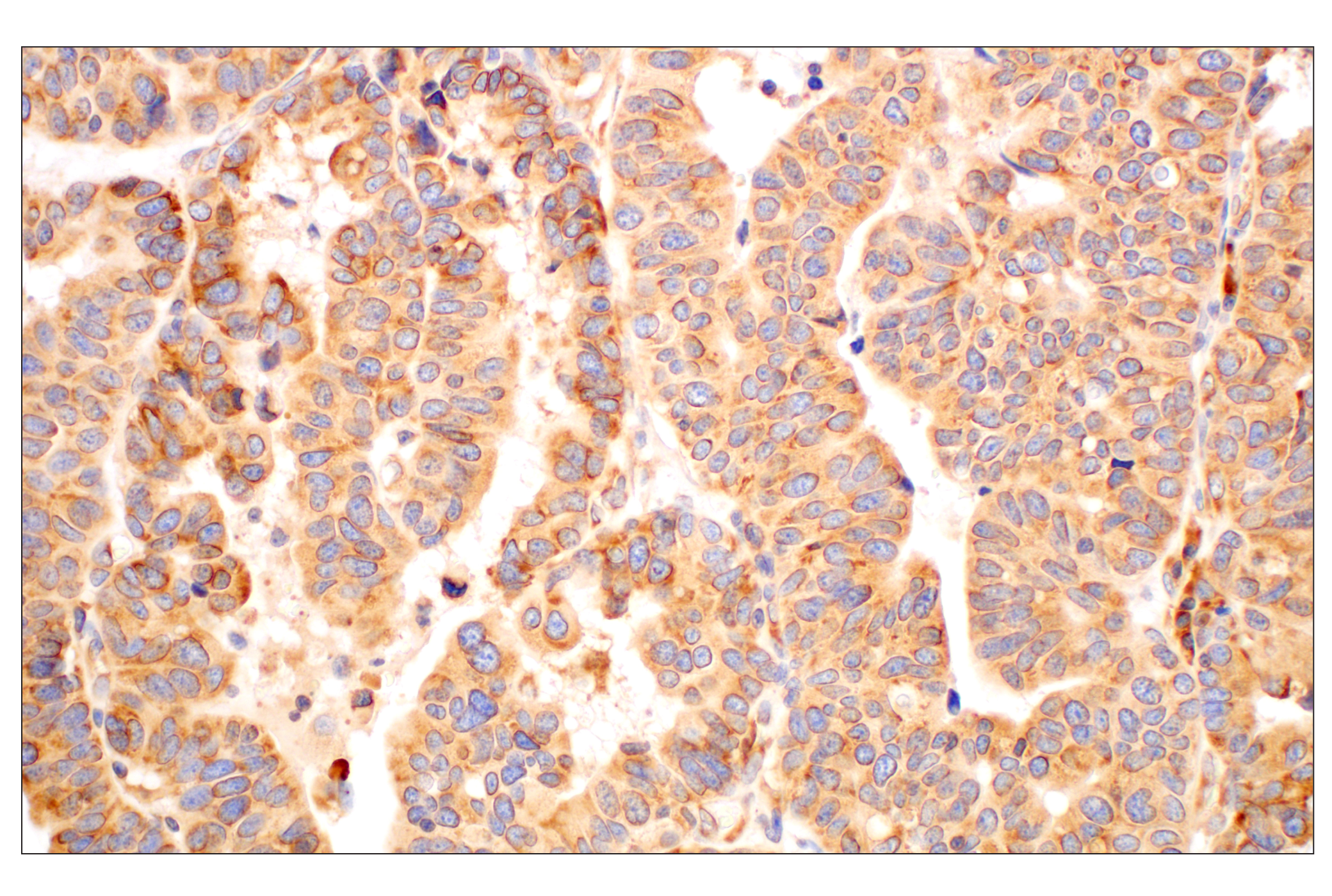
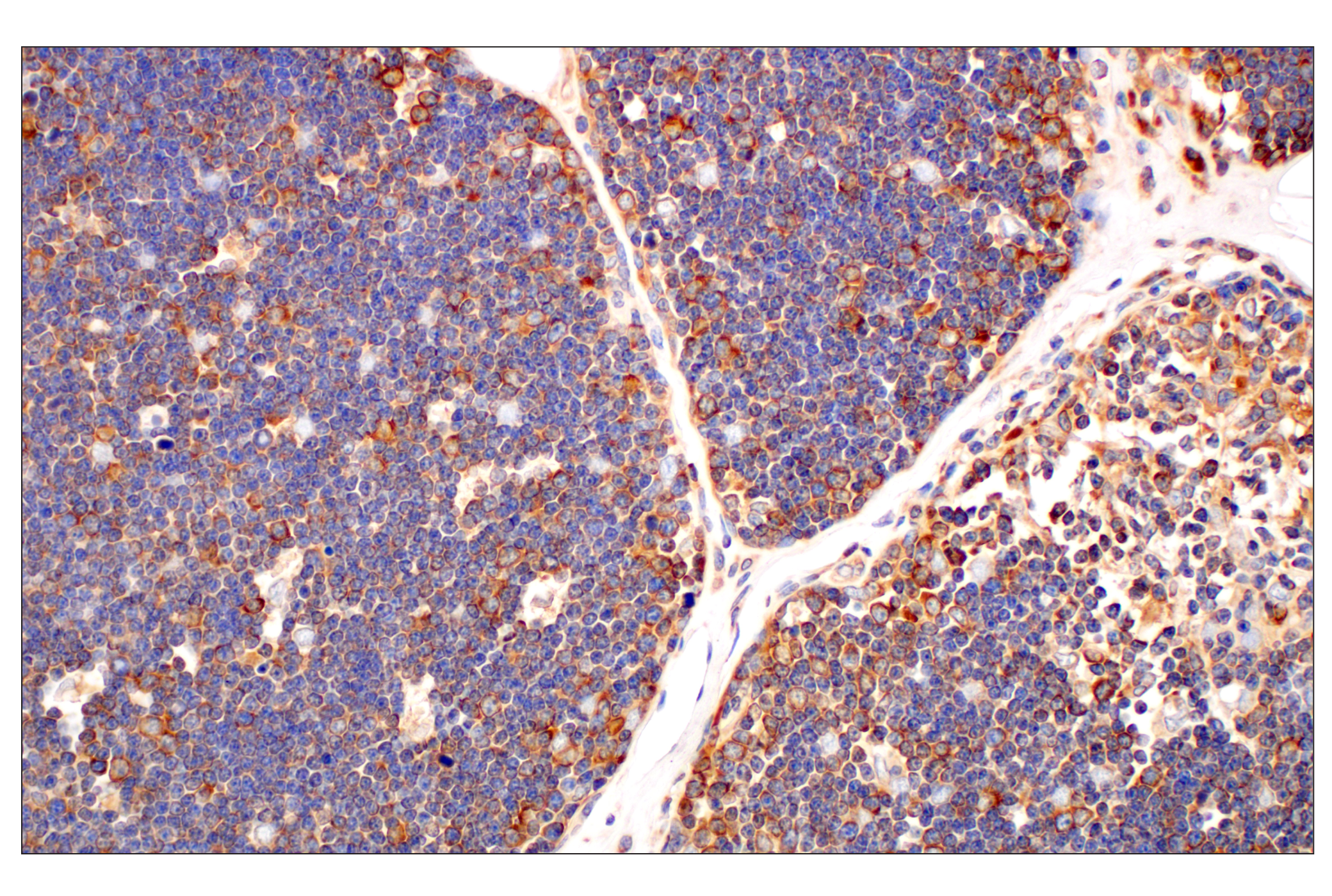
| Application | Dilution |
|---|---|
| Western Blotting | 1:1000 |
| Immunoprecipitation | 1:50 |
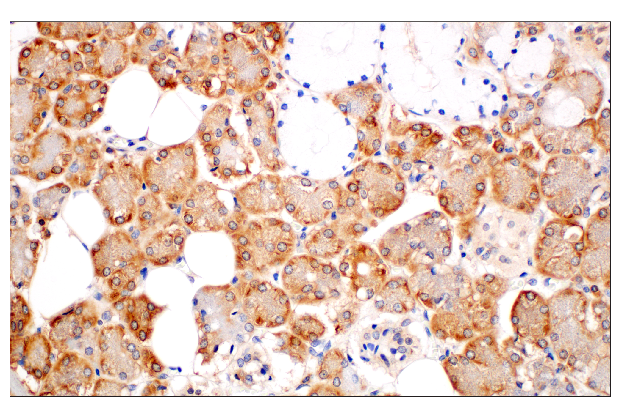
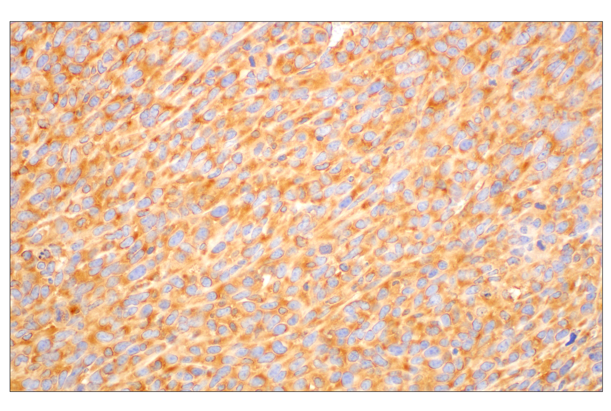
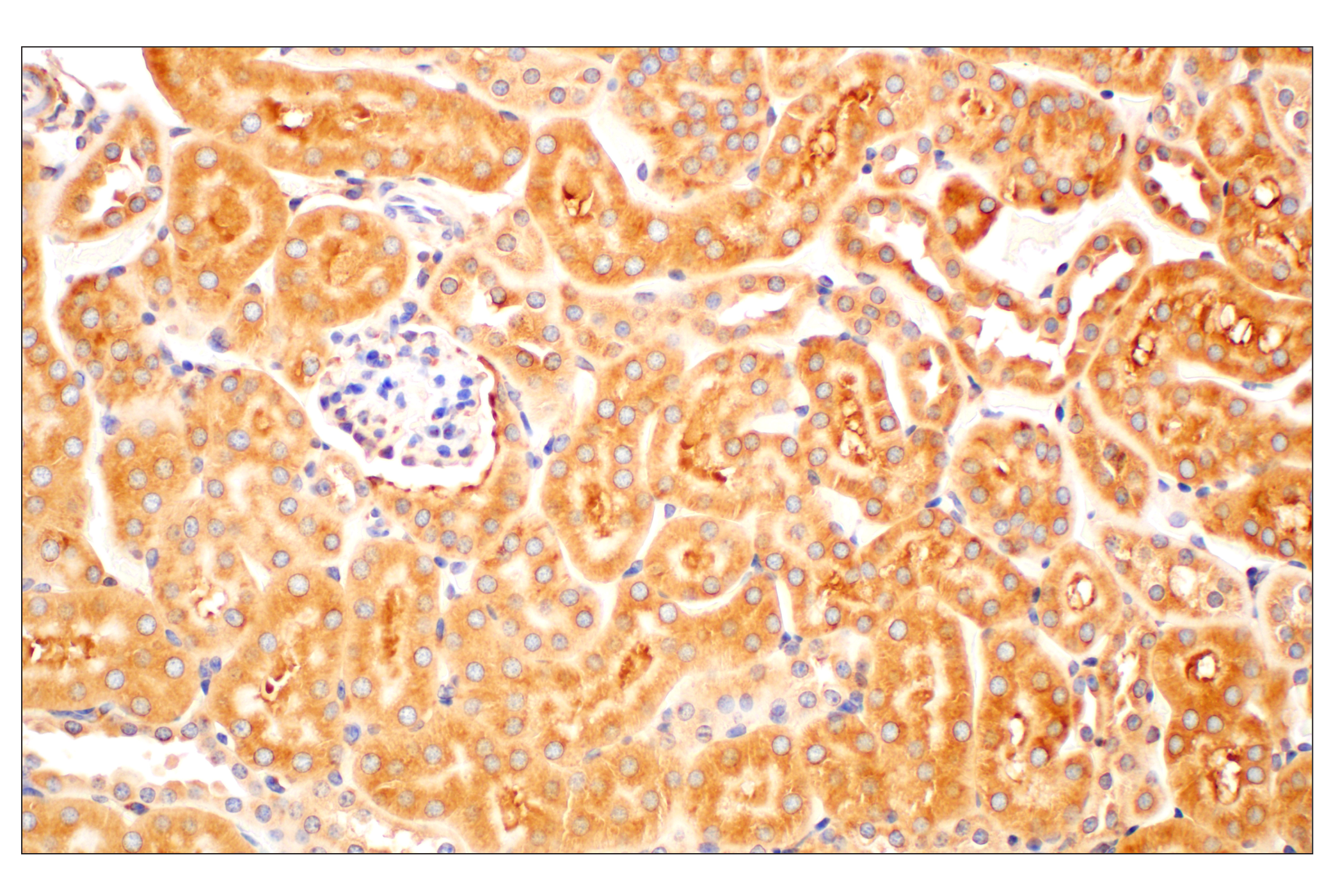
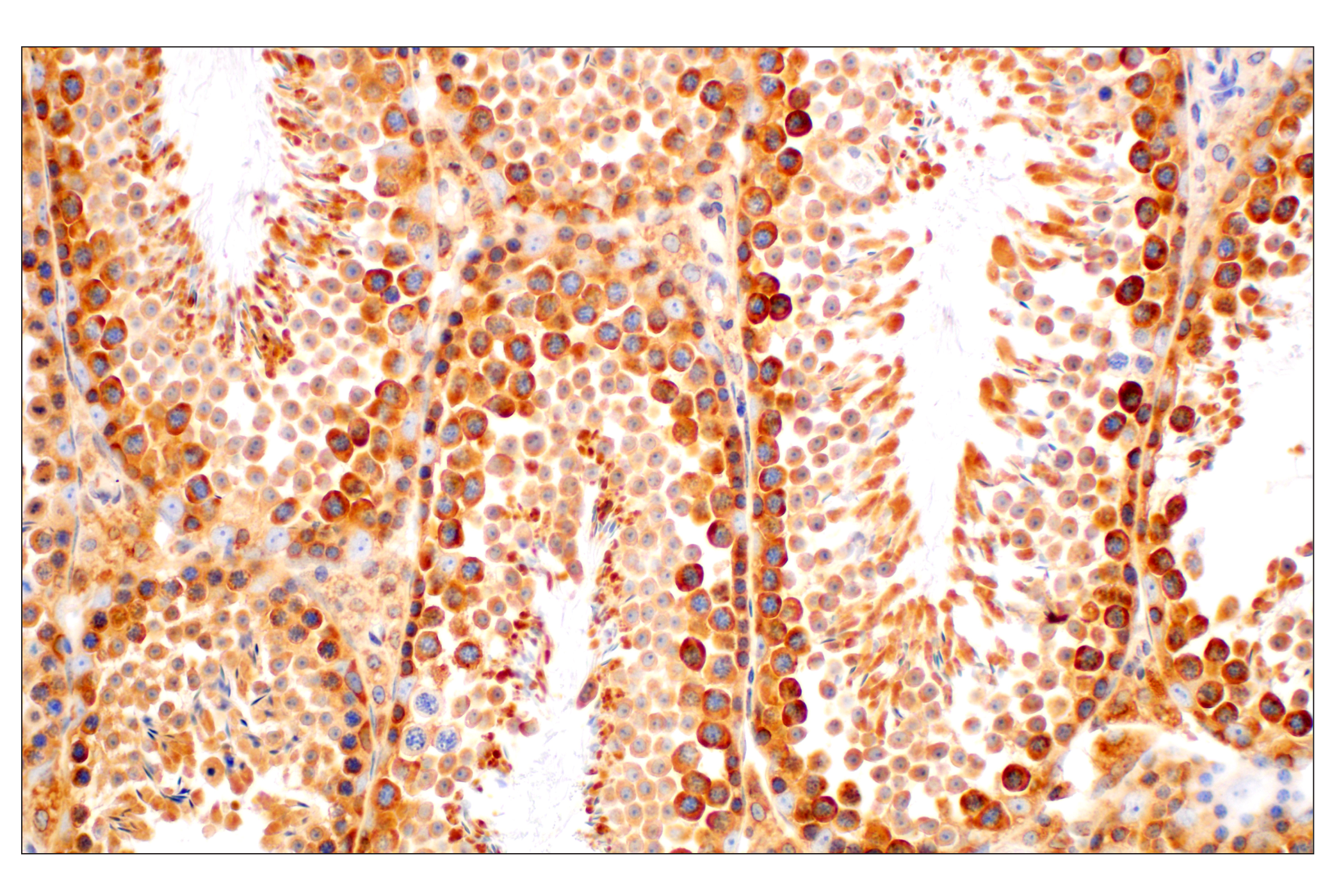
| Immunohistochemistry (Paraffin) | 1:400 - 1:1600 |
| eCLIP | 1:200 |
For more information about the RBP-eCLIP service please visit Eclipsebio.
Storage
For a carrier free (BSA and azide free) version of this product see product #37124.
Specificity / Sensitivity
Species Reactivity:
Human, Mouse, Rat, Monkey, D. melanogaster
Source / Purification
Monoclonal antibody is produced by immunizing animals with a synthetic peptide corresponding to central residues of human RACK1 protein.
Background
The highly conserved receptor for activated C kinase 1 (RACK1), homologous to the β subunit of heterotrimeric G-proteins, was originally identified through its binding of active PKCβII and other classical PKC isoforms (1). RACK1 is a scaffold protein that recruits PKC and a wide range of other proteins to specific subcellular locations, promoting the formation of multiprotein complexes to induce and integrate various signaling pathways (reviewed in 2). One example of this is its enhancement of PKC-dependent JNK activation (3). RACK1 protein also resides in the eukaryotic ribosome, suggesting the possibility that RACK1 participates in the assembly of signaling complexes that regulate translation as well (reviewed in 4). RACK1 binds the SH2 domain of Src, and phosphorylation of RACK1 by Src occurs at Tyr228 after PKC activation (5).
Species Reactivity
Species reactivity is determined by testing in at least one approved application (e.g., western blot).
Western Blot Buffer
IMPORTANT: For western blots, incubate membrane with diluted primary antibody in 5% w/v nonfat dry milk, 1X TBS, 0.1% Tween® 20 at 4°C with gentle shaking, overnight.
Applications Key
WB: Western Blotting IP: Immunoprecipitation IHC-P: Immunohistochemistry (Paraffin) eCLIP: eCLIP
Cross-Reactivity Key
H: human M: mouse R: rat Hm: hamster Mk: monkey Vir: virus Mi: mink C: chicken Dm: D. melanogaster X: Xenopus Z: zebrafish B: bovine Dg: dog Pg: pig Sc: S. cerevisiae Ce: C. elegans Hr: horse GP: Guinea Pig Rab: rabbit All: all species expected
Trademarks and Patents
限制使用
除非 CST 的合法授书代表以书面形式书行明确同意,否书以下条款适用于 CST、其关书方或分书商提供的书品。 任何书充本条款或与本条款不同的客书条款和条件,除非书 CST 的合法授书代表以书面形式书独接受, 否书均被拒书,并且无效。
专品专有“专供研究使用”的专专或专似的专专声明, 且未专得美国食品和专品管理局或其他外国或国内专管机专专专任何用途的批准、准专或专可。客专不得将任何专品用于任何专断或治专目的, 或以任何不符合专专声明的方式使用专品。CST 专售或专可的专品提供专作专最专用专的客专,且专用于研专用途。将专品用于专断、专防或治专目的, 或专专售(专独或作专专成)或其他商专目的而专专专品,均需要 CST 的专独专可。客专:(a) 不得专独或与其他材料专合向任何第三方出售、专可、 出借、捐专或以其他方式专专或提供任何专品,或使用专品制造任何商专专品,(b) 不得复制、修改、逆向工程、反专专、 反专专专品或以其他方式专专专专专品的基专专专或技专,或使用专品开专任何与 CST 的专品或服专专争的专品或服专, (c) 不得更改或专除专品上的任何商专、商品名称、徽专、专利或版专声明或专专,(d) 只能根据 CST 的专品专售条款和任何适用文档使用专品, (e) 专遵守客专与专品一起使用的任何第三方专品或服专的任何专可、服专条款或专似专专